


Rabbit PGAM5 Polyclonal Antibody | anti-PGAM5 antibody
PGAM5 (CT) Antibody
Antibody validated: Western Blot in human, mouse, and rat samples; Immunofluorescence in human, mouse, and rat samples. All other applications and species not yet tested.
The immunogen is located within the last 50 amino acids of PGAM5.
IF (Immunofluorescence)
(Figure 7 Immunofluorescence Validation of PGAM5 in Rat TestisImmunofluorescent analysis of 4% paraformaldehyde-fixed rat testis labeling PGAM5 with AAA11041 at 20ug/mL, followed by goat anti-rabbit IgG secondary antibody at 1/500 dilution (green) and DAPI staining (blue).)
IF (Immunofluorescence)
(Figure 6 Immunofluorescence Validation of PGAM5 in Mouse TestisImmunofluorescent analysis of 4% paraformaldehyde-fixed mouse testis labeling PGAM5 with AAA11041 at 10ug/mL, followed by goat anti-rabbit IgG secondary antibody at 1/500 dilution (green) and DAPI staining (blue).)
IF (Immunofluorescence)
(Figure 5 Immunofluorescence Validation of PGAM5 in Human LiverImmunofluorescent analysis of 4% paraformaldehyde-fixed human liver labeling PGAM5 with AAA11041 at 20ug/mL, followed by goat anti-rabbit IgG secondary antibody at 1/500 dilution (green) and DAPI staining (blue).)
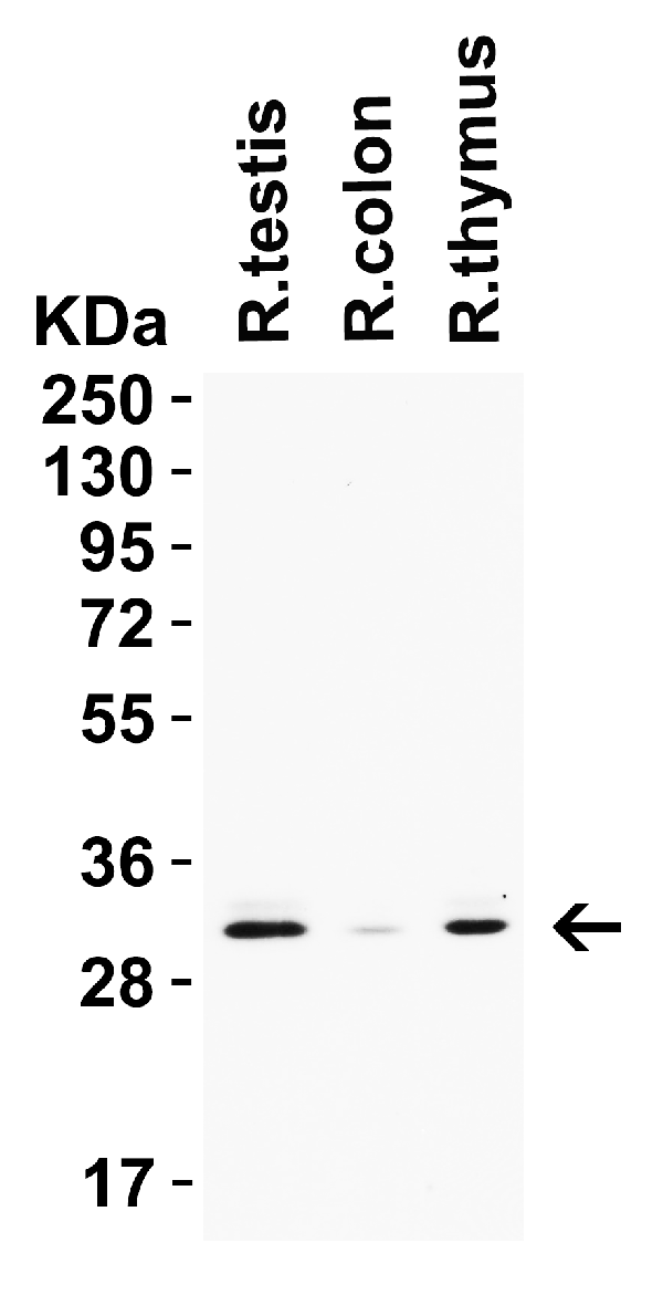
WB (Western Blot)
(Figure 4 Western Blot Validation in Rat Tissues Loading: 15ug of lysates per lane. Antibodies: PGAM5, AAA11041 0.5ug/mL, 1h incubation at RT in 5% NFDM/TBST. Secondary: Goat anti-rabbit IgG HRP conjugate at 1:10,000 dilution.)
WB (Western Blot)
(Figure 3 Western Blot Validation in Mouse Tissues Loading: 15ug of lysates per lane. Antibodies: PGAM5, AAA11041 0.5ug/mL, 1h incubation at RT in 5% NFDM/TBST. Secondary: Goat anti-rabbit IgG HRP conjugate at 1:10,000 dilution.)
WB (Western Blot)
(Figure 2 Western Blot Validation in Human Cell Lines Loading: 15ug of lysates per lane. Antibodies: PGAM5, AAA11041 0.5ug/mL, 1h incubation at RT in 5% NFDM/TBST. Secondary: Goat anti-rabbit IgG HRP conjugate at 1:10,000 dilution.)
WB (Western Blot)
(Figure 1 Western Blot Validation in Mouse and Rat Cell Lines Loading: 15ug of lysates per lane. Antibodies: PGAM5, AAA11041 0.5ug/mL, 1h incubation at RT in 5% NFDM/TBST. Secondary: Goat anti-rabbit IgG HRP conjugate at 1:10,000 dilution.)
Takeda et al. PNAS 2009; 106(30): 12301-12305
Want et al. Cell 2012; 148(1-2); 228-243