Filters
▼Clonality
▼Type
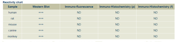
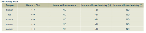
▼Reactivity
▼Gene Name
▼Isotype
▼Host
▼Application
▼Clone
▼Polyclonal Antibodies
At AAA Biotech also known as AAA Bio or AAABio, we provide a broad range of purified polyclonal antibodies (pAbs) that are able to all be browsed online through our website. Due to their high specificity and strong binding affinity, these antibodies are ideal for wide swathes of research and experimental applications.
Our polyclonal antibodies can easily support your work, whether you use them for Western Blotting, Immunocytochemistry (with or without Immunofluorescence used in conjunction), Immunohistochemistry, Immunoprecipitation, and ELISA tests. We highly encourage you to browse our range of pAbs and choose the one that best suits your experimental model.
Viewing 1-50 of 118597 product results
 Application Data
Goat Random Ab, Polyclonal Antibody (Cat# AAA63073)
 IF (Immunofluorescence)
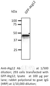
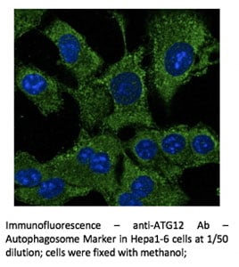
ATG12, Polyclonal Antibody (Cat# AAA63087)
 Application Data
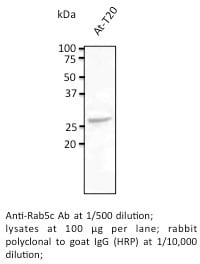
Rab5c, Polyclonal Antibody (Cat# AAA63090)
 Application Data
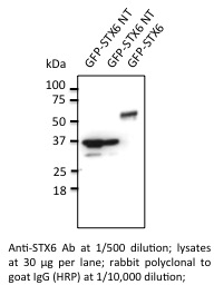
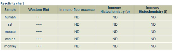
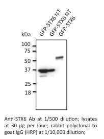
Syntaxin-6, Polyclonal Antibody (Cat# AAA63102)
 Application Data
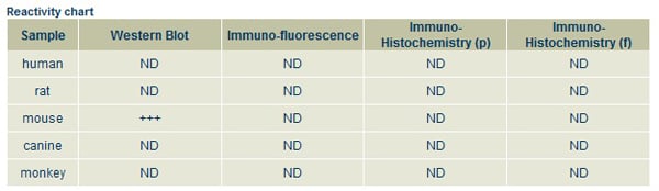
Mup20, Polyclonal Antibody (Cat# AAA63109)
 Application Data
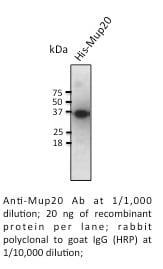
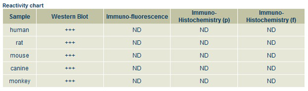
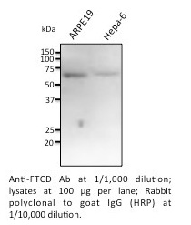
FTCD, Polyclonal Antibody (Cat# AAA63119)
 Application Data
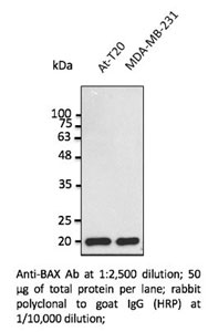
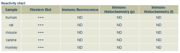
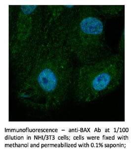
BAX, Polyclonal Antibody (Cat# AAA63121)
 Application Data
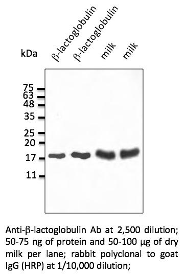
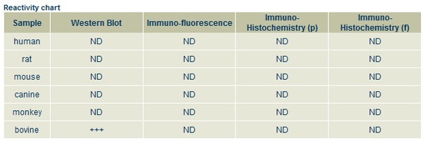
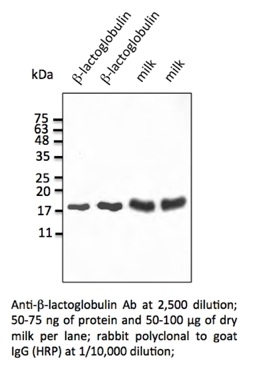
Lactoglobulin-beta, Polyclonal Antibody (Cat# AAA63123)
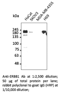 WB (Western Blot)
ERBB1, Polyclonal Antibody (Cat# AAA63134)
 WB (Western Blot)
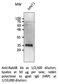
Rab38, Polyclonal Antibody (Cat# AAA63140)
 WB (Western Blot)
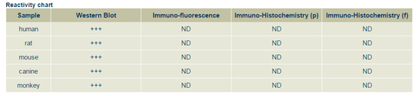
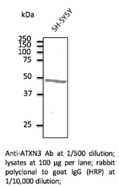
ATXN3, Polyclonal Antibody (Cat# AAA63143)

 Application Data
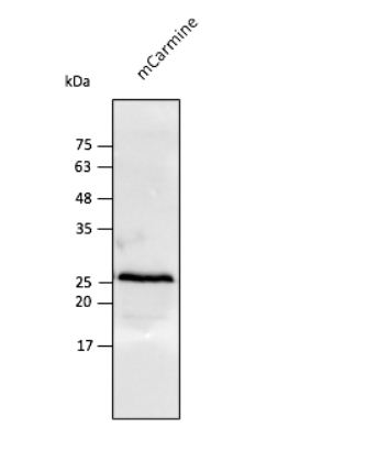
(Anti-mCarmine Ab at 1/2,500 dilution using HEK293 transfected cell lysates at 50 ug per lane; rabbit polyclonal to goat IgG (HRP) at 1/10,000 dilution;)
Application Data
(Anti-mCarmine Ab at 1/2,500 dilution using HEK293 transfected cell lysates at 50 ug per lane; rabbit polyclonal to goat IgG (HRP) at 1/10,000 dilution;)
mCarmine, Polyclonal Antibody (Cat# AAA63243)
 Application Data
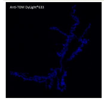
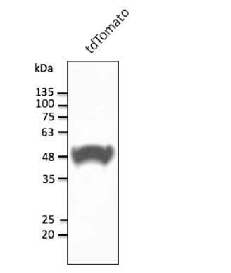
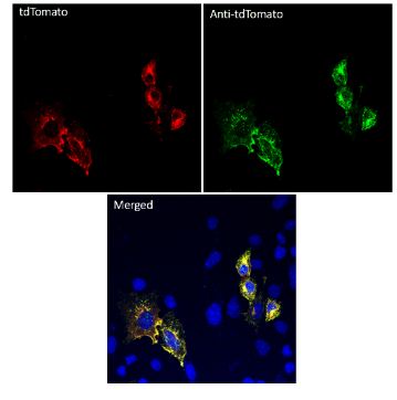
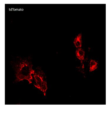
(Anti-tdTomato Ab conjugated to DyLight 633 at 1/2,500 dilution using HEK293 transfected cell lysates at 50 ug per lane;)
Application Data
(Anti-tdTomato Ab conjugated to DyLight 633 at 1/2,500 dilution using HEK293 transfected cell lysates at 50 ug per lane;)
tdTomato, Polyclonal Antibody (Cat# AAA63209)
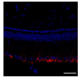
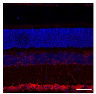 IS (Immunostaining)
(Immunostaining - C57BL/6J mouse retina label with: Red - 1st Ab GRP78 BiP (1/250); 2nd Ab goat anti-rat (AB28550, 1/1000), Blue - Nuclear staining (DAPI), scale bar = 40 um;)
IS (Immunostaining)
(Immunostaining - C57BL/6J mouse retina label with: Red - 1st Ab GRP78 BiP (1/250); 2nd Ab goat anti-rat (AB28550, 1/1000), Blue - Nuclear staining (DAPI), scale bar = 40 um;)
IgG, Polyclonal Secondary Antibody (Cat# AAA63222)
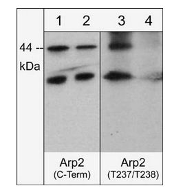
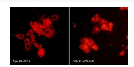 ICC (Immunocytochemistry)
(Immunocytochemical labeling of Arp2 phosphorylation in rat PC12 cells differentiated with NGF. The cells were probed with Arp2 (C-terminal region) and Arp2 (Thr-237/Thr-238) rabbit polyclonal antibodies, then the antibodies were detected using appropriate secondary antibody conjugated to Cy3.)
ICC (Immunocytochemistry)
(Immunocytochemical labeling of Arp2 phosphorylation in rat PC12 cells differentiated with NGF. The cells were probed with Arp2 (C-terminal region) and Arp2 (Thr-237/Thr-238) rabbit polyclonal antibodies, then the antibodies were detected using appropriate secondary antibody conjugated to Cy3.)
Arp2, Polyclonal Antibody (Cat# AAA71568)
 Application Data
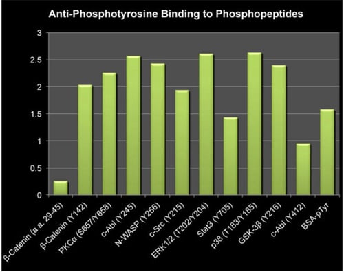
(Bar graph showing rabbit polyclonal anti-Phosphotyrosine binding to a variety of phosphotyrosine containing peptides, but no binding to unphosphorylated peptide (beta-Catenin (a.a. 29-45).)
Application Data
(Bar graph showing rabbit polyclonal anti-Phosphotyrosine binding to a variety of phosphotyrosine containing peptides, but no binding to unphosphorylated peptide (beta-Catenin (a.a. 29-45).)
Phosphotyrosine, Polyclonal Antibody (Cat# AAA71527)
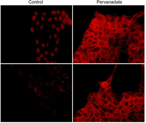
 ICC (Immunocytochemistry)
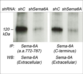
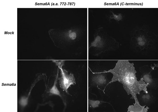
(Immunocytochemical labeling of Sema-6A in COS7 cells that were mock transfected (top images) or Sema-6A transfected (bottom images). The cells were labeled with anti-Sema-6A (a.a. 772-787) (Left top and bottom image) or anti-Sema-6A (C-terminus) (Right top and bottom image). The antibodies were detected using anti-rabbit fluorescent secondary antibody. (Images provided by Dr. Luca Tamagnone from the IRCC, University of Torino, Italy).)
ICC (Immunocytochemistry)
(Immunocytochemical labeling of Sema-6A in COS7 cells that were mock transfected (top images) or Sema-6A transfected (bottom images). The cells were labeled with anti-Sema-6A (a.a. 772-787) (Left top and bottom image) or anti-Sema-6A (C-terminus) (Right top and bottom image). The antibodies were detected using anti-rabbit fluorescent secondary antibody. (Images provided by Dr. Luca Tamagnone from the IRCC, University of Torino, Italy).)
Semaphorin-6A, Polyclonal Antibody (Cat# AAA71553)
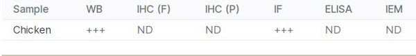
H5N1, Polyclonal Antibody (Cat# AAA71330)
 WB (Western Blot)
(WB: The highly purified Ovotranferrin from chicken egg white was immunoblotted by Rabbit anti Ovotransferrin antibody (AAA71336) at 1:500. An immunoreactive band is observed at ~80kDa.)
WB (Western Blot)
(WB: The highly purified Ovotranferrin from chicken egg white was immunoblotted by Rabbit anti Ovotransferrin antibody (AAA71336) at 1:500. An immunoreactive band is observed at ~80kDa.)
Ovotransferrin, Polyclonal Antibody (Cat# AAA71336)

 ICC (Immunocytochemistry)
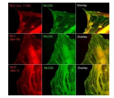
(Immunocytochemical labeling of phosphorylated MLC in paraformaldehyde fixed A7r5 cells. The cells were dual-labeled with anti-MLC (MM3441; middle) and anti-MLC (MP4201; top left), anti-MLC (Ser-19) (MP4221; middle left) and anti-MLC (Ser-1) (MP3461; bottom left). Goat anti-Mouse DyLight 488 and Goat anti-Rabbit DyLight 594 were used for detection of primary antibodies. The overlay of staining patterns are shown to the right.)
ICC (Immunocytochemistry)
(Immunocytochemical labeling of phosphorylated MLC in paraformaldehyde fixed A7r5 cells. The cells were dual-labeled with anti-MLC (MM3441; middle) and anti-MLC (MP4201; top left), anti-MLC (Ser-19) (MP4221; middle left) and anti-MLC (Ser-1) (MP3461; bottom left). Goat anti-Mouse DyLight 488 and Goat anti-Rabbit DyLight 594 were used for detection of primary antibodies. The overlay of staining patterns are shown to the right.)
Myosin, Polyclonal Antibody (Cat# AAA71663)
 ICC (Immunocytochemistry)
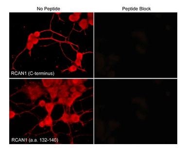
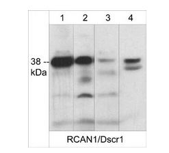
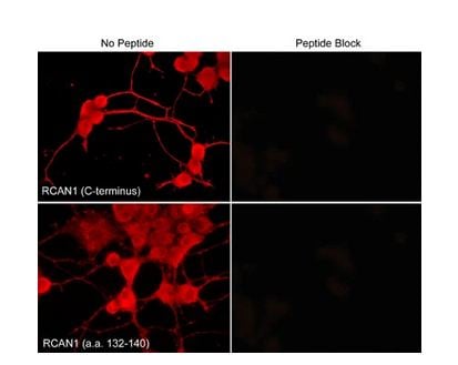
(Immunocytochemical labeling of RCAN1 in aldehyde-fixed NGF-differentiated PC12 cells. The cells were labeled with rabbit polyclonal anti-RCAN1 (C-terminus) (RP3941) and anti-RCAN1 (a.a. 132-140) (RP3961) antibodies (Left side). These antibodies were also used in the presence (Right side) of blocking peptide RX3945 and RX3965, respectively. The antibodies were detected using appropriate secondary antibody conjugated to DyLight 594.)
ICC (Immunocytochemistry)
(Immunocytochemical labeling of RCAN1 in aldehyde-fixed NGF-differentiated PC12 cells. The cells were labeled with rabbit polyclonal anti-RCAN1 (C-terminus) (RP3941) and anti-RCAN1 (a.a. 132-140) (RP3961) antibodies (Left side). These antibodies were also used in the presence (Right side) of blocking peptide RX3945 and RX3965, respectively. The antibodies were detected using appropriate secondary antibody conjugated to DyLight 594.)
RCAN1/Dscr1, Polyclonal Antibody (Cat# AAA71700)
 IF (Immunofluorescence)
(Immunofluorescence - anti-tdTomato Ab using hCEC cells transduced with tdTomato-CAV1; cells were fixed with methanol and anti-tdTomato at 1/250;)
IF (Immunofluorescence)
(Immunofluorescence - anti-tdTomato Ab using hCEC cells transduced with tdTomato-CAV1; cells were fixed with methanol and anti-tdTomato at 1/250;)
tdTomato, Polyclonal Antibody (Cat# AAA63250)
 Application Data
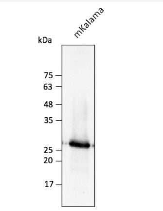
(Anti-mKalama Ab at 1/2,500 dilution using HEK293 transfected cell lysates at 30 ug per lane; rabbit polyclonal to goat IgG (HRP) at 1/10,000 dilution;)
Application Data
(Anti-mKalama Ab at 1/2,500 dilution using HEK293 transfected cell lysates at 30 ug per lane; rabbit polyclonal to goat IgG (HRP) at 1/10,000 dilution;)
mKalama, Polyclonal Antibody (Cat# AAA63261)
 Application Data
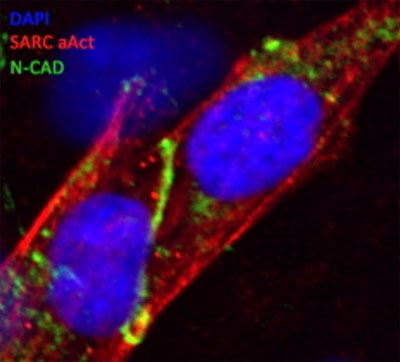
(N-Cadherin antibody marking the intercalated risk (distal cell junctions in heart tissue-green line in the middle) of Human iPS-dervived cardiomycocytes; Methanol fixation; DAPI: DNA (nuclear) stain, SARC aAct: Sacromeric a-Actinin, N_CAD: N-Cadherin)
Application Data
(N-Cadherin antibody marking the intercalated risk (distal cell junctions in heart tissue-green line in the middle) of Human iPS-dervived cardiomycocytes; Methanol fixation; DAPI: DNA (nuclear) stain, SARC aAct: Sacromeric a-Actinin, N_CAD: N-Cadherin)
CDH2, Polyclonal Antibody (Cat# AAA63053)
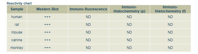
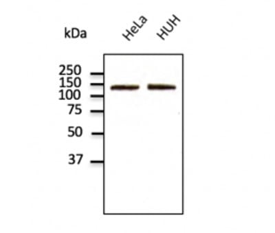
 WB (Western Blot)
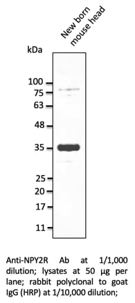
NPY2R, Polyclonal Antibody (Cat# AAA63163)
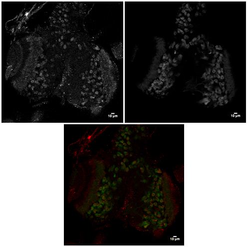
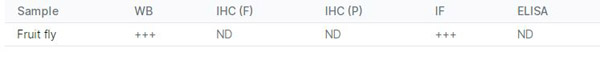
 IF (Immunofluorescence)
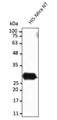
(Immunofluorescence - anti-Miranda Ab in fly cells at 1/100 dilution; InsGAL4 UASCD8 GFP wandering L3; cells were fixed with 4% of PFA;)
IF (Immunofluorescence)
(Immunofluorescence - anti-Miranda Ab in fly cells at 1/100 dilution; InsGAL4 UASCD8 GFP wandering L3; cells were fixed with 4% of PFA;)
Miranda, Polyclonal Antibody (Cat# AAA63170)
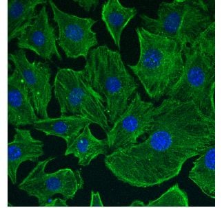 IF (Immunofluorescence)
(Immunofluorescence using hCEC cells, 1st Ab (anti-beta-Actin at 1/250) and 2nd Ab (anti-mouse AB27488 at 1/1,000); cells were fixed with methanol;)
IF (Immunofluorescence)
(Immunofluorescence using hCEC cells, 1st Ab (anti-beta-Actin at 1/250) and 2nd Ab (anti-mouse AB27488 at 1/1,000); cells were fixed with methanol;)
IgG, Polyclonal Secondary Antibody (Cat# AAA63218)
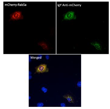
 IF (Immunofluorescence)
(Immunofluorescence - anti-mCherry Ab (AB9770) using hCEC cells transduced with mCherry-Rab5a; cells were fixed with methanol and mCherry Ab at 1/250; 2nd Ab goat anti-IgY (AB307488) at 1:1,000;)
IF (Immunofluorescence)
(Immunofluorescence - anti-mCherry Ab (AB9770) using hCEC cells transduced with mCherry-Rab5a; cells were fixed with methanol and mCherry Ab at 1/250; 2nd Ab goat anti-IgY (AB307488) at 1:1,000;)
IgY, Polyclonal Secondary Antibody (Cat# AAA63224)
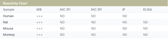
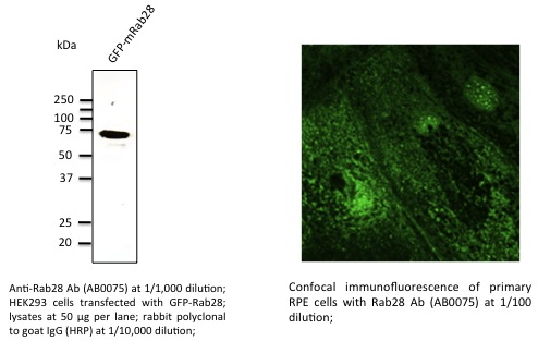 Application Data
Rab28, Polyclonal Antibody (Cat# AAA63069)
 Application Data
(Anti-TLR2 Ab at 1/500 dilution; Rabbit polyclonal to goat IgG (HRP) at 1/10,000 dilution.)
Application Data
(Anti-TLR2 Ab at 1/500 dilution; Rabbit polyclonal to goat IgG (HRP) at 1/10,000 dilution.)
TLR2, Polyclonal Antibody (Cat# AAA63076)
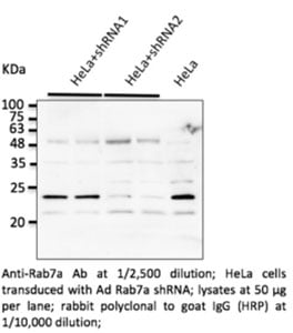
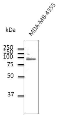
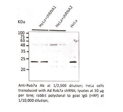 WB (Western Blot)
Rab7a, Polyclonal Antibody (Cat# AAA63091)
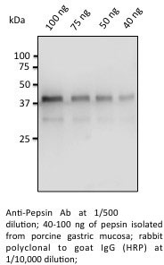
 Application Data
Pepsin, Polyclonal Antibody (Cat# AAA63097)
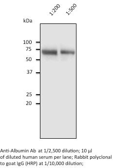
 Application Data
Albumin, Polyclonal Antibody (Cat# AAA63098)
 Application Data
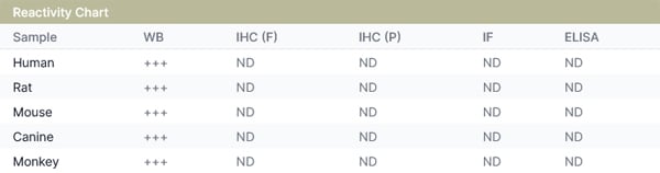
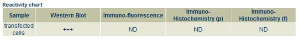
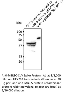
MERSC-CoV, Polyclonal Antibody (Cat# AAA63120)
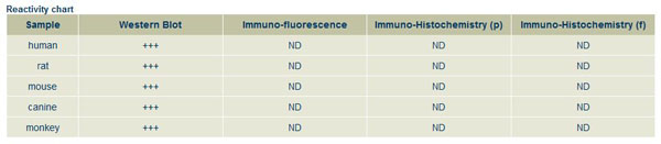
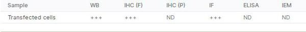
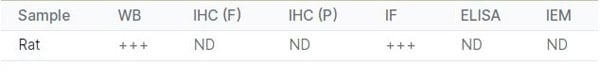
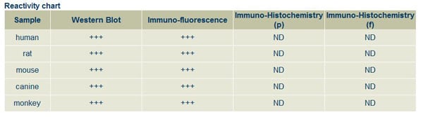
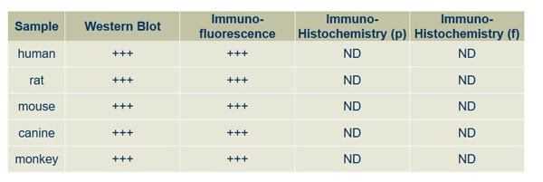
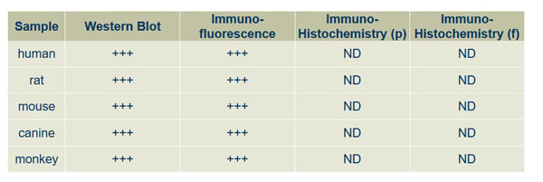
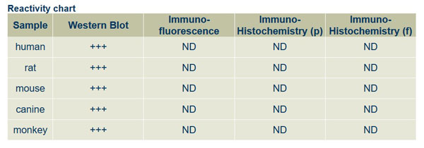
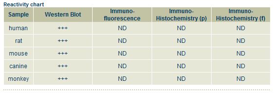
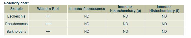
 Reactivity Data
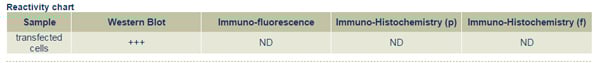
VSV-G Tag, Polyclonal Antibody (Cat# AAA63135)
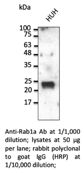 WB (Western Blot)
Rab1a, Polyclonal Antibody (Cat# AAA63146)
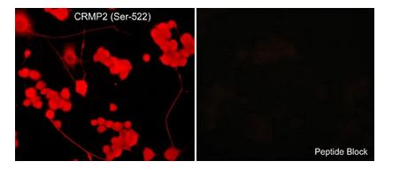 ICC (Immunocytochemistry)
(Immunocytochemical labeling of CRMP2 phosphorylation in aldehyde-fixed and NP-40-permeabilized NGF-differentiated PC12 cells. The cells were labeled with rabbit polyclonal anti-CRMP2 (Ser-522) (CP2191) antibody in the absence (Left) or presence (Right) of blocking peptide (CX2195). The antibody was detected using appropriate secondary antibody conjugated to DyLight 594.)
ICC (Immunocytochemistry)
(Immunocytochemical labeling of CRMP2 phosphorylation in aldehyde-fixed and NP-40-permeabilized NGF-differentiated PC12 cells. The cells were labeled with rabbit polyclonal anti-CRMP2 (Ser-522) (CP2191) antibody in the absence (Left) or presence (Right) of blocking peptide (CX2195). The antibody was detected using appropriate secondary antibody conjugated to DyLight 594.)
CRMP2, Polyclonal Antibody (Cat# AAA71604)
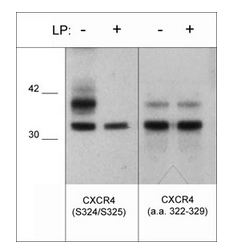
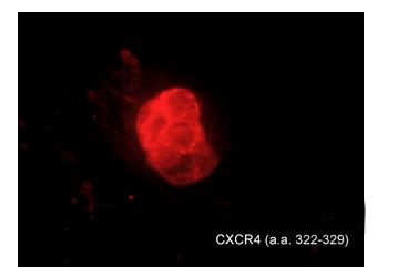 ICC (Immunocytochemistry)
(Immunocytochemical labeling of CXCR4 in chick pluripotent cells. The cells were labeled with rabbit polyclonal CXCR4 (a.a. 322-329) antibody (CP4211), then detected using appropriate secondary antibody (Red). (Image provided by Dr. Yangqing Lu at the Regenerative Bioscience Center, University of Georgia).)
ICC (Immunocytochemistry)
(Immunocytochemical labeling of CXCR4 in chick pluripotent cells. The cells were labeled with rabbit polyclonal CXCR4 (a.a. 322-329) antibody (CP4211), then detected using appropriate secondary antibody (Red). (Image provided by Dr. Yangqing Lu at the Regenerative Bioscience Center, University of Georgia).)
CXCR4, Polyclonal Antibody (Cat# AAA71613)
 ICC (Immunocytochemistry)
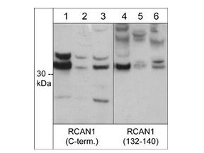
(Immunocytochemical labeling of RCAN1 in aldehyde-fixed NGF-differentiated PC12 cells. The cells were labeled with rabbit polyclonal anti-RCAN1 (C-terminus) (RP3941) and anti-RCAN1 (a.a. 132-140) (RP3961) antibodies (Left side). These antibodies were also used in the presence (Right side) of blocking peptide RX3945 and RX3965, respectively. The antibodies were detected using appropriate secondary antibody conjugated to DyLight 594.)
ICC (Immunocytochemistry)
(Immunocytochemical labeling of RCAN1 in aldehyde-fixed NGF-differentiated PC12 cells. The cells were labeled with rabbit polyclonal anti-RCAN1 (C-terminus) (RP3941) and anti-RCAN1 (a.a. 132-140) (RP3961) antibodies (Left side). These antibodies were also used in the presence (Right side) of blocking peptide RX3945 and RX3965, respectively. The antibodies were detected using appropriate secondary antibody conjugated to DyLight 594.)
RCAN1/Dscr1, Polyclonal Antibody (Cat# AAA71699)
 Application Data
EGFR, Polyclonal Antibody (Cat# AAA71518)
 Application Data
Plexin A1 (Sema Domain), Polyclonal Antibody (Cat# AAA71529)
Epoxide Hydrolase, Polyclonal Antibody (Cat# AAA71784)

 IF (Immunofluorescence)
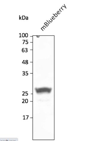
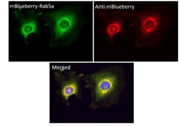
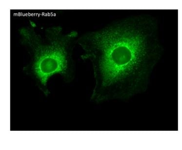
(Immunofluorescence -anti-mBlueberry Ab using hCEC cells transduced with mBlueberry-Rab5a; cells were fixed with methanol and anti-mBlueberry at 1/250;)
IF (Immunofluorescence)
(Immunofluorescence -anti-mBlueberry Ab using hCEC cells transduced with mBlueberry-Rab5a; cells were fixed with methanol and anti-mBlueberry at 1/250;)
mBlueberry, Polyclonal Antibody (Cat# AAA63242)
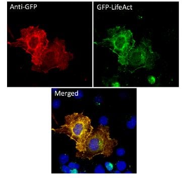
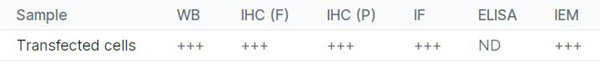
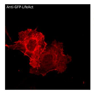 IF (Immunofluorescence)
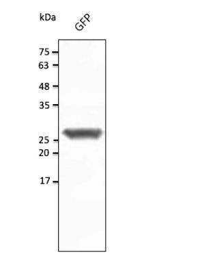
(Immunofluorescence - anti-GFP Ab using COS7 cells transduced with GFP; cells were fixed with methanol and anti-GFP at 1/250;)
IF (Immunofluorescence)
(Immunofluorescence - anti-GFP Ab using COS7 cells transduced with GFP; cells were fixed with methanol and anti-GFP at 1/250;)
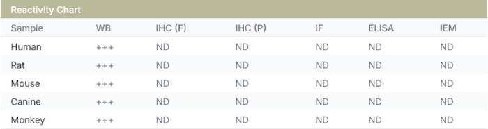
GFP, Polyclonal Antibody (Cat# AAA63257)
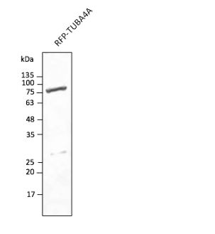
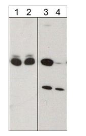
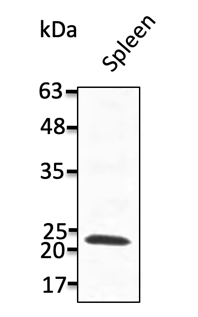 WB (Western Blot)
(Anti-TMEM170A Ab at 1/1,000 dilution; lysates at 50 ug per lane; rabbit polyclonal to goat IgG (HRP) at 1/10,000 dilution;)
WB (Western Blot)
(Anti-TMEM170A Ab at 1/1,000 dilution; lysates at 50 ug per lane; rabbit polyclonal to goat IgG (HRP) at 1/10,000 dilution;)
TMEM170A, Polyclonal Antibody (Cat# AAA63262)
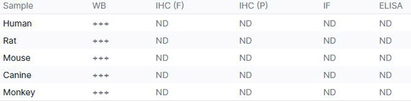
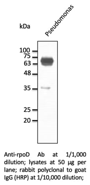 WB (Western Blot)
rpoD, Polyclonal Antibody (Cat# AAA63158)
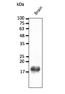 Application Data
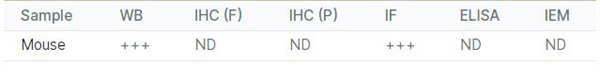
(Anti-SNCB Ab at 1/1,000 dilution; mouse brain lysate at 50 ug per lane; chicken polyclonal to goat IgG (HRP) at 1/10,000 dilution.)
Application Data
(Anti-SNCB Ab at 1/1,000 dilution; mouse brain lysate at 50 ug per lane; chicken polyclonal to goat IgG (HRP) at 1/10,000 dilution.)
SNCB, Polyclonal Antibody (Cat# AAA63166)
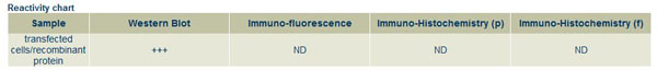
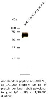
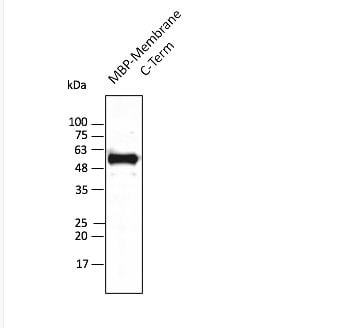 Application Data
(Anti-Membrane protein Ab at 1/2,500 dilution; lane with 30 ng of recombinant fusion protein (103-stop aa); rabbit polyclonal to goat IgG (HRP) at 1/10,000 dilution;)
Application Data
(Anti-Membrane protein Ab at 1/2,500 dilution; lane with 30 ng of recombinant fusion protein (103-stop aa); rabbit polyclonal to goat IgG (HRP) at 1/10,000 dilution;)
COVID 19 Membrane Protein Coronavirus, Polyclonal Antibody (Cat# AAA63193)
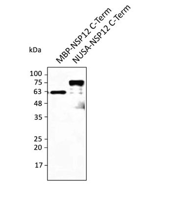 Application Data
(Anti-NSP12 Ab at 1/2,500 dilution; lanes with 30 ng of recombinant fusion protein (820 aa - stop); rabbit polyclonal to goat IgG (HRP) at 1/10,000 dilution)
Application Data
(Anti-NSP12 Ab at 1/2,500 dilution; lanes with 30 ng of recombinant fusion protein (820 aa - stop); rabbit polyclonal to goat IgG (HRP) at 1/10,000 dilution)
COVID 19 NSP12 Coronavirus, Polyclonal Antibody (Cat# AAA63196)
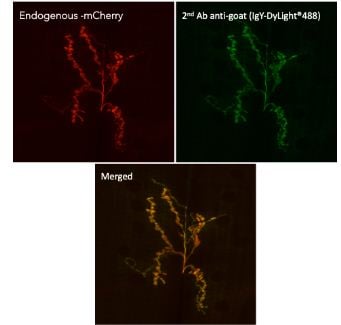 IF (Immunofluorescence)
(Immunofluorescence in Drosophila larvaenmJ muscle 6/7 expressing mCherry in neurons (DyGlutmcherry) using 1st AbAnti-mCherry at 1/1,000 and 2nd AbAnti-goat IgY conjugated to DyLight488 at 1/500;)
IF (Immunofluorescence)
(Immunofluorescence in Drosophila larvaenmJ muscle 6/7 expressing mCherry in neurons (DyGlutmcherry) using 1st AbAnti-mCherry at 1/1,000 and 2nd AbAnti-goat IgY conjugated to DyLight488 at 1/500;)
IgG, Polyclonal Secondary Antibody (Cat# AAA63210)
What are Polyclonal Antibodies?
Polyclonal antibodies are antibodies that come from multiple B cell clones of a host animal. The typical hosts used for the majority of polyclonal antibody production are rabbits, goats, sheep, and donkeys. These polyclonal antibodies, once having identified their target, will bind to different epitopes located at different regions or sequences on the same protein/antigen. This ability to bind multiple epitopes is what makes polyclonal antibodies highly sensitive, as explained in our detailed guide on polyclonal antibodies and why they matter.
As a result, they are ideal at locating and binding to the target, even if the target is in very low concentrations (due to many different antibodies being able to bind to the same target molecule, which allows for significant amplification of a downstream signal).
Polyclonal antibodies are typically produced by injecting an antigen into a host animal, which causes the animal’s immune system to attack the foreign antigen by mass generating antibodies against it. After a period of time, serum is collected from the animal and purified using physicochemical fractionation, class-specific affinity purification, and/or antigen-affinity purification.
Key Uses of Polyclonal Antibodies
- Western Blotting: This method is used to find specific proteins in biological samples after separating them by size.
- Immunohistochemistry: IHC helps visualize the location of proteins in tissue sections using various staining techniques.
- ELISA: (Enzyme-Linked Immunosorbent Assay) is typically used to identify specific protein quantities in a sample. ELISAs can be either “Quantitative” or “Qualitative”.
- Flow Cytometry: technique that identifies and measures the specific protein on the surface or inside the cells in a fluid suspension.
- Immunoprecipitation: IP isolates and studies a specific protein from a complex mixture using antibodies.
Why Buy Polyclonal Antibodies from AAA Biotech?
1. Ideal for Various Applications
Our antibodies are generally going to be validated for use in multiple types of assays, including ELISA, Western Blotting, Immunohistochemistry, Immunoprecipitation, amongst others. They are ideal for a wide range of research applications.
2. Rigorous Quality Control
All of the antibodies in our catalog undergo strict quality testing to ensure specificity, sensitivity, and consistent performance. We are confident in the ability of our antibodies to provide you with accurate results.
3. Wide Assortment of Antibodies
Antibodies in our catalog can be found for both common and exotic species, and these antibodies are also available in both conjugated and recombinant forms to suit many diverse experimental needs.
4. Highly Purified
Our antibodies are available in purified forms with over 85% purity, as confirmed by SDS-PAGE. They are also available with tags such as His, Flag, GST, or MBP. We cater to customers worldwide.
FAQ
1. How are polyclonal antibodies produced?
Traditionally, polyclonal antibodies are produced by injecting an antigen into a host animal (such as a rabbit or goat), which then triggers an immune response from the host animal. The animal’s B cells produce antibodies that will recognize different parts of the injected antigen. These antibodies are then collected from the animal’s blood and purified for use.
2. How do polyclonal antibodies differ from monoclonal antibodies?
Polyclonal antibodies are a mix of antibodies that bind to different locations (epitopes) of the same antigen, while monoclonal antibodies are identical and bind to just one specific epitope. This makes polyclonal antibodies more versatile and better at detecting proteins that may be present in low quantities or in altered/modified forms.
3. How should I store polyclonal antibodies?
Polyclonal antibodies should be stored at 4°C for short-term use (up to a few weeks) and at -20°C or -80°C for long-term storage. Avoid repeated freeze-thaw cycles by dividing them into small aliquots. Always check the datasheet for specific storage instructions.