
Filters
▼Clonality
▼Type
▼Reactivity
▼Gene Name
▼Isotype
▼Host
▼Application
▼Clone
▼Recombinant Antibodies
Also available for purchase in our catalog, we have flexible recombinant antibodies that are excellent tools in many different types of experiments, and well-suited for those that are large-scale or long-term. Our collection of recombinant antibodies includes different forms/formats of said antibodies, such as labeled or purified versions, and can be made utilizing different species’ isotypes to suit various experimental requirements.
Viewing 1-50 of 3337 product results
 IF (Immunofluorescence)
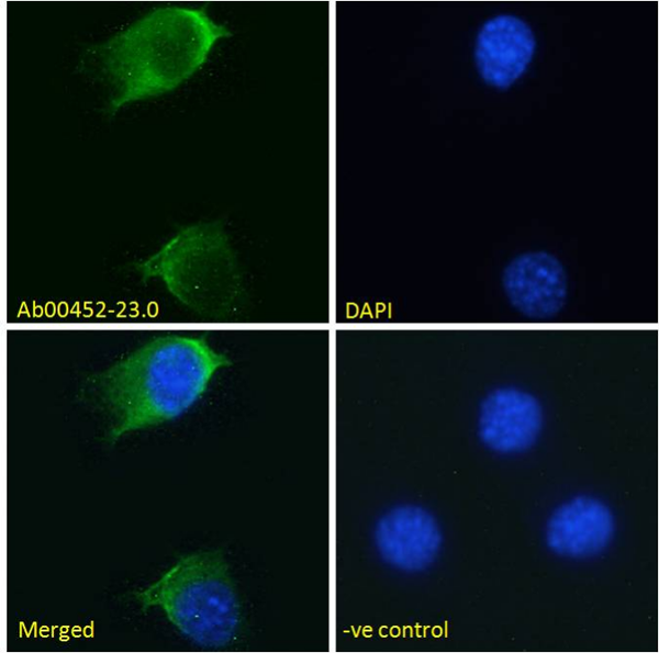
( Immunofluorescence staining of fixed NIH3T3 cells with anti-EGFR antibody S3 12D Immunofluorescence analysis of unpermeabilisd paraformaldehyde fixed NIH3T3 cells on Shi-fix coverslips stained with the chimeric rabbit version of S3 12D at 10 ug/ml for 1h followed by Alexa Fluor 488 secondary antibody (1 ug/ml), showing membrane staining. The nuclear stain is DAPI (blue). Panels show from left-right, top-bottom DAPI, merged channels and an isotype control. The isotype control was stained with unimmunised rabbit IgG followed by Alexa Fluor 488 secondary antibody.)
IF (Immunofluorescence)
( Immunofluorescence staining of fixed NIH3T3 cells with anti-EGFR antibody S3 12D Immunofluorescence analysis of unpermeabilisd paraformaldehyde fixed NIH3T3 cells on Shi-fix coverslips stained with the chimeric rabbit version of S3 12D at 10 ug/ml for 1h followed by Alexa Fluor 488 secondary antibody (1 ug/ml), showing membrane staining. The nuclear stain is DAPI (blue). Panels show from left-right, top-bottom DAPI, merged channels and an isotype control. The isotype control was stained with unimmunised rabbit IgG followed by Alexa Fluor 488 secondary antibody.)
EGFR, Monoclonal Recombinant Antibody (Cat# AAA71995)
 IF (Immunofluorescence)
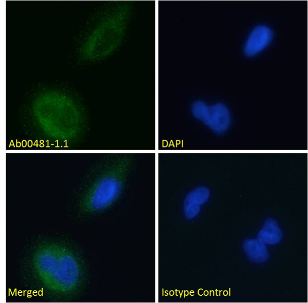
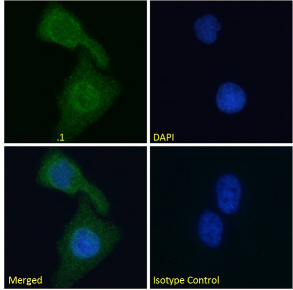
( Immunofluorescence staining of fixed U251 cells with anti-BACE2 antibody 1/9 (Ab00481) Immunofluorescence analysis of paraformaldehyde fixed U251 cells permeabilized with 0.15% Triton and stained with the chimeric mouse IgG1 version of 1/9 (Ab00481-1.1) at 10 ug/ml for 1h followed by Alexa Fluor® 488 secondary antibody (2 ug/ml), showing cytoplasmic staining. The nuclear stain is DAPI (blue). Panels show from left-right, top-bottom Ab00481-1.1, DAPI, merged channels and an isotype control. The isotype control was stained with an anti-unknown specificity antibody (Ab178-1.1) followed by Alexa Fluor® 488 secondary antibody.)
IF (Immunofluorescence)
( Immunofluorescence staining of fixed U251 cells with anti-BACE2 antibody 1/9 (Ab00481) Immunofluorescence analysis of paraformaldehyde fixed U251 cells permeabilized with 0.15% Triton and stained with the chimeric mouse IgG1 version of 1/9 (Ab00481-1.1) at 10 ug/ml for 1h followed by Alexa Fluor® 488 secondary antibody (2 ug/ml), showing cytoplasmic staining. The nuclear stain is DAPI (blue). Panels show from left-right, top-bottom Ab00481-1.1, DAPI, merged channels and an isotype control. The isotype control was stained with an anti-unknown specificity antibody (Ab178-1.1) followed by Alexa Fluor® 488 secondary antibody.)
BACE2, Monoclonal Recombinant Antibody (Cat# AAA71996)



 WB (Western Blot)
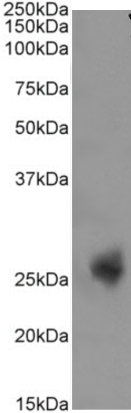
( Western Blot using anti-CD3E antibody YTH 12.5 Human spleen sample (35ug protein in RIPA buffer) were resolved on a 10% SDS PAGE gel and blots probed with the chimeric rabbit version of YTH 12.5 at 0.3 ug/ml before detection using an anti-rabbit secondary antibody. A primary incubation of 1h was used and protein was detected by chemiluminescence. The expected band size for CD3E is 23.1 kDa. successfully detected human CD3E.)
WB (Western Blot)
( Western Blot using anti-CD3E antibody YTH 12.5 Human spleen sample (35ug protein in RIPA buffer) were resolved on a 10% SDS PAGE gel and blots probed with the chimeric rabbit version of YTH 12.5 at 0.3 ug/ml before detection using an anti-rabbit secondary antibody. A primary incubation of 1h was used and protein was detected by chemiluminescence. The expected band size for CD3E is 23.1 kDa. successfully detected human CD3E.)
CD3 epsilon, Monoclonal Recombinant Antibody (Cat# AAA71963)
Protein A Affinity Purified
CD22, Monoclonal Recombinant Antibody (Cat# AAA71979)
 IF (Immunofluorescence)
( Immunofluorescence staining of fixed mouse splenocytes with anti-PDL1 (CD274) antibody YDC 127.1.1 Immunofluorescence analysis of paraformaldehyde fixed mouse (Mus musculus) splenocytes on Shi-fix coverslips, permeabilized with 0.15% Triton stained with the chimeric rabbit version of YDC 127.1.1 at 10 ug/ml for 1h followed by Alexa Fluor 488 secondary antibody (1 ug/ml), showing membrane and some cytoplasmic staining in a subset of cells. The nuclear stain is DAPI (blue). Panels show from left-right, top-bottom DAPI, merged channels and an isotype control. The isotype control was stained with an anti-Fluorescein antibody followed by Alexa Fluor 488 secondary antibody.)
IF (Immunofluorescence)
( Immunofluorescence staining of fixed mouse splenocytes with anti-PDL1 (CD274) antibody YDC 127.1.1 Immunofluorescence analysis of paraformaldehyde fixed mouse (Mus musculus) splenocytes on Shi-fix coverslips, permeabilized with 0.15% Triton stained with the chimeric rabbit version of YDC 127.1.1 at 10 ug/ml for 1h followed by Alexa Fluor 488 secondary antibody (1 ug/ml), showing membrane and some cytoplasmic staining in a subset of cells. The nuclear stain is DAPI (blue). Panels show from left-right, top-bottom DAPI, merged channels and an isotype control. The isotype control was stained with an anti-Fluorescein antibody followed by Alexa Fluor 488 secondary antibody.)
PDL1, Monoclonal Recombinant Antibody (Cat# AAA71990)
 FCM/FACS (Flow Cytometry)
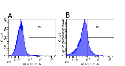
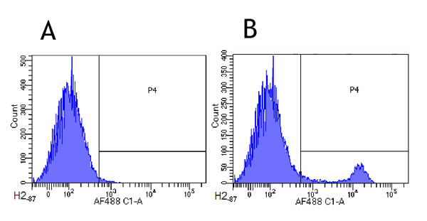
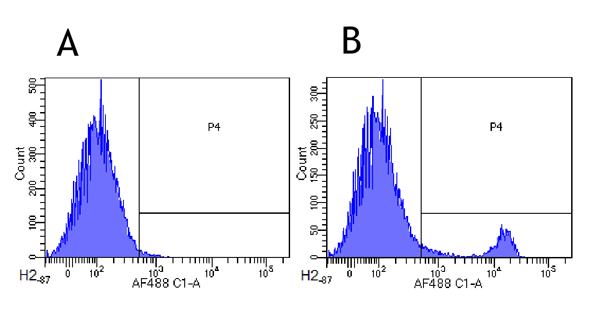
(Flow-cytometry using anti-CD8alpha/Lyt-2 antibody YTS 169.4 Mouse lymphocytes were stained with an anti-CD44 control or the rabbit-chimeric version of YTS 169.4 (AAA72017, panel B) at a concentration of 1 ug/ml for 30 mins at RT. After washing, bound antibody was detected using a AF488 conjugated donkey anti-rabbit antibody and cells analysed on a FACSCanto flow-cytometer.)
FCM/FACS (Flow Cytometry)
(Flow-cytometry using anti-CD8alpha/Lyt-2 antibody YTS 169.4 Mouse lymphocytes were stained with an anti-CD44 control or the rabbit-chimeric version of YTS 169.4 (AAA72017, panel B) at a concentration of 1 ug/ml for 30 mins at RT. After washing, bound antibody was detected using a AF488 conjugated donkey anti-rabbit antibody and cells analysed on a FACSCanto flow-cytometer.)
CD8 alpha/Lyt-2, Monoclonal Recombinant Antibody (Cat# AAA72017)
 WB (Western Blot)
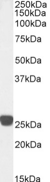
(Western Blot using anti-Cardiac Troponin I antibody scFv 180 Human heart lysate (35ug protein in RIPA buffer) was resolved on a 10% SDS PAGE gel and blots probed with the chimeric mouse IgG1 version of scFv 180 at 0.001 ug/ml before detection using an anti-mouse secondary antibody. A primary incubation of 1h was used and protein was detected by chemiluminescence. The predicted running size for Cardiac Troponin I is 24.0 kDa. It successfully detected Cardiac Troponin I in human heart lysate.)
WB (Western Blot)
(Western Blot using anti-Cardiac Troponin I antibody scFv 180 Human heart lysate (35ug protein in RIPA buffer) was resolved on a 10% SDS PAGE gel and blots probed with the chimeric mouse IgG1 version of scFv 180 at 0.001 ug/ml before detection using an anti-mouse secondary antibody. A primary incubation of 1h was used and protein was detected by chemiluminescence. The predicted running size for Cardiac Troponin I is 24.0 kDa. It successfully detected Cardiac Troponin I in human heart lysate.)
Cardiac Troponin I, Monoclonal Recombinant Antibody (Cat# AAA72031)

 Application Data
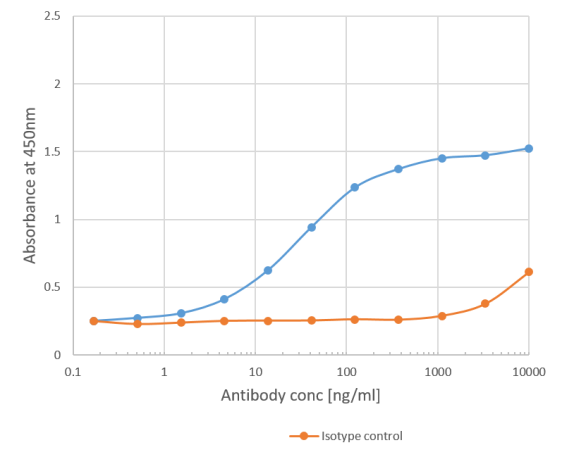
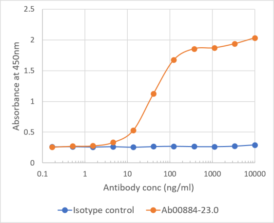
(Binding curve of anti-Phl p 1 antibody Clone 25 to Phl p 1 ELISA Plate coated with Phl p 1 (RayBiotech, 228-22412) at a concentration of 2 ug/ml. A 3-fold serial dilution from 10,000 to 0.1 ng/ml was performed using AAA72039. For detection, a 1:4000 dilution of HRP-labelled goat anti-human kappa antibody (Bio-Rad) was used.)
Application Data
(Binding curve of anti-Phl p 1 antibody Clone 25 to Phl p 1 ELISA Plate coated with Phl p 1 (RayBiotech, 228-22412) at a concentration of 2 ug/ml. A 3-fold serial dilution from 10,000 to 0.1 ng/ml was performed using AAA72039. For detection, a 1:4000 dilution of HRP-labelled goat anti-human kappa antibody (Bio-Rad) was used.)
Phl p 1, Monoclonal Recombinant Antibody (Cat# AAA72039)
Muscle Acetylcholine Receptor, Monoclonal Recombinant Antibody (Cat# AAA71993)
 IF (Immunofluorescence)
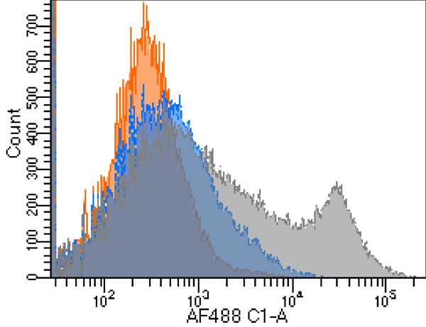
( Immunofluorescence staining of fixed mouse splenocytes with anti-GITR antibody Immunofluorescence analysis of paraformaldehyde fixed mouse (Mus musculus) splenocytes on Shi-fix coverslips, permeabilized with 0.15% Triton stained with the chimeric rabbit version at 10 ug/ml for 1h followed by Alexa Fluor 488 secondary antibody (1 ug/ml), showing membrane staining in a subset of cells. The nuclear stain is DAPI (blue). Panels show from left-right, top-bottom DAPI, merged channels and an isotype control. The isotype control was stained with an anti-Fluorescein antibody followed by Alexa Fluor 488 secondary antibody.)
IF (Immunofluorescence)
( Immunofluorescence staining of fixed mouse splenocytes with anti-GITR antibody Immunofluorescence analysis of paraformaldehyde fixed mouse (Mus musculus) splenocytes on Shi-fix coverslips, permeabilized with 0.15% Triton stained with the chimeric rabbit version at 10 ug/ml for 1h followed by Alexa Fluor 488 secondary antibody (1 ug/ml), showing membrane staining in a subset of cells. The nuclear stain is DAPI (blue). Panels show from left-right, top-bottom DAPI, merged channels and an isotype control. The isotype control was stained with an anti-Fluorescein antibody followed by Alexa Fluor 488 secondary antibody.)
GITR, Monoclonal Recombinant Antibody (Cat# AAA71998)
 IF (Immunofluorescence)
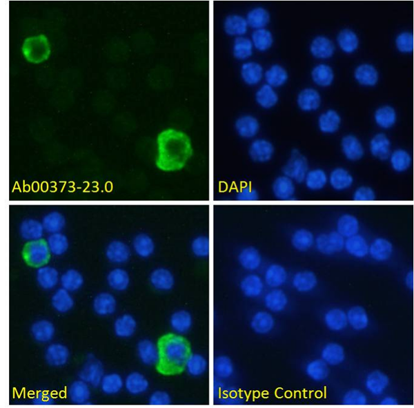
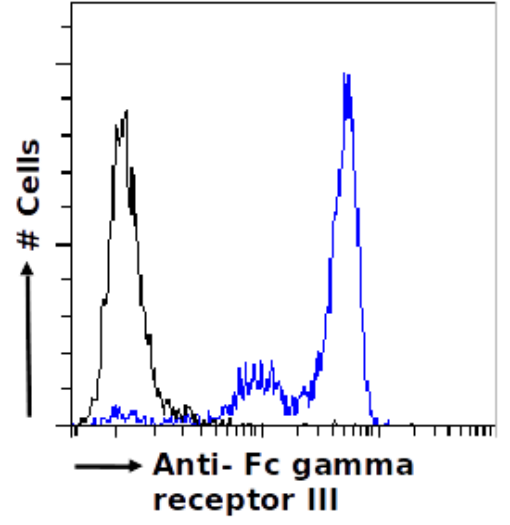
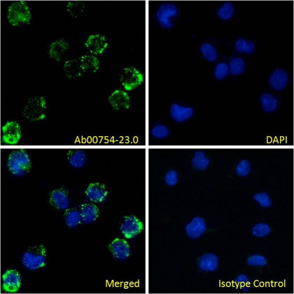
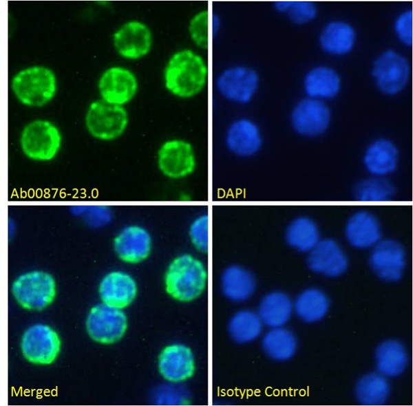
( Immunofluorescence staining of fixed U937 cells with anti-Fc gamma receptor III antibody 3G8 (Ab00754) Immunofluorescence analysis of paraformaldehyde fixed U937 cells permeabilized with 0.15% Triton and stained with the chimeric mouse IgG1 version of 3G8 (Ab00754-23.0) at 10 ug/ml for 1h followed by Alexa Fluor® 488 secondary antibody (2 ug/ml), showing membrane staining. The nuclear stain is DAPI (blue). Panels show from left-right, top-bottom Ab00754-23.0, DAPI, merged channels and an isotype control. The isotype control was stained with an anti-unknown specificity antibody (Ab178-23.0) followed by Alexa Fluor® 488 secondary antibody.)
IF (Immunofluorescence)
( Immunofluorescence staining of fixed U937 cells with anti-Fc gamma receptor III antibody 3G8 (Ab00754) Immunofluorescence analysis of paraformaldehyde fixed U937 cells permeabilized with 0.15% Triton and stained with the chimeric mouse IgG1 version of 3G8 (Ab00754-23.0) at 10 ug/ml for 1h followed by Alexa Fluor® 488 secondary antibody (2 ug/ml), showing membrane staining. The nuclear stain is DAPI (blue). Panels show from left-right, top-bottom Ab00754-23.0, DAPI, merged channels and an isotype control. The isotype control was stained with an anti-unknown specificity antibody (Ab178-23.0) followed by Alexa Fluor® 488 secondary antibody.)
Fc gamma receptor III, Monoclonal Recombinant Antibody (Cat# AAA72003)
 FCM/FACS (Flow Cytometry)
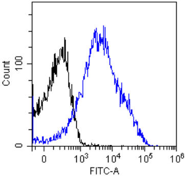
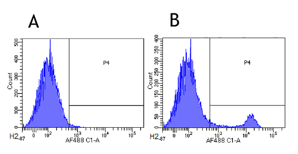
(Flow-cytometry using anti-CD8alpha/Lyt-2 antibody YTS 169.4 Mouse lymphocytes were stained with an anti-CD44 control or the rabbit-chimeric version of YTS 169.4 at a concentration of 1 ug/ml for 30 mins at RT. After washing, bound antibody was detected using a AF488 conjugated donkey anti-rabbit antibody and cells analysed on a FACSCanto flow-cytometer.)
FCM/FACS (Flow Cytometry)
(Flow-cytometry using anti-CD8alpha/Lyt-2 antibody YTS 169.4 Mouse lymphocytes were stained with an anti-CD44 control or the rabbit-chimeric version of YTS 169.4 at a concentration of 1 ug/ml for 30 mins at RT. After washing, bound antibody was detected using a AF488 conjugated donkey anti-rabbit antibody and cells analysed on a FACSCanto flow-cytometer.)
CD8 alpha/Lyt-2, Monoclonal Recombinant Antibody (Cat# AAA72016)
Integrin beta-7, Monoclonal Recombinant Antibody (Cat# AAA72037)
 IF (Immunofluorescence)
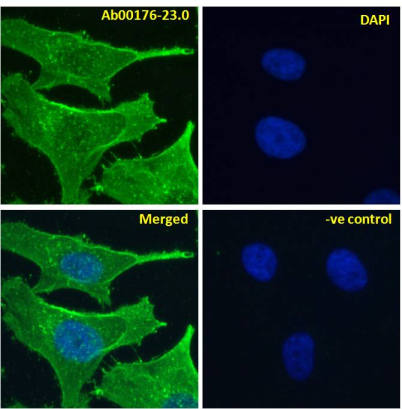
( Immunofluoresence staining of fixed HeLa cells with anti-Notch 2 antibody B6 Immunofluorescence analysis of paraformaldehyde fixed HeLa cells, permeabilized with 0.15% Triton stained with the chimeric rabbit IgG version of B6 at 10 ug/ml for 1h followed by Alexa Fluor 488 secondary antibody (1 ug/ml), showing membrane staining. The nuclear stain is DAPI (blue). Panels show from left-right, top-bottom DAPI, merged channels and a negative control. The negative control was stained with unimmunized rabbit IgG followed by Alexa Fluor 488 secondary antibody.)
IF (Immunofluorescence)
( Immunofluoresence staining of fixed HeLa cells with anti-Notch 2 antibody B6 Immunofluorescence analysis of paraformaldehyde fixed HeLa cells, permeabilized with 0.15% Triton stained with the chimeric rabbit IgG version of B6 at 10 ug/ml for 1h followed by Alexa Fluor 488 secondary antibody (1 ug/ml), showing membrane staining. The nuclear stain is DAPI (blue). Panels show from left-right, top-bottom DAPI, merged channels and a negative control. The negative control was stained with unimmunized rabbit IgG followed by Alexa Fluor 488 secondary antibody.)
Notch 2, Monoclonal Recombinant Antibody (Cat# AAA71965)
IL-6 receptor, Monoclonal Recombinant Antibody (Cat# AAA71981)
Protein A Affinity Purified
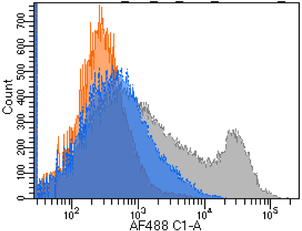
 IF (Immunofluorescence)
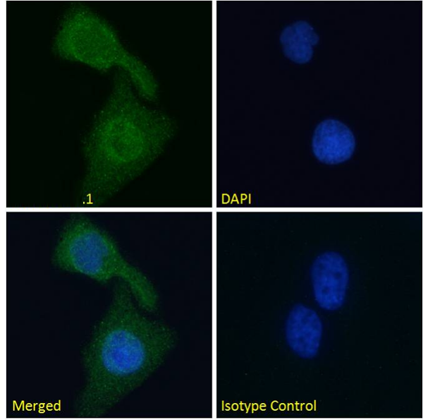
( Immunofluorescence staining of fixed mouse splenocytes with anti-GITR (Tumor necrosis factor ligand superfamily member 18) antibody YGITR 860.103.5 Immunofluorescence analysis of paraformaldehyde fixed mouse (Mus musculus) splenocytes on Shi-fix coverslips, permeabilized with 0.15% Triton stained with the chimeric rabbit version of YGITR 860.103.5 at 10 ug/ml for 1h followed by Alexa Fluor 488 secondary antibody (1 ug/ml), showing membrane and some cytoplasmic staining in a subset of cells. The nuclear stain is DAPI (blue). Panels show from left-right, top-bottom DAPI, merged channels and an isotype control. The isotype control was stained with an anti-Fluorescein antibody followed by Alexa Fluor 488 secondary antibody.)
IF (Immunofluorescence)
( Immunofluorescence staining of fixed mouse splenocytes with anti-GITR (Tumor necrosis factor ligand superfamily member 18) antibody YGITR 860.103.5 Immunofluorescence analysis of paraformaldehyde fixed mouse (Mus musculus) splenocytes on Shi-fix coverslips, permeabilized with 0.15% Triton stained with the chimeric rabbit version of YGITR 860.103.5 at 10 ug/ml for 1h followed by Alexa Fluor 488 secondary antibody (1 ug/ml), showing membrane and some cytoplasmic staining in a subset of cells. The nuclear stain is DAPI (blue). Panels show from left-right, top-bottom DAPI, merged channels and an isotype control. The isotype control was stained with an anti-Fluorescein antibody followed by Alexa Fluor 488 secondary antibody.)
GITR, Monoclonal Recombinant Antibody (Cat# AAA71991)
 IF (Immunofluorescence)
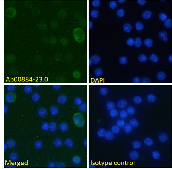
( Immunofluorescence staining of fixed mouse splenocytes with anti-B7-H3 (CD276) antibody MJ18 Immunofluorescence analysis of paraformaldehyde fixed mouse (Mus musculus) splenocytes on Shi-fix coverslips, permeabilized with 0.15% Triton stained with the chimeric rabbit version of MJ18 at 10 ug/ml for 1h followed by Alexa Fluor 488 secondary antibody (1 ug/ml), showing weak membrane staining of a subset of cells. The nuclear stain is DAPI (blue). Panels show from left-right, top-bottom DAPI, merged channels and an isotype control. The isotype control was stained with an anti-Fluorescein antibody followed by Alexa Fluor 488 secondary antibody)
IF (Immunofluorescence)
( Immunofluorescence staining of fixed mouse splenocytes with anti-B7-H3 (CD276) antibody MJ18 Immunofluorescence analysis of paraformaldehyde fixed mouse (Mus musculus) splenocytes on Shi-fix coverslips, permeabilized with 0.15% Triton stained with the chimeric rabbit version of MJ18 at 10 ug/ml for 1h followed by Alexa Fluor 488 secondary antibody (1 ug/ml), showing weak membrane staining of a subset of cells. The nuclear stain is DAPI (blue). Panels show from left-right, top-bottom DAPI, merged channels and an isotype control. The isotype control was stained with an anti-Fluorescein antibody followed by Alexa Fluor 488 secondary antibody)
B7-H3, Monoclonal Recombinant Antibody (Cat# AAA72012)
 WB (Western Blot)
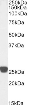
(Western Blot using anti-Cardiac Troponin I antibody scFv 180 Human heart lysate (35ug protein in RIPA buffer) was resolved on a 10% SDS PAGE gel and blots probed with the chimeric mouse IgG1 version of scFv 180 (AAA72032) at 0.001 ug/ml before detection using an anti-mouse secondary antibody. A primary incubation of 1h was used and protein was detected by chemiluminescence. The predicted running size for Cardiac Troponin I is 24.0 kDa. AAA72032 successfully detected Cardiac Troponin I in human heart lysate.)
WB (Western Blot)
(Western Blot using anti-Cardiac Troponin I antibody scFv 180 Human heart lysate (35ug protein in RIPA buffer) was resolved on a 10% SDS PAGE gel and blots probed with the chimeric mouse IgG1 version of scFv 180 (AAA72032) at 0.001 ug/ml before detection using an anti-mouse secondary antibody. A primary incubation of 1h was used and protein was detected by chemiluminescence. The predicted running size for Cardiac Troponin I is 24.0 kDa. AAA72032 successfully detected Cardiac Troponin I in human heart lysate.)
Cardiac Troponin I, Monoclonal Recombinant Antibody (Cat# AAA72032)
 WB (Western Blot)
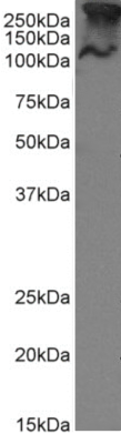
( Western Blot using anti-MUC1 antibody SM3 MCF-7 cell lysate (35ug protein in RIPA buffer) were resolved on a 10% SDS PAGE gel and blots probed with the chimeric rabbit version of SM3 at 1 ug/ml before detection using an anti-rabbit secondary antibody. A primary incubation of 1h was used and protein was detected by chemiluminescence. The predicted band size for unmodified MUC1 is 122.1kDa, though in breast cancer cell lines like MCF-7 MUC1 can be up to 90% glycosylated (c.f. Mueller et al. PMID: 10373415; T47D cells) and expected band sizes are ~250-300kDa. Thus the two bands likely represent processed (>250kDa) and unprocessed (~121kDa) populations of the protein. successfully detected human MUC1 in MCF-7 breast cancer cells.)
WB (Western Blot)
( Western Blot using anti-MUC1 antibody SM3 MCF-7 cell lysate (35ug protein in RIPA buffer) were resolved on a 10% SDS PAGE gel and blots probed with the chimeric rabbit version of SM3 at 1 ug/ml before detection using an anti-rabbit secondary antibody. A primary incubation of 1h was used and protein was detected by chemiluminescence. The predicted band size for unmodified MUC1 is 122.1kDa, though in breast cancer cell lines like MCF-7 MUC1 can be up to 90% glycosylated (c.f. Mueller et al. PMID: 10373415; T47D cells) and expected band sizes are ~250-300kDa. Thus the two bands likely represent processed (>250kDa) and unprocessed (~121kDa) populations of the protein. successfully detected human MUC1 in MCF-7 breast cancer cells.)
MUC1, Monoclonal Recombinant Antibody (Cat# AAA71969)
Protein A Affinity Purified
 WB (Western Blot)
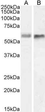
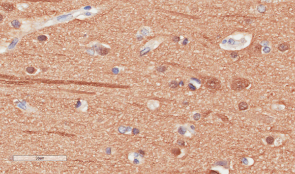
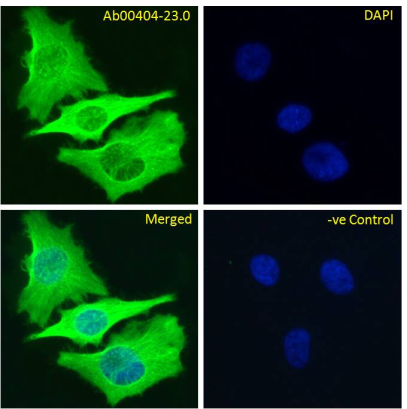
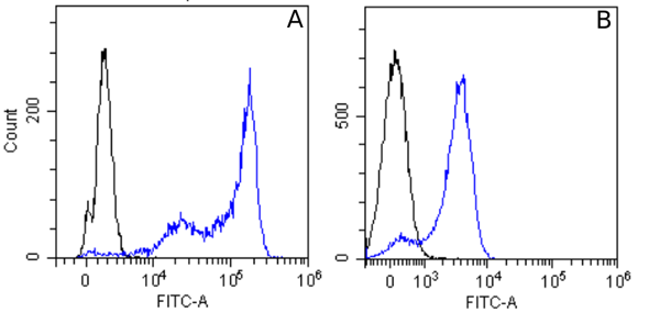
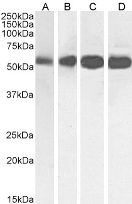
( Western Blot using anti-Beta-Tubulin antibody S11B HeLa (A), A431 (B), HEK293 (C) and MCF-7 (D) cell lysate samples (35ug protein in RIPA buffer) were resolved on a 10% SDS PAGE gel and blots probed with the chimeric rabbit version of S11B at 0.01 ug/ml before detection using an anti-rabbit secondary antibody. A primary incubation of 1h was used and protein was detected by chemiluminescence. The expected band size for Beta-Tubulin is ~54kDa. A431, HEK293 and MCF-7 cell lysate samples.)
WB (Western Blot)
( Western Blot using anti-Beta-Tubulin antibody S11B HeLa (A), A431 (B), HEK293 (C) and MCF-7 (D) cell lysate samples (35ug protein in RIPA buffer) were resolved on a 10% SDS PAGE gel and blots probed with the chimeric rabbit version of S11B at 0.01 ug/ml before detection using an anti-rabbit secondary antibody. A primary incubation of 1h was used and protein was detected by chemiluminescence. The expected band size for Beta-Tubulin is ~54kDa. A431, HEK293 and MCF-7 cell lysate samples.)
Beta-Tubulin, Monoclonal Recombinant Antibody (Cat# AAA71972)
Protein A Affinity Purified
 FCM/FACS (Flow Cytometry)
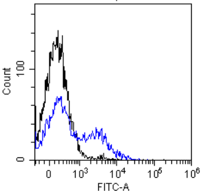
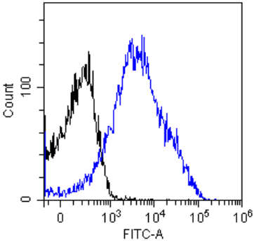
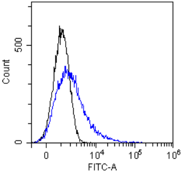
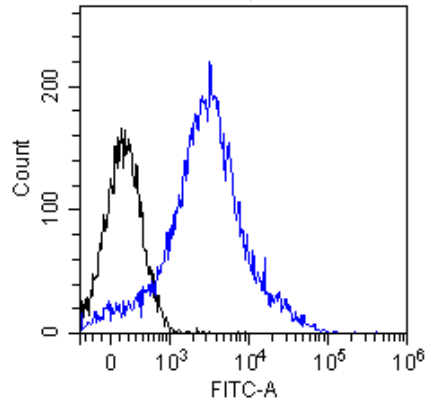
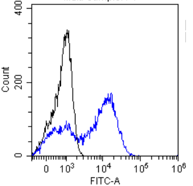
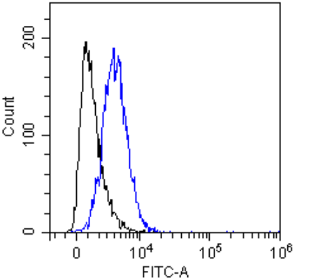
( Flow-cytometry using the anti-CD8 beta antibody YTS 156.7.7 Mouse thymocytes were stained with unimmunized rabbit IgG antibody (black line) or the rabbit-chimeric version of YTS 156.7.7 at a concentration of 10 ug/ml for 30 mins at RT. After washing, bound antibody was detected using an anti-rabbit IgG JK (FITC-conjugate) antibody (129936) at 2 ug/ml and cells analyzed on a FACSCanto flow-cytometer.)
FCM/FACS (Flow Cytometry)
( Flow-cytometry using the anti-CD8 beta antibody YTS 156.7.7 Mouse thymocytes were stained with unimmunized rabbit IgG antibody (black line) or the rabbit-chimeric version of YTS 156.7.7 at a concentration of 10 ug/ml for 30 mins at RT. After washing, bound antibody was detected using an anti-rabbit IgG JK (FITC-conjugate) antibody (129936) at 2 ug/ml and cells analyzed on a FACSCanto flow-cytometer.)
CD8 beta, Monoclonal Recombinant Antibody (Cat# AAA71974)
Protein A Affinity Purified
 WB (Western Blot)
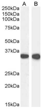
( Western Blot using anti-CD74 antibody In-1 Mouse spleen (A) and mouse lymph node (B) lysate samples (35ug protein in RIPA buffer) were resolved on a 10% SDS PAGE gel and blots probed with the chimeric rabbit version of In-1 at 0.0001 ug/ml before detection using an anti-rabbit secondary antibody. A primary incubation of 1h was used and protein was detected by chemiluminescence. The expected running size for unmodified CD74 is 31.6kDa, and this protein is glycosylated at several residues. successfully detected CD74 in mouse spleen and mouse lymph node lysates.)
WB (Western Blot)
( Western Blot using anti-CD74 antibody In-1 Mouse spleen (A) and mouse lymph node (B) lysate samples (35ug protein in RIPA buffer) were resolved on a 10% SDS PAGE gel and blots probed with the chimeric rabbit version of In-1 at 0.0001 ug/ml before detection using an anti-rabbit secondary antibody. A primary incubation of 1h was used and protein was detected by chemiluminescence. The expected running size for unmodified CD74 is 31.6kDa, and this protein is glycosylated at several residues. successfully detected CD74 in mouse spleen and mouse lymph node lysates.)
CD74, Monoclonal Recombinant Antibody (Cat# AAA72009)
 IF (Immunofluorescence)
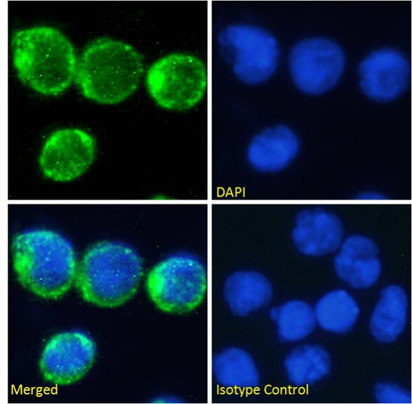
(Immunofluorescence staining of fixed Molt4 cells with anti-CD2 antibody YTH 655 Immunofluorescence analysis of paraformaldehyde fixed Molt4 cells on Shi-fix coverslips, permeabilized with 0.15% Triton and stained with the chimeric rabbit IgG version of YTH 655 (AAA72014) at 10 ug/ml for 1h followed by Alexa Fluor 488 secondary antibody (1 ug/ml), showing membrane, and some cytoplasmic staining. The nuclear stain is DAPI (blue). Panels show from left-right, top-bottom AAA72014, DAPI, merged channels and an isotype control. The isotype control was stained with an anti-Fluorescein antibody followed by Alexa Fluor 488 secondary antibody.)
IF (Immunofluorescence)
(Immunofluorescence staining of fixed Molt4 cells with anti-CD2 antibody YTH 655 Immunofluorescence analysis of paraformaldehyde fixed Molt4 cells on Shi-fix coverslips, permeabilized with 0.15% Triton and stained with the chimeric rabbit IgG version of YTH 655 (AAA72014) at 10 ug/ml for 1h followed by Alexa Fluor 488 secondary antibody (1 ug/ml), showing membrane, and some cytoplasmic staining. The nuclear stain is DAPI (blue). Panels show from left-right, top-bottom AAA72014, DAPI, merged channels and an isotype control. The isotype control was stained with an anti-Fluorescein antibody followed by Alexa Fluor 488 secondary antibody.)
CD2, Monoclonal Recombinant Antibody (Cat# AAA72014)
 IF (Immunofluorescence)
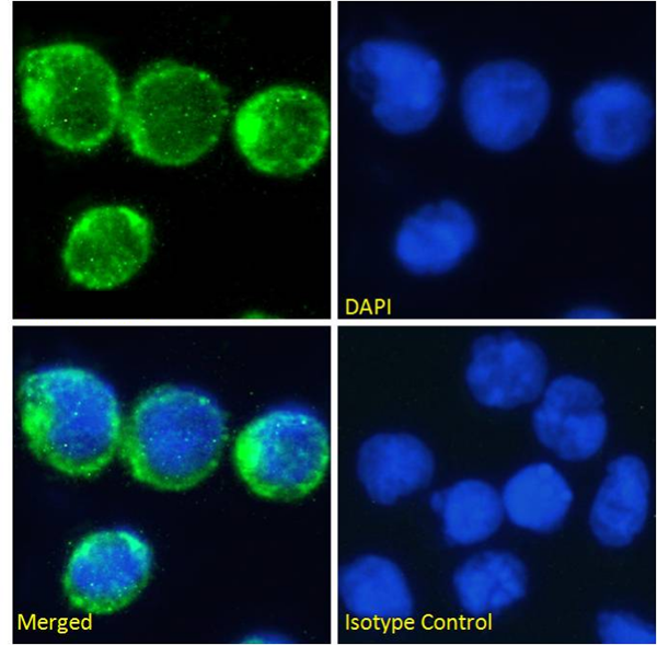
(Immunofluorescence staining of fixed Molt4 cells with anti-CD2 antibody YTH 655 Immunofluorescence analysis of paraformaldehyde fixed Molt4 cells on Shi-fix coverslips, permeabilized with 0.15% Triton and stained with the chimeric rabbit IgG version of YTH 655 at 10 ug/ml for 1h followed by Alexa Fluor 488 secondary antibody (1 ug/ml), showing membrane, and some cytoplasmic staining. The nuclear stain is DAPI (blue). Panels show from left-right, top-bottom, DAPI, merged channels and an isotype control. The isotype control was stained with an anti-Fluorescein antibody followed by Alexa Fluor 488 secondary antibody.)
IF (Immunofluorescence)
(Immunofluorescence staining of fixed Molt4 cells with anti-CD2 antibody YTH 655 Immunofluorescence analysis of paraformaldehyde fixed Molt4 cells on Shi-fix coverslips, permeabilized with 0.15% Triton and stained with the chimeric rabbit IgG version of YTH 655 at 10 ug/ml for 1h followed by Alexa Fluor 488 secondary antibody (1 ug/ml), showing membrane, and some cytoplasmic staining. The nuclear stain is DAPI (blue). Panels show from left-right, top-bottom, DAPI, merged channels and an isotype control. The isotype control was stained with an anti-Fluorescein antibody followed by Alexa Fluor 488 secondary antibody.)
CD2, Monoclonal Recombinant Antibody (Cat# AAA72015)
 IF (Immunofluorescence)
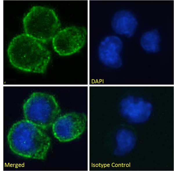
(Immunofluorescence staining of fixed Daudi cells with anti-CD40 antibody A54 scFv Immunofluorescence analysis of paraformaldehyde fixed Daudi cells on Shi-fix coverslips, permeabilized with 0.15% Triton and stained with the chimeric rabbit IgG version of A54 scFv (AAA72042) at 10 ug/ml for 1h followed by Alexa Fluor 488 secondary antibody (1 ug/ml), showing membrane staining. The nuclear stain is DAPI (blue). Panels show from left-right, top-bottom AAA72042, DAPI, merged channels and an isotype control. The isotype control was stained with an anti-Fluorescein antibody followed by Alexa Fluor 488 secondary antibody.)
IF (Immunofluorescence)
(Immunofluorescence staining of fixed Daudi cells with anti-CD40 antibody A54 scFv Immunofluorescence analysis of paraformaldehyde fixed Daudi cells on Shi-fix coverslips, permeabilized with 0.15% Triton and stained with the chimeric rabbit IgG version of A54 scFv (AAA72042) at 10 ug/ml for 1h followed by Alexa Fluor 488 secondary antibody (1 ug/ml), showing membrane staining. The nuclear stain is DAPI (blue). Panels show from left-right, top-bottom AAA72042, DAPI, merged channels and an isotype control. The isotype control was stained with an anti-Fluorescein antibody followed by Alexa Fluor 488 secondary antibody.)
CD40, Monoclonal Recombinant Antibody (Cat# AAA72042)
CD33, Monoclonal Recombinant Antibody (Cat# AAA71970)
Protein A Affinity Purified
 FCM/FACS (Flow Cytometry)
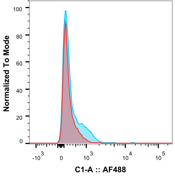
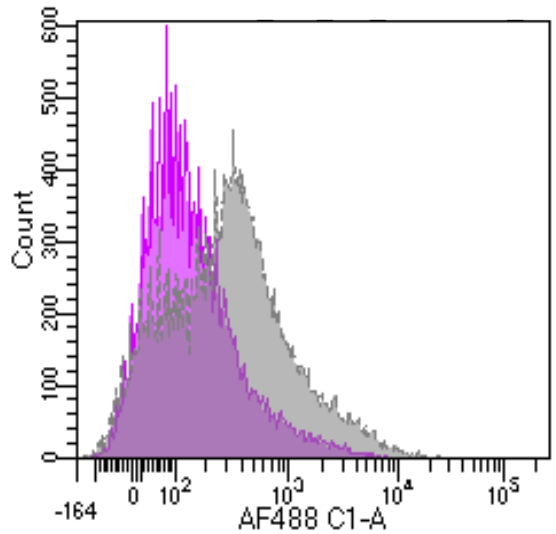
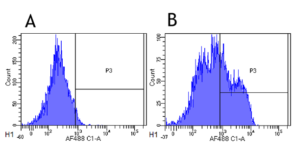
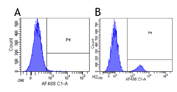
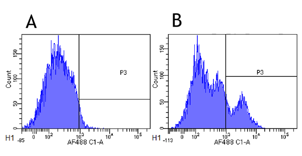
( Flow-cytometry using anti-CD22 antibody Epratuzumab Cynomolgus monkey lymphocytes were stained with an isotype control or the rabbit-chimeric version of Epratuzumab at a concentration of 1 ug/ml for 30 mins at RT. After washing, bound antibody was detected using a AF488 conjugated donkey anti-rabbit antibody and cells analysed on a FACSCanto flow-cytometer.)
FCM/FACS (Flow Cytometry)
( Flow-cytometry using anti-CD22 antibody Epratuzumab Cynomolgus monkey lymphocytes were stained with an isotype control or the rabbit-chimeric version of Epratuzumab at a concentration of 1 ug/ml for 30 mins at RT. After washing, bound antibody was detected using a AF488 conjugated donkey anti-rabbit antibody and cells analysed on a FACSCanto flow-cytometer.)
CD22, Monoclonal Recombinant Antibody (Cat# AAA71980)
 WB (Western Blot)
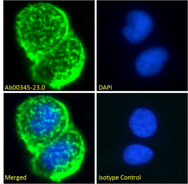
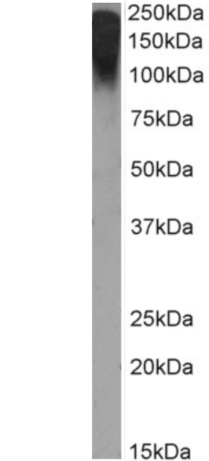
( Western Blot using anti-breast epithelial mucin antibody Mc5 (Ab00345) MCF7 cell lysate (35ug protein in RIPA buffer) was resolved on a 10% SDS PAGE gel and blots probed with the chimeric rabbit IgG version of Mc5 (Ab00345-23.0) at 2 ug/ml before detection using an anti-rabbit secondary antibody. A primary incubation of 1h was used and protein was detected by chemiluminescence. The expected running size for unmodified breast epithelial mucin is 122.1kDa, but this protein is heavily glycosylated and lipidated at several positions leading to the observed running size.)
WB (Western Blot)
( Western Blot using anti-breast epithelial mucin antibody Mc5 (Ab00345) MCF7 cell lysate (35ug protein in RIPA buffer) was resolved on a 10% SDS PAGE gel and blots probed with the chimeric rabbit IgG version of Mc5 (Ab00345-23.0) at 2 ug/ml before detection using an anti-rabbit secondary antibody. A primary incubation of 1h was used and protein was detected by chemiluminescence. The expected running size for unmodified breast epithelial mucin is 122.1kDa, but this protein is heavily glycosylated and lipidated at several positions leading to the observed running size.)
Breast Epithelial Mucin, Monoclonal Recombinant Antibody (Cat# AAA71988)
CD11c, Monoclonal Recombinant Antibody (Cat# AAA71992)
TrkA, Monoclonal Recombinant Antibody (Cat# AAA71994)
CD40, Monoclonal Recombinant Antibody (Cat# AAA72004)
 WB (Western Blot)
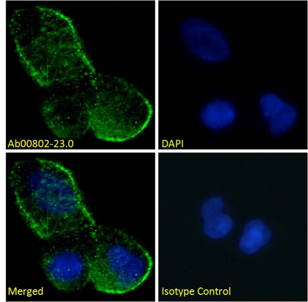
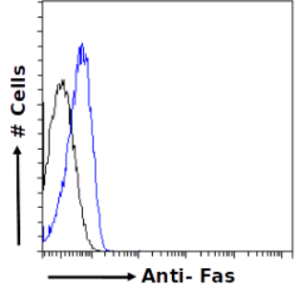
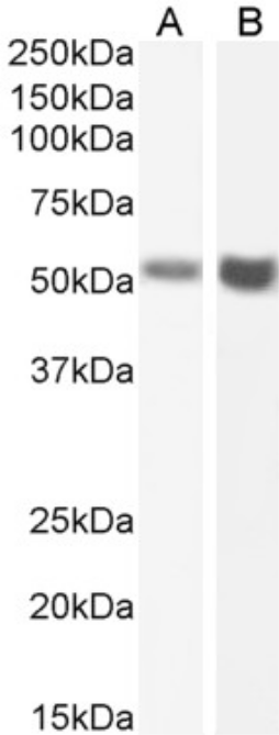
( Western Blot using anti-Fas antibody R-125224 (Ab00802) Human testis (A) and human ovary (B) lysate samples (35ug protein in RIPA buffer) were resolved on a 10% SDS PAGE gel and blots probed with the chimeric rabbit IgG version of R-125224 (Ab00802-23.0) at 2 ug/ml before detection using an anti-rabbit secondary antibody. A primary incubation of 1h was used and protein was detected by chemiluminescence. The expected running size for unmodified Fas is 37.7kDa, but this protein is glycosylated at several positions leading to the observed running size.)
WB (Western Blot)
( Western Blot using anti-Fas antibody R-125224 (Ab00802) Human testis (A) and human ovary (B) lysate samples (35ug protein in RIPA buffer) were resolved on a 10% SDS PAGE gel and blots probed with the chimeric rabbit IgG version of R-125224 (Ab00802-23.0) at 2 ug/ml before detection using an anti-rabbit secondary antibody. A primary incubation of 1h was used and protein was detected by chemiluminescence. The expected running size for unmodified Fas is 37.7kDa, but this protein is glycosylated at several positions leading to the observed running size.)
Fas, Monoclonal Recombinant Antibody (Cat# AAA72007)
 IF (Immunofluorescence)
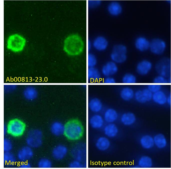
( Immunofluorescence staining of fixed mouse splenocytes with anti-PD-1 (programmed cell death protein 1) antibody RMP1-14 Immunofluorescence analysis of paraformaldehyde fixed mouse (Mus musculus) splenocytes on Shi-fix coverslips, permeabilized with 0.15% Triton stained with the chimeric rabbit version of RMP1-14 at 10 ug/ml for 1h followed by Alexa Fluor 488 secondary antibody (1 ug/ml), showing membrane staining of a subset of cells. The nuclear stain is DAPI (blue). Panels show from left-right, top-bottom DAPI, merged channels and an isotype control. The isotype control was stained with an anti-Fluorescein antibody followed by Alexa Fluor 488 secondary antibody.)
IF (Immunofluorescence)
( Immunofluorescence staining of fixed mouse splenocytes with anti-PD-1 (programmed cell death protein 1) antibody RMP1-14 Immunofluorescence analysis of paraformaldehyde fixed mouse (Mus musculus) splenocytes on Shi-fix coverslips, permeabilized with 0.15% Triton stained with the chimeric rabbit version of RMP1-14 at 10 ug/ml for 1h followed by Alexa Fluor 488 secondary antibody (1 ug/ml), showing membrane staining of a subset of cells. The nuclear stain is DAPI (blue). Panels show from left-right, top-bottom DAPI, merged channels and an isotype control. The isotype control was stained with an anti-Fluorescein antibody followed by Alexa Fluor 488 secondary antibody.)
PD-1, Monoclonal Recombinant Antibody (Cat# AAA72008)
 WB (Western Blot)
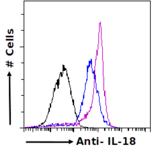
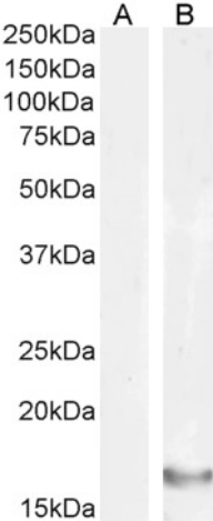
(Western Blot using anti-IL-18 antibody SAIC-24B-4 Untreated human peripheral blood monocyte (PBM; A) and PMA/lonomycin-treated PBM lysates (35ug protein in RIPA buffer) were resolved on a 15% SDS PAGE gel and blots probed with the rabbit IgG-chimeric version of SAIC-24B-4 at 0.3 ug/ml before detection using an anti-rabbit secondary antibody. Protein was detected by chemiluminescence. The expected running size for IL-18 is 22.3kDa. successfully detected IL-18 in untreated human peripheral blood monocyte samples.)
WB (Western Blot)
(Western Blot using anti-IL-18 antibody SAIC-24B-4 Untreated human peripheral blood monocyte (PBM; A) and PMA/lonomycin-treated PBM lysates (35ug protein in RIPA buffer) were resolved on a 15% SDS PAGE gel and blots probed with the rabbit IgG-chimeric version of SAIC-24B-4 at 0.3 ug/ml before detection using an anti-rabbit secondary antibody. Protein was detected by chemiluminescence. The expected running size for IL-18 is 22.3kDa. successfully detected IL-18 in untreated human peripheral blood monocyte samples.)
IL-18, Monoclonal Recombinant Antibody (Cat# AAA72030)
 FCM/FACS (Flow Cytometry)
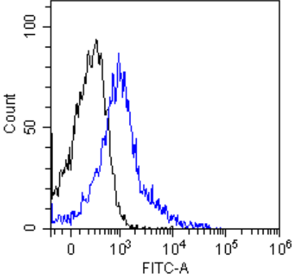
(Flow-cytometry using anti-CD62L antibody MEL-14 Mouse lymphocytes were stained with an anti-CD44 control or the rabbit-chimeric version of MEL-14 at a concentration of 1 ug/ml for 30 mins at RT. After washing, bound antibody was detected using a AF488 conjugated donkey anti-rabbit antibody and cells analysed on a FACSCanto flow-cytometer.)
FCM/FACS (Flow Cytometry)
(Flow-cytometry using anti-CD62L antibody MEL-14 Mouse lymphocytes were stained with an anti-CD44 control or the rabbit-chimeric version of MEL-14 at a concentration of 1 ug/ml for 30 mins at RT. After washing, bound antibody was detected using a AF488 conjugated donkey anti-rabbit antibody and cells analysed on a FACSCanto flow-cytometer.)
CD62L, Monoclonal Recombinant Antibody (Cat# AAA72035)
 FCM/FACS (Flow Cytometry)
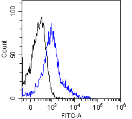
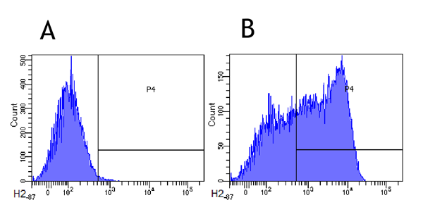
(Flow-cytometry using anti-CD62L antibody MEL-14 Mouse lymphocytes were stained with an anti-CD44 control or the rabbit-chimeric version of MEL-14 (AAA72036, panel B) at a concentration of 1 ug/ml for 30 mins at RT. After washing, bound antibody was detected using a AF488 conjugated donkey anti-rabbit antibody and cells analysed on a FACSCanto flow-cytometer.)
FCM/FACS (Flow Cytometry)
(Flow-cytometry using anti-CD62L antibody MEL-14 Mouse lymphocytes were stained with an anti-CD44 control or the rabbit-chimeric version of MEL-14 (AAA72036, panel B) at a concentration of 1 ug/ml for 30 mins at RT. After washing, bound antibody was detected using a AF488 conjugated donkey anti-rabbit antibody and cells analysed on a FACSCanto flow-cytometer.)
CD62L, Monoclonal Recombinant Antibody (Cat# AAA72036)
 IF (Immunofluorescence)
(Immunofluorescence staining of fixed MCF7 cells with anti-GM2 ganglioside antibody L55-81 Immunofluorescence analysis of paraformaldehyde fixed MCF7 cells on Shi-fix coverslips, permeabilized with 0.15% Triton and stained with the chimeric rabbit IgG version of L55-81 at 10 ug/ml for 1h followed by Alexa Fluor 488 secondary antibody (1 ug/ml), showing diffuse membrane and cytoplasmic staining. The nuclear stain is DAPI (blue). Panels show from left-right, top-bottom, DAPI, merged channels and an isotype control. The isotype control was stained with an anti-Fluorescein antibody followed by Alexa Fluor 488 secondary antibody.)
IF (Immunofluorescence)
(Immunofluorescence staining of fixed MCF7 cells with anti-GM2 ganglioside antibody L55-81 Immunofluorescence analysis of paraformaldehyde fixed MCF7 cells on Shi-fix coverslips, permeabilized with 0.15% Triton and stained with the chimeric rabbit IgG version of L55-81 at 10 ug/ml for 1h followed by Alexa Fluor 488 secondary antibody (1 ug/ml), showing diffuse membrane and cytoplasmic staining. The nuclear stain is DAPI (blue). Panels show from left-right, top-bottom, DAPI, merged channels and an isotype control. The isotype control was stained with an anti-Fluorescein antibody followed by Alexa Fluor 488 secondary antibody.)
GM2 Ganglioside, Monoclonal Recombinant Antibody (Cat# AAA72043)
 ELISA
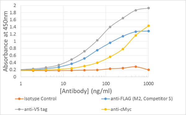
(ELISA of anti-epitope tag antibodies on multi-tag protein Plate coated with 5ug/ml of Multitag protein)
ELISA
(ELISA of anti-epitope tag antibodies on multi-tag protein Plate coated with 5ug/ml of Multitag protein)
V5 epitope tag, Recombinant Antibody (Cat# AAA71935)
 WB (Western Blot)
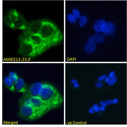
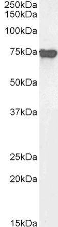
( Western Blot using anti-Alpha-fetoprotein antibody AF5 HepG2 cell lysate (35ug protein in RIPA buffer) was resolved on a 10% SDS PAGE gel and blots probed with the chimeric rabbit version of AF5 at 1 ug/ml before detection using an anti-rabbit secondary antibody. A primary incubation of 1h was used and protein was detected by chemiluminescence. The expected band size for Alpha-fetoprotein is 68.7kDa, though this protein is also glycosylated (Uniprot ID: P02771). successfully detected human Alpha-fetoprotein in HepG2 cell lysates.)
WB (Western Blot)
( Western Blot using anti-Alpha-fetoprotein antibody AF5 HepG2 cell lysate (35ug protein in RIPA buffer) was resolved on a 10% SDS PAGE gel and blots probed with the chimeric rabbit version of AF5 at 1 ug/ml before detection using an anti-rabbit secondary antibody. A primary incubation of 1h was used and protein was detected by chemiluminescence. The expected band size for Alpha-fetoprotein is 68.7kDa, though this protein is also glycosylated (Uniprot ID: P02771). successfully detected human Alpha-fetoprotein in HepG2 cell lysates.)
Alpha-fetoprotein (AFP), Monoclonal Recombinant Antibody (Cat# AAA71967)
Protein A Affinity Purified
amyloid beta, Monoclonal Recombinant Antibody (Cat# AAA71977)
IL-2, Monoclonal Recombinant Antibody (Cat# AAA71984)
 IF (Immunofluorescence)
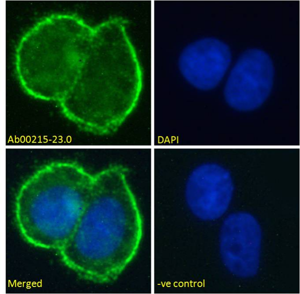
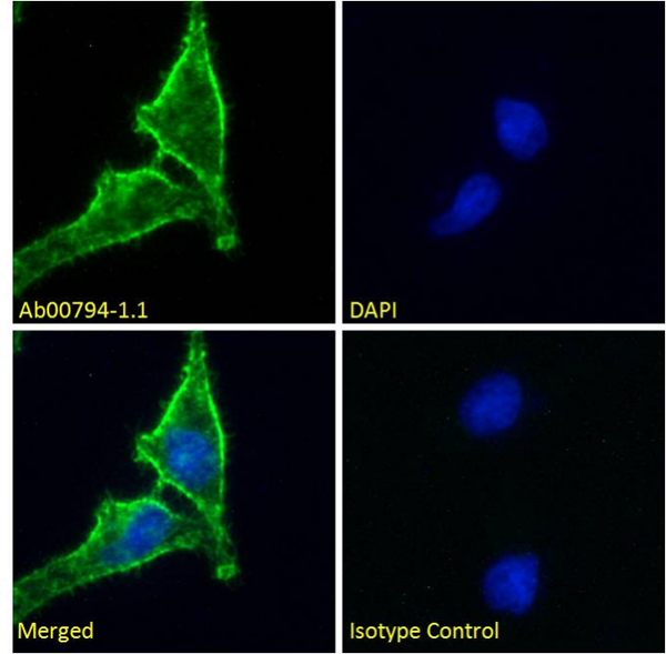
( Immunofluorescence staining of fixed HeLa cells with anti-CD98 heavy chain antibody HBJ127 (Ab00794) Immunofluorescence analysis of paraformaldehyde fixed HeLa cells on Shi-fix™ coverslips, permeabilized with 0.15% Triton and stained with the chimeric mouse IgG1 version of HBJ127 (Ab00794-1.1) at 10 ug/ml for 1h followed by Alexa Fluor® 488 secondary antibody (2 ug/ml), showing membrane staining. The nuclear stain is DAPI (blue). Panels show from left-right, top-bottom Ab00794-1.1, DAPI, merged channels and an isotype control. The isotype control was stained with an unknown specificity antibody (Ab00178-1.1) followed by Alexa Fluor® 488 secondary antibody.)
IF (Immunofluorescence)
( Immunofluorescence staining of fixed HeLa cells with anti-CD98 heavy chain antibody HBJ127 (Ab00794) Immunofluorescence analysis of paraformaldehyde fixed HeLa cells on Shi-fix™ coverslips, permeabilized with 0.15% Triton and stained with the chimeric mouse IgG1 version of HBJ127 (Ab00794-1.1) at 10 ug/ml for 1h followed by Alexa Fluor® 488 secondary antibody (2 ug/ml), showing membrane staining. The nuclear stain is DAPI (blue). Panels show from left-right, top-bottom Ab00794-1.1, DAPI, merged channels and an isotype control. The isotype control was stained with an unknown specificity antibody (Ab00178-1.1) followed by Alexa Fluor® 488 secondary antibody.)
CD98 heavy chain, Monoclonal Recombinant Antibody (Cat# AAA72006)
 WB (Western Blot)
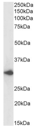
( Western Blot using anti-DARC antibody 2C3 Human liver lysate samples (35ug protein in RIPA buffer) were resolved on a 10% SDS PAGE gel and blots probed with the chimeric rabbit version of 2C3 at 0.03 ug/ml before detection using an anti-rabbit secondary antibody. A primary incubation of 1h was used and protein was detected by chemiluminescence. The expected running size for DARC is 35.6kDa. successfully detected DARC in human liver lysate.)
WB (Western Blot)
( Western Blot using anti-DARC antibody 2C3 Human liver lysate samples (35ug protein in RIPA buffer) were resolved on a 10% SDS PAGE gel and blots probed with the chimeric rabbit version of 2C3 at 0.03 ug/ml before detection using an anti-rabbit secondary antibody. A primary incubation of 1h was used and protein was detected by chemiluminescence. The expected running size for DARC is 35.6kDa. successfully detected DARC in human liver lysate.)
DARC, Monoclonal Recombinant Antibody (Cat# AAA72013)

 WB (Western Blot)
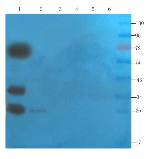
(Western Blot using anti-SNAP25 antibody AAA72025 Mouse brain (lane 1), mouse rectum (lane 2), mouse pancreas (lane 3), human lung cancer (lane 4), human breast cancer (lane 5) and Hela cell(lane 6) samples were resolved on a 12% SDS PAGE gel and blots probed with AAA72025 at 2 ug/ml before being detected by a secondary antibody. Bands correspondig to the expected 23 kDa SNAP25 protein were detected in mouse brain (along with likely oligomers) as well as in mouse rectum (faint). In this experiment no SNAP25 was dected in the other samples.)
WB (Western Blot)
(Western Blot using anti-SNAP25 antibody AAA72025 Mouse brain (lane 1), mouse rectum (lane 2), mouse pancreas (lane 3), human lung cancer (lane 4), human breast cancer (lane 5) and Hela cell(lane 6) samples were resolved on a 12% SDS PAGE gel and blots probed with AAA72025 at 2 ug/ml before being detected by a secondary antibody. Bands correspondig to the expected 23 kDa SNAP25 protein were detected in mouse brain (along with likely oligomers) as well as in mouse rectum (faint). In this experiment no SNAP25 was dected in the other samples.)
SNAP-25, Monoclonal Recombinant Antibody (Cat# AAA72025)
 WB (Western Blot)
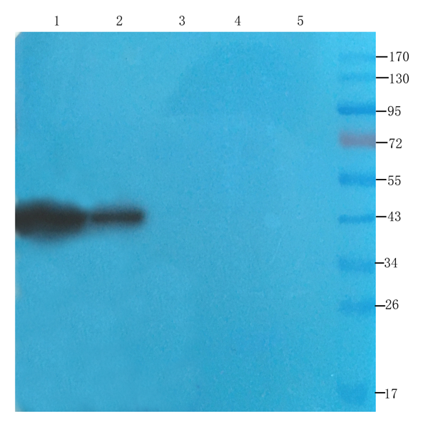
(Western Blot using anti-CDCrel-1 antibody AAA72028 Rat brain (lane 1), mouse spinal cord (lane 2), rat testis (lane 3), human lung cancer (lane 4) and human breast cancer (lane 5) samples were resolved on a 12% SDS PAGE gel and blots probed with AAA72028 at 2 ug/ml before being detected by a secondary antibody. Bands correspondig to the expected 43 kDa CDCrel-1 protein were detected in rat brain and mouse spinal chord. In this experiment no CDCrel-1 was dected in the other samples.)
WB (Western Blot)
(Western Blot using anti-CDCrel-1 antibody AAA72028 Rat brain (lane 1), mouse spinal cord (lane 2), rat testis (lane 3), human lung cancer (lane 4) and human breast cancer (lane 5) samples were resolved on a 12% SDS PAGE gel and blots probed with AAA72028 at 2 ug/ml before being detected by a secondary antibody. Bands correspondig to the expected 43 kDa CDCrel-1 protein were detected in rat brain and mouse spinal chord. In this experiment no CDCrel-1 was dected in the other samples.)
CDCrel-1, Monoclonal Recombinant Antibody (Cat# AAA72028)
 FCM/FACS (Flow Cytometry)
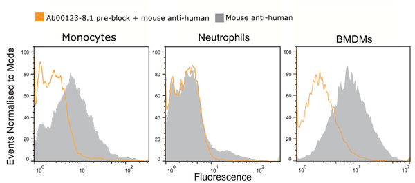
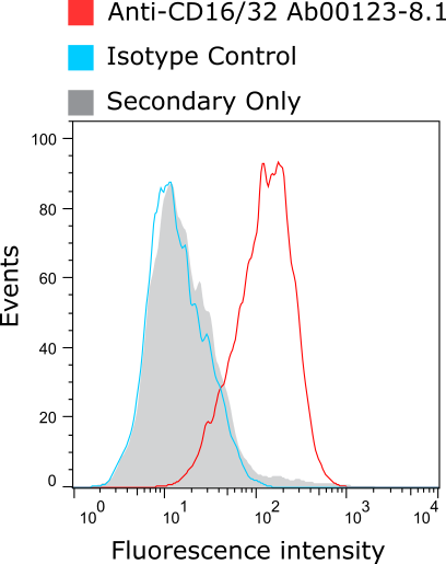
( Anti-CD16/32 staining of murine macrophages Murine bone marrow-derived macrophages (BMDMs) were stained with anti-CD16/32 antibody (as well as isotype control) and bound antibody detected using goat IgG anti-rat IgG (H&L-chain) polyclonal antibody directly conjugated to Alexa Fluor 647 commercially available from a competitor. Compared to the isotype control and secondary-only control, a CD16/32-positive population is apparent.)
FCM/FACS (Flow Cytometry)
( Anti-CD16/32 staining of murine macrophages Murine bone marrow-derived macrophages (BMDMs) were stained with anti-CD16/32 antibody (as well as isotype control) and bound antibody detected using goat IgG anti-rat IgG (H&L-chain) polyclonal antibody directly conjugated to Alexa Fluor 647 commercially available from a competitor. Compared to the isotype control and secondary-only control, a CD16/32-positive population is apparent.)
CD16/CD32, Recombinant Antibody (Cat# AAA71928)
 IHC (Immunohiostchemistry)
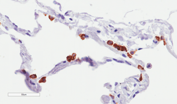
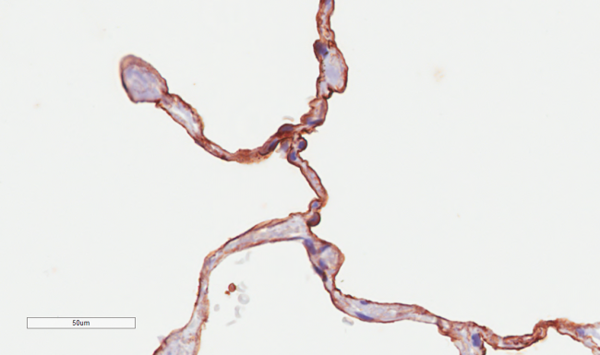
( Immunohistochemical staining of human lung tissue using anti-CD55 antibody LU30 Anti-CD55 (Complement decay-accelerating factor)staining of paraffin embedded human lung tissue using the rabbit-chimeric version of LU30 for 20 mins followed by DAB (3,3'-diaminobenzidine), and counter staining with haematoxylin. Strong staining of the epithelial surface of alveoli may be observed. Recommended concentration, 2-4 ug/ml.)
IHC (Immunohiostchemistry)
( Immunohistochemical staining of human lung tissue using anti-CD55 antibody LU30 Anti-CD55 (Complement decay-accelerating factor)staining of paraffin embedded human lung tissue using the rabbit-chimeric version of LU30 for 20 mins followed by DAB (3,3'-diaminobenzidine), and counter staining with haematoxylin. Strong staining of the epithelial surface of alveoli may be observed. Recommended concentration, 2-4 ug/ml.)
CD55, Monoclonal Recombinant Antibody (Cat# AAA71973)
Protein A Affinity Purified
 IF (Immunofluorescence)
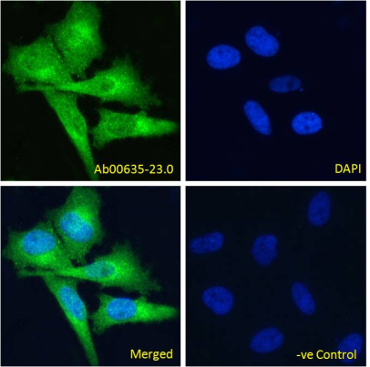
( Immunofluoresence staining of fixed HeLa cells with anti-ED-B domain of fibronectin antibody G4 Immunofluorescence analysis of paraformaldehyde fixed HeLa cells, permeabilized with 0.15% Triton stained with the chimeric rabbit IgG version of G4 at 10 ug/ml for 1h followed by Alexa Fluor 488 secondary antibody (1 ug/ml), showing cytoplasmic staining. The nuclear stain is DAPI (blue). Panels show from left-right, top-bottom DAPI, merged channels and a negative control. The negative control was stained with unimmunized rabbit IgG followed by Alexa Fluor 488 secondary antibody.)
IF (Immunofluorescence)
( Immunofluoresence staining of fixed HeLa cells with anti-ED-B domain of fibronectin antibody G4 Immunofluorescence analysis of paraformaldehyde fixed HeLa cells, permeabilized with 0.15% Triton stained with the chimeric rabbit IgG version of G4 at 10 ug/ml for 1h followed by Alexa Fluor 488 secondary antibody (1 ug/ml), showing cytoplasmic staining. The nuclear stain is DAPI (blue). Panels show from left-right, top-bottom DAPI, merged channels and a negative control. The negative control was stained with unimmunized rabbit IgG followed by Alexa Fluor 488 secondary antibody.)
ED-B domain of fibronectin, Monoclonal Recombinant Antibody (Cat# AAA71975)
Protein A Affinity Purified
CD74, Monoclonal Recombinant Antibody (Cat# AAA72010)
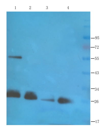
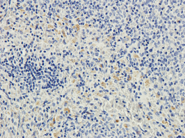 IHC (Immunohiostchemistry)
(Immunohistochemical staining of rat spleen using anti-CD28 antibody Formalin fixed rat spleen slices were were stained at 3 ug/ml.)
IHC (Immunohiostchemistry)
(Immunohistochemical staining of rat spleen using anti-CD28 antibody Formalin fixed rat spleen slices were were stained at 3 ug/ml.)
CD28, Monoclonal Recombinant Antibody (Cat# AAA72022)
What are recombinant antibodies?
Recombinant antibodies are produced in the lab by utilizing a known genetic sequence (an amino acid and nucleotide sequence). This allows for high consistency in how each new lot will perform and how specific they will be for the target, regardless of how many times they are produced/reproduced.
They help solve the problems observed when traditional non-recombinant techniques are utilized, such as changes in cell behavior over time, differences between production batches, and accuracy in complex biological systems.
A powerful feature of recombinant antibodies is that they can also be modified through progressive genetic engineering and subsequent testing in order to make them perform better and/or more reliably.
Applications For Recombinant Antibodies
1. Basic Research
Recombinant antibodies are primarily utilized in laboratory experiments to study the function of various proteins in cells. They are helpful in finding and tracking specific molecules through their use in techniques such as ELISA, Western blotting, immunofluorescence, and flow cytometry.
2. Diagnostics
They are also key components in diagnostic tests used to detect diseases and infections. As recombinant antibodies are produced in a consistent and controlled way, they are able to provide highly reliable results across batches. Though recombinant antibodies are indeed quite useful in diagnostic applications, all of the products listed in AAA Biotech’s also known as AAA Bio or AAABio catalog are strictly for research-use only (RUO).
3. Therapeutics (Medical Treatments)
Please note that all of the products listed in AAA Biotech’s catalog are strictly for research-use only (RUO).
- Chimeric Antibodies: Commonly used in therapeutic treatment. They combine different pieces of mouse and human antibodies in order to reduce immune reactions.
- Humanized Antibodies: Modified non-human antibodies that are made to look the same as actual native human antibodies, making them safer and more effective.
- Antibody Fragments (Fab, scFv, VHH): Smaller pieces of antibody fragments that can reach deep into tissues and tumors. They are useful in cancer treatments and imaging.
- Bispecific Antibodies: Specially-engineered antibodies that can bind to two different target molecules at once. Each antibody has two “arms” that allow for two places where the target antigen can bind (both “arms” will recognize the same exact epitope). These bispecific antibodies are engineered so that each arm can detect a completely different epitope than the other. Used in many of the treatments for cancer, diabetes, Alzheimer’s disease, and more.
- Fc-Fusion Proteins: Used as drugs and research tools. They combine parts of antibodies with other proteins to improve stability and effectiveness.
4. Imaging and Detection
Small antibody fragments are also used as imaging agents in medical scans. Their small size helps them reach and report on the status of tumors quickly.
5. Drug Development and Delivery
Recombinant antibodies can also be designed and made to deliver drugs directly to diseased cells, especially in targeted cancer therapy.
6. Immunotherapy
These antibodies can aid the immune system recognize and fight diseases more effectively, which is often their role in cancer treatment.
Please note that all of the products listed in AAA Biotech’s catalog are strictly for research-use only (RUO).
Advantages of Recombinant Antibodies
- Reliable Results Every Time: Recombinant antibodies are exceptionally consistent from batch to batch. This means researchers can trust that results will remain the same each time they purchase and use them.
- More Specific and Accurate: Compared to polyclonal antibodies, recombinant antibodies are significantly less likely to exhibit non-specific binding, which promotes more accurate and clearer results.
- Easier to Develop: Making recombinant antibodies is simpler and quicker than the traditional method of producing monoclonal antibodies via hybridomas.
- No Genetic Drift: Over time, hybridoma cells can change or become unstable, which can result in the antibodies progressively losing their function/specificity. Recombinant antibodies avoid this problem because they are produced from a fixed template DNA sequence.
- Truly Monoclonal: Studies have shown that many hybridomas thought to be monoclonal actually are not so, which can lead to, in some cases, unreliable results. Recombinant antibodies are made using precise genetic sequence templates, ensuring they are truly specific to one target.
- Long-Term Stability and Safety: Since their amino acid and genetic sequences are known and stored, recombinant antibodies can be reproduced at any time with the same quality. This method protects against contamination, genetic changes over time, or even accidental loss/deterioration of cell lines.
Why Buy Recombinant Antibody from AAA Biotech?
- Highly Validated: The vast majority of our antibodies are thoroughly tested to ensure they work reliably across different applications and experimental platforms.
- Versatile Applications: Our antibodies can be used in a wide range of research techniques, including (but not limited to) immunocytochemistry (ICC), ELISA, immunofluorescence (IF), immunohistochemistry (IHC), flow cytometry (FC), immunoprecipitation (IP), and Western blotting (WB).
- Support for Rare Species: We offer antibodies specifically designed for rare or less commonly used laboratory species — something many other suppliers do not provide.
- Wide Selection of Conjugated and Recombinant Forms: Choose from various labeled (conjugated) options and browse our high-quality recombinant antibodies – we are confident that we will have materials to meet your research needs.
Please note that all of the products listed in AAA Biotech’s catalog are strictly for research-use only (RUO).
FAQ
1. What are recombinant antibodies used for?
Research (e.g., ELISA, Western blot, flow cytometry)
Disease diagnostics
Therapeutic treatments (e.g., cancer, autoimmune diseases)
Medical imaging
Targeted drug delivery
Vaccine development
Please note that all of the products listed in AAA Biotech’s catalog are strictly for research-use only (RUO).
2. What is the difference between traditional and recombinant antibodies?
Traditional antibodies: Produced using animal hosts (hybridomas are typically made from an original animal inoculation), slower, less consistent, risk of genetic drift
Recombinant antibodies: Produced using DNA template sequences via laboratory cell lines, faster, highly consistent, animal-free, easily customizable
3. What are antibodies used to treat?
Please note that all of the products listed in AAA Biotech’s catalog are strictly for research-use only (RUO).
Antibodies are proteins that your body makes to protect you from infections, allergies, and other potentially harmful substances. Scientists have used the intrinsic properties of antibodies to develop them into tools for scientific advancement, and can now create special lab-grown antibodies that aid in the treatment of diseases such as cancer, heart-related issues, and arthritis.
4. Why do we need recombinant proteins?
Recombinant proteins are lab-made proteins that have become essential in the advancement of science and medicine. They’re used in the lab for innumerable purposes, such as to help grow and support cells, especially for tasks like expanding cell colonies, helping them develop into other types, or turning them into stem cells.