Filters
▼Clonality
▼Type
▼Reactivity
▼Gene Name
▼Isotype
▼Host
▼Application
▼Clone
▼Phospho Antibodies
Phospho-specific antibodies’ typical purpose is to enable researchers to detect changes in proteins. They will exclusively bind to the amino acid sequence on a protein that has been phosphorylated (which is both a physical & chemical change) and do not bind to the same amino acid sequence on said protein if it lacks said phosphorylation. This aids in being able to clearly see and understand the data produced from this particular protein modification.
Viewing 1-50 of 7206 product results
 ICC (Immunocytochemistry)
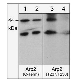
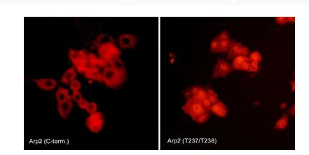
(Immunocytochemical labeling of Arp2 phosphorylation in rat PC12 cells differentiated with NGF. The cells were probed with Arp2 (C-terminal region) and Arp2 (Thr-237/Thr-238) rabbit polyclonal antibodies, then the antibodies were detected using appropriate secondary antibody conjugated to Cy3.)
ICC (Immunocytochemistry)
(Immunocytochemical labeling of Arp2 phosphorylation in rat PC12 cells differentiated with NGF. The cells were probed with Arp2 (C-terminal region) and Arp2 (Thr-237/Thr-238) rabbit polyclonal antibodies, then the antibodies were detected using appropriate secondary antibody conjugated to Cy3.)
Arp2, Polyclonal Antibody (Cat# AAA71568)
 Application Data
IkBa (pS32/36) (NFKB1A), Antibody (Cat# AAA71434)
 ICC (Immunocytochemistry)
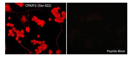
(Immunocytochemical labeling of CRMP2 phosphorylation in aldehyde-fixed and NP-40-permeabilized NGF-differentiated PC12 cells. The cells were labeled with rabbit polyclonal anti-CRMP2 (Ser-522) (CP2191) antibody in the absence (Left) or presence (Right) of blocking peptide (CX2195). The antibody was detected using appropriate secondary antibody conjugated to DyLight 594.)
ICC (Immunocytochemistry)
(Immunocytochemical labeling of CRMP2 phosphorylation in aldehyde-fixed and NP-40-permeabilized NGF-differentiated PC12 cells. The cells were labeled with rabbit polyclonal anti-CRMP2 (Ser-522) (CP2191) antibody in the absence (Left) or presence (Right) of blocking peptide (CX2195). The antibody was detected using appropriate secondary antibody conjugated to DyLight 594.)
CRMP2, Polyclonal Antibody (Cat# AAA71604)
 Application Data
GSK3b (Paired S9), Antibody (Cat# AAA71385)
 Application Data
JAK2 (pY1007/Y1008) (Biotinylated), Antibody (Cat# AAA71402)

 ICC (Immunocytochemistry)
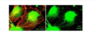
(Immunocytochemical labeling of Talin phosphorylation relative to F-actin in chick fibroblasts. The cells were labeled with rabbit polyclonal Talin (Ser-425) antibody (TP4171), then the antibody was detected using appropriate secondary antibody (Green). This labeling is compared to F-actin staining (Red, Left). (Image provided by Dr. Gianluca Gallo at Drexel University).)
ICC (Immunocytochemistry)
(Immunocytochemical labeling of Talin phosphorylation relative to F-actin in chick fibroblasts. The cells were labeled with rabbit polyclonal Talin (Ser-425) antibody (TP4171), then the antibody was detected using appropriate secondary antibody (Green). This labeling is compared to F-actin staining (Red, Left). (Image provided by Dr. Gianluca Gallo at Drexel University).)
Talin, Polyclonal Antibody (Cat# AAA71722)
 ICC (Immunocytochemistry)
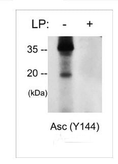
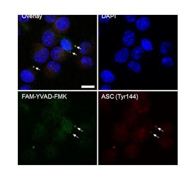
(Immunocytochemical labeling of Asc (Tyr-144) in inflammasomes. Paraformaldehyde fixed J774 cells were primed with LPS and treated with nigericin. Cells were co-labeled with DAPI, a caspase-1 inhibitor (FAM-YVAD-FMK), and anti-Asc (Tyr-144) phosphospecific antibody detected with AlexaFluor 568 secondary. (Image provided by Jordan Yaron, Center for Biosignatures Discovery Automation, Arizona State University))
ICC (Immunocytochemistry)
(Immunocytochemical labeling of Asc (Tyr-144) in inflammasomes. Paraformaldehyde fixed J774 cells were primed with LPS and treated with nigericin. Cells were co-labeled with DAPI, a caspase-1 inhibitor (FAM-YVAD-FMK), and anti-Asc (Tyr-144) phosphospecific antibody detected with AlexaFluor 568 secondary. (Image provided by Jordan Yaron, Center for Biosignatures Discovery Automation, Arizona State University))
Asc, Polyclonal Antibody (Cat# AAA71570)
 ICC (Immunocytochemistry)
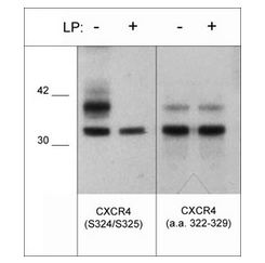
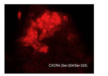
(Immunocytochemical labeling of CXCR4 in chick pluripotent cells. The cells were labeled with rabbit polyclonal CXCR4 (S324/S325) antibody (CP4251), then detected using appropriate secondary antibody (Red). (Image provided by Dr. Yangqing Lu at the Regenerative Bioscience Center, University of Georgia).)
ICC (Immunocytochemistry)
(Immunocytochemical labeling of CXCR4 in chick pluripotent cells. The cells were labeled with rabbit polyclonal CXCR4 (S324/S325) antibody (CP4251), then detected using appropriate secondary antibody (Red). (Image provided by Dr. Yangqing Lu at the Regenerative Bioscience Center, University of Georgia).)
CXCR4, Polyclonal Antibody (Cat# AAA71615)
 Application Data
PLCg1 (Tyr-775), Polyclonal Antibody (Cat# AAA71528)
 WB (Western Blot)
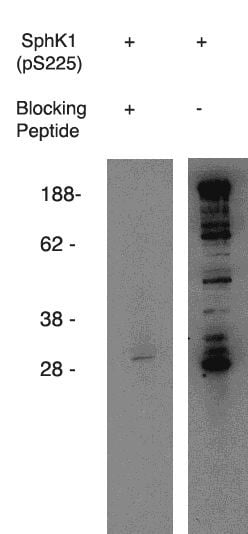
(Western blot using anti-mouse SphK1 (pS225) on mouse B-cell lysate. Antibody used at 1 ug/ml with phosphorylated blocking peptide (lane A) and without (laneB).)
WB (Western Blot)
(Western blot using anti-mouse SphK1 (pS225) on mouse B-cell lysate. Antibody used at 1 ug/ml with phosphorylated blocking peptide (lane A) and without (laneB).)
phospho-Sphingosine Kinase 1,(pS225), Mouse Reactive, Polyclonal Antibody (Cat# AAA60497)
Purification: Antigen Immunoaffiinity Purification
 WB (Western Blot)
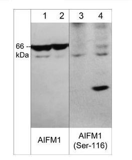
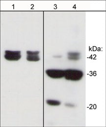
(Western blot image of human jurkat cells untreated (lanes 1 & 3) or treated with calyculin A (100nM, 30 min.) (lanes 2 and 4). The blot was probed with rabbit polyclonals anti-AIFM1 (C-terminal region) at 1:500 (lanes 1 & 2) and anti-AIFM1 (Ser-116) phospho-specific antibody at 1:1000 (lanes 3 & 4).)
WB (Western Blot)
(Western blot image of human jurkat cells untreated (lanes 1 & 3) or treated with calyculin A (100nM, 30 min.) (lanes 2 and 4). The blot was probed with rabbit polyclonals anti-AIFM1 (C-terminal region) at 1:500 (lanes 1 & 2) and anti-AIFM1 (Ser-116) phospho-specific antibody at 1:1000 (lanes 3 & 4).)
AIFM1, Polyclonal Antibody (Cat# AAA71569)
 ICC (Immunocytochemistry)
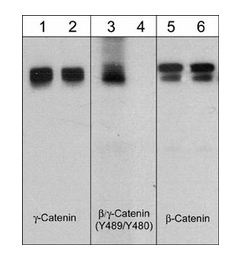
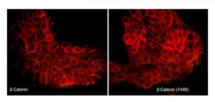
(Immunocytochemical labeling of beta-Catenin in pervanadate-treated A431 cells. The cells were labeled with mouse monoclonal beta-Catenin (CM1181) or rabbit polyclonal beta-Catenin (Tyr-489) antibodies, then the antibodies were detected using appropriate secondary antibodies conjugated to Cy3.)
ICC (Immunocytochemistry)
(Immunocytochemical labeling of beta-Catenin in pervanadate-treated A431 cells. The cells were labeled with mouse monoclonal beta-Catenin (CM1181) or rabbit polyclonal beta-Catenin (Tyr-489) antibodies, then the antibodies were detected using appropriate secondary antibodies conjugated to Cy3.)
beta-Catenin, Polyclonal Antibody (Cat# AAA71609)
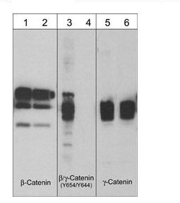
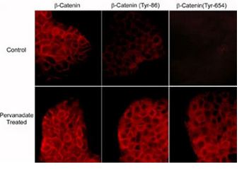 ICC (Immunocytochemistry)
(Immunocytochemical labeling of phosphorylated beta-Catenin in control and pervanadate-treated A431 cells. The cells were labeled with mouse monoclonal beta-Catenin (CM1181) or rabbit polyclonal beta-Catenin (Tyr-86) or beta-Catenin (Y654) antibodies, then the antibodies were detected using appropriate secondary antibodies conjugated to Cy3.)
ICC (Immunocytochemistry)
(Immunocytochemical labeling of phosphorylated beta-Catenin in control and pervanadate-treated A431 cells. The cells were labeled with mouse monoclonal beta-Catenin (CM1181) or rabbit polyclonal beta-Catenin (Tyr-86) or beta-Catenin (Y654) antibodies, then the antibodies were detected using appropriate secondary antibodies conjugated to Cy3.)
beta-Catenin, Polyclonal Antibody (Cat# AAA71610)
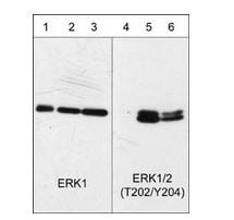
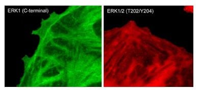 ICC (Immunocytochemistry)
(Immunocytochemical labeling of phosphorylated ERK1 in paraformaldehyde-fixed and NP-40-permeabilized rat A7r5 cells treated with calyculin A. The fixed cells were labeled with mouse monoclonal antibodies to anti-ERK1 (EM2331) and anti-ERK1/2 (Thr-202/Tyr-204) (EM2061). The antibodies were detected using Goat anti-Mouse secondary antibodies conjugated to DyLight 488 (left) and DyLight 594 (right).)
ICC (Immunocytochemistry)
(Immunocytochemical labeling of phosphorylated ERK1 in paraformaldehyde-fixed and NP-40-permeabilized rat A7r5 cells treated with calyculin A. The fixed cells were labeled with mouse monoclonal antibodies to anti-ERK1 (EM2331) and anti-ERK1/2 (Thr-202/Tyr-204) (EM2061). The antibodies were detected using Goat anti-Mouse secondary antibodies conjugated to DyLight 488 (left) and DyLight 594 (right).)
ERK1, Monoclonal Antibody (Cat# AAA71624)
 Application Data
Cofilin 1 (Ser-3), Polyclonal Antibody (Cat# AAA71517)
 ICC (Immunocytochemistry)
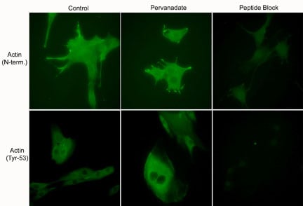
(Immunocytochemical labeling using anti-Actin (N-terminal) and anti-Actin (Tyr-53) polyclonal antibodies in C2C12 cells control (left) or treated with pervanadate (1 mM) for 30 min (middle). The cells were fixed in paraformaldehyde and permeabilized in acetone. Both antibodies were used in the presence of blocking peptide: Actin (N-terminal) peptide or phospho-Actin (Tyr-53) peptide, respectively (right).)
ICC (Immunocytochemistry)
(Immunocytochemical labeling using anti-Actin (N-terminal) and anti-Actin (Tyr-53) polyclonal antibodies in C2C12 cells control (left) or treated with pervanadate (1 mM) for 30 min (middle). The cells were fixed in paraformaldehyde and permeabilized in acetone. Both antibodies were used in the presence of blocking peptide: Actin (N-terminal) peptide or phospho-Actin (Tyr-53) peptide, respectively (right).)
Actin, Polyclonal Antibody (Cat# AAA71548)
 Application Data
IRS-1 (pS312),, Antibody (Cat# AAA71386)
 WB (Western Blot)
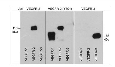
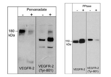
(Left: Western blot image of HUVEC cells untreated (-) or treated with pervanadate (1mM) for 30 min. (+). Right: Western blot image of GST-recombinant VEGFR-2 kinase without (-) or with (+) akaline phosphatase treatment. Both sets of blots were probed with rabbit polyclonal anti-VEGFR-2 (a.a. 1304-1317) or anti-VEGFR-2 (Tyr-801).)
WB (Western Blot)
(Left: Western blot image of HUVEC cells untreated (-) or treated with pervanadate (1mM) for 30 min. (+). Right: Western blot image of GST-recombinant VEGFR-2 kinase without (-) or with (+) akaline phosphatase treatment. Both sets of blots were probed with rabbit polyclonal anti-VEGFR-2 (a.a. 1304-1317) or anti-VEGFR-2 (Tyr-801).)
VEGFR-2, Polyclonal Antibody (Cat# AAA71731)
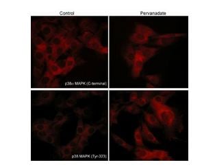 ICC (Immunocytochemistry)
(Immunocytochemical labeling of p38 MAPK in pervanadate-treated mouse C2C12. The cells were labeled with mouse monoclonal p38alpha MAPK and rabbit polyclonal p38 MAPK (Tyr-323) antibodies, then the antibodies were detected using appropriate secondary antibodies conjugated to Cy3.)
ICC (Immunocytochemistry)
(Immunocytochemical labeling of p38 MAPK in pervanadate-treated mouse C2C12. The cells were labeled with mouse monoclonal p38alpha MAPK and rabbit polyclonal p38 MAPK (Tyr-323) antibodies, then the antibodies were detected using appropriate secondary antibodies conjugated to Cy3.)
p38alpha MAP Kinase, Polyclonal Antibody (Cat# AAA71695)
 WB (Western Blot)
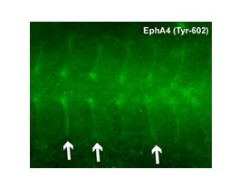
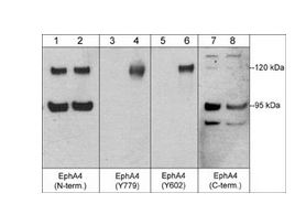
(Western blot analysis of humanuMbilical vein endothelial cells untreated (lanes 1, 3, 5, & 7) or treated with pervanadate (1mM) for 30 min. (lanes 2, 4, 6, & 8). The blot was probed with anti-EphA4 (N-terminal region) (lanes 1 & 2), anti-EphA4 (Tyr-779) (lanes 3 & 4), anti-EphA4 (Tyr-602) (lanes 5 & 6), or anti-EphA4 (C-terminal region) (lanes 7 & 8).)
WB (Western Blot)
(Western blot analysis of humanuMbilical vein endothelial cells untreated (lanes 1, 3, 5, & 7) or treated with pervanadate (1mM) for 30 min. (lanes 2, 4, 6, & 8). The blot was probed with anti-EphA4 (N-terminal region) (lanes 1 & 2), anti-EphA4 (Tyr-779) (lanes 3 & 4), anti-EphA4 (Tyr-602) (lanes 5 & 6), or anti-EphA4 (C-terminal region) (lanes 7 & 8).)
EphA4, Polyclonal Antibody (Cat# AAA71630)
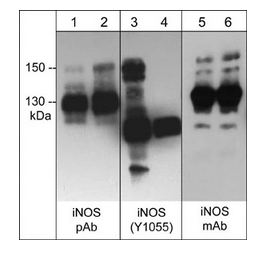
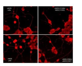 ICC (Immunocytochemistry)
(Immunocytochemical labeling of nNOS phosphorylation in rat PC12 cells differentiated with NGF. The cells were probed with mouse monoclonal (mAb) nNOS (NM4011), and rabbit polyclonal (pAb) nNOS (C-terminal region), nNOS (Tyr-895)/eNOS (Tyr-657), and nNOS (Tyr-1326)/iNOS (Tyr-1055). The antibodies were detected using appropriate secondary antibody conjugated to DyLight 594.)
ICC (Immunocytochemistry)
(Immunocytochemical labeling of nNOS phosphorylation in rat PC12 cells differentiated with NGF. The cells were probed with mouse monoclonal (mAb) nNOS (NM4011), and rabbit polyclonal (pAb) nNOS (C-terminal region), nNOS (Tyr-895)/eNOS (Tyr-657), and nNOS (Tyr-1326)/iNOS (Tyr-1055). The antibodies were detected using appropriate secondary antibody conjugated to DyLight 594.)
iNOS, Polyclonal Antibody (Cat# AAA71676)
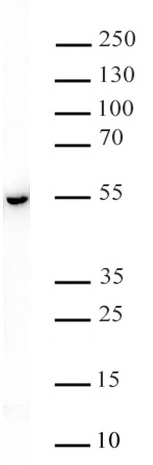
 WB (Western Blot)
EGFR (Tyr-1101), Monoclonal Antibody (Cat# AAA71505)
 Application Data
EGFR (Ser-967), Polyclonal Antibody (Cat# AAA71520)
 WB (Western Blot)
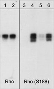
(Western blot analysis of human RhoA GST fusion recombinant unphosphorylated (lanes 1 & 3) or phosphorylated with PKA (lanes 2, 4, 5 & 6). The blots were probed with anti-Rho ( lanes 1 & 2) or with anti-RhoA (Ser-188) (AAA71530; lanes 3-6). The latter antibody was used in the presence of no peptide (lanes 3 & 4), phospho-RhoA (Ser-188) peptide (lane 5), or a non-specific phosphoserine peptide (lane 6).)
WB (Western Blot)
(Western blot analysis of human RhoA GST fusion recombinant unphosphorylated (lanes 1 & 3) or phosphorylated with PKA (lanes 2, 4, 5 & 6). The blots were probed with anti-Rho ( lanes 1 & 2) or with anti-RhoA (Ser-188) (AAA71530; lanes 3-6). The latter antibody was used in the presence of no peptide (lanes 3 & 4), phospho-RhoA (Ser-188) peptide (lane 5), or a non-specific phosphoserine peptide (lane 6).)
RhoA (Ser-188), Polyclonal Antibody (Cat# AAA71530)
 Application Data
SHP1 (Ser-591), Polyclonal Antibody (Cat# AAA71532)
 WB (Western Blot)
CD136 (RON) (pY1239), Antibody (Cat# AAA71376)
 Application Data
IRS-2 (pS731), Antibody (Cat# AAA71387)
 Application Data
Rho Kinase/ROCKII (pT396), Antibody (Cat# AAA71409)
 DB (Dot Blot)
(STAT3 phospho Ser727 pAb tested by dot blot analysis. Dot blot analysis was used to confirm the specificity of STAT3 phospho Ser727 pAb for STAT2 phospho Ser727. Phosphorylated peptides corresponding to the immunogen and related peptides were spotted onto PVDF and probed with the antibody at 1:30,000. The amount of peptide (picomoles) spotted is indicated next to each row. Lane 1: Unmodified Ser727 STAT1 peptide. Lane 2: Phospho Ser727 STAT1 peptide. Lane 3: Unmodified Tyr689 STAT2 peptide. Lane 4: Phospho Tyr689 STAT2 peptide. Lane 5: Unmodified Ser727 STAT3 peptide. Lane 6: Phospho Ser727 STAT3 peptide. Lane 7: Unmodified Tyr705 STAT3 peptide. Lane 8: Phospho Tyr705 STAT3 peptide. Lane 9: Unmodified Ser726 STAT5A/Ser731 STAT5B peptide. Lane 10: Phospho Ser726 STAT5A/Ser731 STAT5B peptide. Lane 11: Unmodified Tyr694 STAT5A/Tyr699 STAT5B peptide. Lane 12: Phospho Tyr694 STAT5A/Tyr699 STAT5B peptide.)
DB (Dot Blot)
(STAT3 phospho Ser727 pAb tested by dot blot analysis. Dot blot analysis was used to confirm the specificity of STAT3 phospho Ser727 pAb for STAT2 phospho Ser727. Phosphorylated peptides corresponding to the immunogen and related peptides were spotted onto PVDF and probed with the antibody at 1:30,000. The amount of peptide (picomoles) spotted is indicated next to each row. Lane 1: Unmodified Ser727 STAT1 peptide. Lane 2: Phospho Ser727 STAT1 peptide. Lane 3: Unmodified Tyr689 STAT2 peptide. Lane 4: Phospho Tyr689 STAT2 peptide. Lane 5: Unmodified Ser727 STAT3 peptide. Lane 6: Phospho Ser727 STAT3 peptide. Lane 7: Unmodified Tyr705 STAT3 peptide. Lane 8: Phospho Tyr705 STAT3 peptide. Lane 9: Unmodified Ser726 STAT5A/Ser731 STAT5B peptide. Lane 10: Phospho Ser726 STAT5A/Ser731 STAT5B peptide. Lane 11: Unmodified Tyr694 STAT5A/Tyr699 STAT5B peptide. Lane 12: Phospho Tyr694 STAT5A/Tyr699 STAT5B peptide.)
STAT3 phospho Ser727, Polyclonal Antibody (Cat# AAA59894)
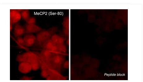 ICC (Immunocytochemistry)
(Immunocytochemical labeling of MeCP2 phosphorylation in rat PC12 cells differentiated with NGF. The cells were probed with MeCP2 (Ser-80) rabbit polyclonal antibody (MP4601) in the absence (left) or presence (right) of blocking peptide (MX4605). The antibody was detected using appropriate secondary antibody conjugated to DyLight 594.)
ICC (Immunocytochemistry)
(Immunocytochemical labeling of MeCP2 phosphorylation in rat PC12 cells differentiated with NGF. The cells were probed with MeCP2 (Ser-80) rabbit polyclonal antibody (MP4601) in the absence (left) or presence (right) of blocking peptide (MX4605). The antibody was detected using appropriate secondary antibody conjugated to DyLight 594.)
MeCP2, Polyclonal Antibody (Cat# AAA71669)
 ICC (Immunocytochemistry)
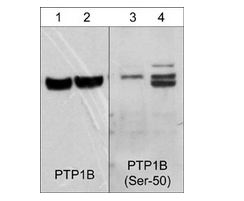
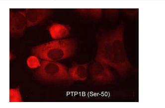
(Immunocytochemical labeling of PTP1B in aldehyde-fixed and NP-40 permeabilized human NCI-H1915 lung carcinoma cells. The cells were labeled with rabbit polyclonal anti-PTP1B (Ser-50) (PP2411) phosphospecific antibody. The antibody was detected using appropriate secondary antibody conjugated to DyLight 594.)
ICC (Immunocytochemistry)
(Immunocytochemical labeling of PTP1B in aldehyde-fixed and NP-40 permeabilized human NCI-H1915 lung carcinoma cells. The cells were labeled with rabbit polyclonal anti-PTP1B (Ser-50) (PP2411) phosphospecific antibody. The antibody was detected using appropriate secondary antibody conjugated to DyLight 594.)
PTP1B, Polyclonal Antibody (Cat# AAA71693)
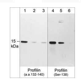
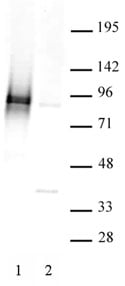
 WB (Western Blot)
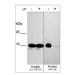
(Western blot of human recombinant Profilin-1 phosphorylated in vitro with PKCalpha kinase then untreated (-) or treated with lambda phosphatase (+). The blots were probed with anti-Profilin (a.a. 126-137) (left panel) or anti-Profilin (Ser-138) phospho-specific (right panel) antibodies at 1:1000.)
WB (Western Blot)
(Western blot of human recombinant Profilin-1 phosphorylated in vitro with PKCalpha kinase then untreated (-) or treated with lambda phosphatase (+). The blots were probed with anti-Profilin (a.a. 126-137) (left panel) or anti-Profilin (Ser-138) phospho-specific (right panel) antibodies at 1:1000.)
Profilin, Polyclonal Antibody (Cat# AAA71697)
 ICC (Immunocytochemistry)
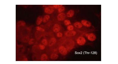
(Immunocytochemical labeling of phosphorylated Sox2 in aldehyde fixed and NP-40 permeabilized human NCI-H446 lung carcinoma cells. The cells were labeled with rabbit polyclonal anti-Sox2 (Thr-128) phospho-specific (SP0381). The antibody was detected using goat anti-rabbit Ig:DyLight 594.)
ICC (Immunocytochemistry)
(Immunocytochemical labeling of phosphorylated Sox2 in aldehyde fixed and NP-40 permeabilized human NCI-H446 lung carcinoma cells. The cells were labeled with rabbit polyclonal anti-Sox2 (Thr-128) phospho-specific (SP0381). The antibody was detected using goat anti-rabbit Ig:DyLight 594.)
Sox2, Polyclonal Antibody (Cat# AAA71709)
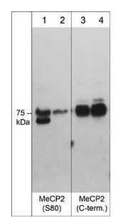
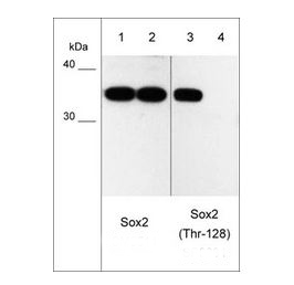
 WB (Western Blot)
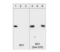
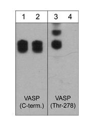
(Western blot of HeLa stimulated with calyculin A (lanes 1-4). The blots were untreated (lane 1 & 3) or treated with lambda phosphatase (lane 2 & 4), then probed with anti-SK1 (Central region) SP1621 (lanes 1 & 2) or anti-SK1 (Ser-225) SP1641 (lanes 3 & 4).)
WB (Western Blot)
(Western blot of HeLa stimulated with calyculin A (lanes 1-4). The blots were untreated (lane 1 & 3) or treated with lambda phosphatase (lane 2 & 4), then probed with anti-SK1 (Central region) SP1621 (lanes 1 & 2) or anti-SK1 (Ser-225) SP1641 (lanes 3 & 4).)
Sphingosine Kinase 1, Polyclonal Antibody (Cat# AAA71711)
 Application Data
Rho Kinase/ROCKII (Paired T249), Antibody (Cat# AAA71407)
 ICC (Immunocytochemistry)
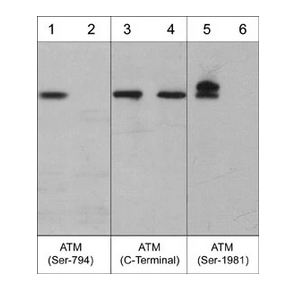
(Immunocytochemical labeling of ATM phosphorylation in control (Top row) or calyculin A-treated A431 cells (Bottom row). The cells were labeled with mouse monoclonal ATM (C-terminal region) (AM3611) and ATM (Ser-1981) (AM3661). The antibodies were detected using goat anti-mouse-DyLight 594.)
ICC (Immunocytochemistry)
(Immunocytochemical labeling of ATM phosphorylation in control (Top row) or calyculin A-treated A431 cells (Bottom row). The cells were labeled with mouse monoclonal ATM (C-terminal region) (AM3611) and ATM (Ser-1981) (AM3661). The antibodies were detected using goat anti-mouse-DyLight 594.)
ATM, Monoclonal Antibody (Cat# AAA71562)
CRMP2 Phospho-Regulation, Antibody Sampler Kit (Cat# AAA71573)
 ICC (Immunocytochemistry)
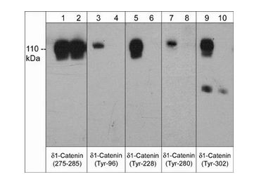
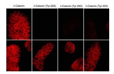
(Immunocytochemical labeling of delta1-Catenin in untreated (Top) or pervanadate-treated (bottom) A431 cells. The cells were labeled with mouse monoclonal delta1-Catenin (a.a. 275-285), delta1-Catenin (Tyr-228), delta1-Catenin (Tyr-280), or delta1-Catenin (Tyr-302) antibodies. The antibodies were detected using donkey anti-mouse secondary antibodies conjugated to Cy3.)
ICC (Immunocytochemistry)
(Immunocytochemical labeling of delta1-Catenin in untreated (Top) or pervanadate-treated (bottom) A431 cells. The cells were labeled with mouse monoclonal delta1-Catenin (a.a. 275-285), delta1-Catenin (Tyr-228), delta1-Catenin (Tyr-280), or delta1-Catenin (Tyr-302) antibodies. The antibodies were detected using donkey anti-mouse secondary antibodies conjugated to Cy3.)
delta1-Catenin, Monoclonal Antibody (Cat# AAA71595)
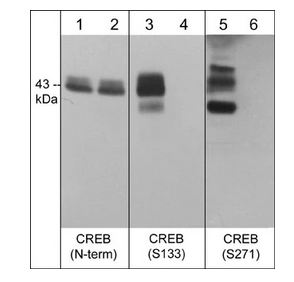
 ICC (Immunocytochemistry)
(Immunocytochemical labeling of phosphorylated CREB in calyculin A-treated A431 cells. The cells were fixed in paraformaldehyde and permeabilized using NP-40 before labeling with mouse monoclonal CREB (Ser-133). The antibody was detected using goat anti-mouse DyLight 594.)
ICC (Immunocytochemistry)
(Immunocytochemical labeling of phosphorylated CREB in calyculin A-treated A431 cells. The cells were fixed in paraformaldehyde and permeabilized using NP-40 before labeling with mouse monoclonal CREB (Ser-133). The antibody was detected using goat anti-mouse DyLight 594.)
CREB, Monoclonal Antibody (Cat# AAA71596)
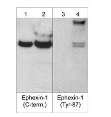
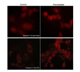 ICC (Immunocytochemistry)
(Immunocytochemical labeling of phosphorylated Exphexin-1 in pervanadate-treated mouse C2C12. The cells were labeled with rabbit polyclonal Ephexin-1 (C-terminal region) and Ephexin-1 (Tyr-87) antibodies, then the antibodies were detected using appropriate secondary antibodies conjugated to Cy3.)
ICC (Immunocytochemistry)
(Immunocytochemical labeling of phosphorylated Exphexin-1 in pervanadate-treated mouse C2C12. The cells were labeled with rabbit polyclonal Ephexin-1 (C-terminal region) and Ephexin-1 (Tyr-87) antibodies, then the antibodies were detected using appropriate secondary antibodies conjugated to Cy3.)
Ephexin-1, Polyclonal Antibody (Cat# AAA71632)
 WB (Western Blot)
(Western blot analysis of A431 cells treated with pervanadate (1 mM) for 30 min. Blots were probed with anti-IKBa (lane 1), anti-IKBa (Tyr-42) (IP1 031; lanes 2-5), or anti-IKBa (Tyr-305) (IP1 041 ; lanes 6-9). In some lanes, the antibodies were used in the absence (lane 2 & 6) or presence of IKBa (Tyr-42) (lane 3 & 8) or IKBa (Tyr-305) (lane 4 & 7) blocking peptides, or BSA conjugated to phospho-tyrosine (lane 5 & 9).)
WB (Western Blot)
(Western blot analysis of A431 cells treated with pervanadate (1 mM) for 30 min. Blots were probed with anti-IKBa (lane 1), anti-IKBa (Tyr-42) (IP1 031; lanes 2-5), or anti-IKBa (Tyr-305) (IP1 041 ; lanes 6-9). In some lanes, the antibodies were used in the absence (lane 2 & 6) or presence of IKBa (Tyr-42) (lane 3 & 8) or IKBa (Tyr-305) (lane 4 & 7) blocking peptides, or BSA conjugated to phospho-tyrosine (lane 5 & 9).)
I-kappa-Ba (Tyr-42), Polyclonal Antibody (Cat# AAA71521)
 Application Data
HDAC3 Phospho Ser424, Polyclonal Antibody (Cat# AAA60353)
 Application Data
c-Cbl (Tyr-700), Monoclonal Antibody (Cat# AAA71501)
 Application Data
Paxillin (Tyr-31), Monoclonal Antibody (Cat# AAA71507)
 Application Data
EGFR (Ser-1142), Polyclonal Antibody (Cat# AAA71519)
 Application Data
Integrin b4 (Tyr-1494), Polyclonal Antibody (Cat# AAA71523)
 Application Data
(Western Blot: The whole cell lysate derived from EGF stimulated A431 immunoblotted by Rabbit anti AMPK alpha 1 (pT172) antibody (Cat#AAA71372) at 1:500 (lane 2).BSA was loaded as a negative control (Lane 3).Observed a major immunoreactive band at molecular weight ~63kDa.)
Application Data
(Western Blot: The whole cell lysate derived from EGF stimulated A431 immunoblotted by Rabbit anti AMPK alpha 1 (pT172) antibody (Cat#AAA71372) at 1:500 (lane 2).BSA was loaded as a negative control (Lane 3).Observed a major immunoreactive band at molecular weight ~63kDa.)
AMPK-alpha (pT172), Antibody (Cat# AAA71372)
 ICC (Immunocytochemistry)
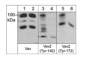
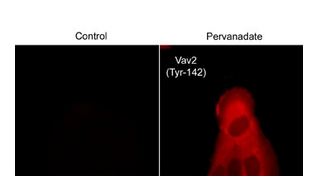
(Immunocytochemical labeling of VAV2 in control and pervanadate-treated human A431 cells. The cells were fixed in paraformaldehyde and permeabilized using NP-40. Then labeled with rabbit polyclonal Vav2 (Tyr-142). The antibody was detected using goat anti-rabbit DyLight 594.)
ICC (Immunocytochemistry)
(Immunocytochemical labeling of VAV2 in control and pervanadate-treated human A431 cells. The cells were fixed in paraformaldehyde and permeabilized using NP-40. Then labeled with rabbit polyclonal Vav2 (Tyr-142). The antibody was detected using goat anti-rabbit DyLight 594.)
Vav2, Polyclonal Antibody (Cat# AAA71727)
 ICC (Immunocytochemistry)
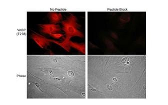
(Immunocytochemical labeling of VASP phosphorylation in rabbit spleen fibroblasts treated with Calyculin A. The cells were labeled with rabbit polyclonal VASP (Thr-278) antibody, then detected using appropriate secondary antibodies conjugated to Cy3. The antibody was used in the absence (top left) or presence (top right) of blocking peptide (VX2785). Corresponding phase images are shown bottom left and right.)
ICC (Immunocytochemistry)
(Immunocytochemical labeling of VASP phosphorylation in rabbit spleen fibroblasts treated with Calyculin A. The cells were labeled with rabbit polyclonal VASP (Thr-278) antibody, then detected using appropriate secondary antibodies conjugated to Cy3. The antibody was used in the absence (top left) or presence (top right) of blocking peptide (VX2785). Corresponding phase images are shown bottom left and right.)
VASP, Polyclonal Antibody (Cat# AAA71729)
 ICC (Immunocytochemistry)
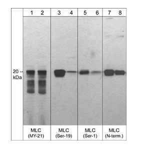
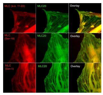
(Immunocytochemical labeling of phosphorylated MLC in paraformaldehyde fixed A7r5 cells. The cells were dual-labeled with anti-MLC (MM3441; middle) and anti-MLC (MP4201; top left), anti-MLC (Ser-19) (MP4221; middle left) and anti-MLC (Ser-1) (MP3461; bottom left). Goat anti-Mouse DyLight 488 and Goat anti-Rabbit DyLight 594 were used for detection of primary antibodies. The overlay of staining patterns are shown to the right.)
ICC (Immunocytochemistry)
(Immunocytochemical labeling of phosphorylated MLC in paraformaldehyde fixed A7r5 cells. The cells were dual-labeled with anti-MLC (MM3441; middle) and anti-MLC (MP4201; top left), anti-MLC (Ser-19) (MP4221; middle left) and anti-MLC (Ser-1) (MP3461; bottom left). Goat anti-Mouse DyLight 488 and Goat anti-Rabbit DyLight 594 were used for detection of primary antibodies. The overlay of staining patterns are shown to the right.)
Myosin, Polyclonal Antibody (Cat# AAA71664)
What Are Phospho Antibodies?
Protein phosphorylation is a process where a phosphate group is added to certain amino acid residues of a protein – usually serine (S), threonine (T), or tyrosine (Y) - by enzymes called kinases. This process is integral in controlling cellular signaling, cellular growth, and other biological functions, as explained in our detailed guide to phospho antibodies.
Our catalog includes a wide range of phospho-specific antibodies that can accurately detect this important marker, including phospho antibodies as well as other formats such as monoclonal antibodies and polyclonal antibodies for different research needs.
They perform strongly in widely used laboratory applications such as Western blot, flow cytometry, immunohistochemistry, and immunofluorescence microscopy. We value your trust in us and are committed to providing top-quality products and services. All of our antibodies are guaranteed to work for the applications and species indicated on our website & associated product pages.
What Are The Key Applications of Phospho Antibodies?
1. Western Blotting
One of the first steps a researcher can take in utilizing these phospho-specific antibodies is to check if the antibody works using a technique referred to as Western blot, learn more in our guide on Western blot roles and uses. For those unfamiliar, Western Blot aids in showing whether the protein that the antibody recognizes is appearing at the correct/expected size. These phospho-specific antibodies should also be able to detect changes in the target protein’s phosphorylation (on/off state) when cells are stimulated in certain ways.
2. Staining of Fixed Cells (Immunocytochemistry)
Another routine use of these phospho-specific antibodies, is to test if the antibody is able to demonstrate similar performance when used on fixed cells (intact cells that have been preserved) as it did in the Western blot tests. It is an important aspect in many cases to confirm that the antibody works in actual intact cell samples. Ideally, the method used for cellular fixation should be the same as what is used in pathology labs (like using 10% formalin). To check if the antibody works well in tissue sections (FFPE), researchers will often test it on fixed cells that are processed similar to tissue samples.
3. Specificity Tests Using Peptides
In order to make sure that the antibody is only binding to the right target:
- Laboratory technicians will mix the antibody with phospho-peptides (short segments of the protein containing the phosphate group modification).
- If the antibody signal disappears, it is confirmation that it is binding to the correct phosphorylated location. Such validation approaches are commonly used across different antibody types, including monoclonal antibodies.
- A more robust test is to use both the phosphorylated and non-phosphorylated (dephosphorylated) versions of the protein. The antibody should react only with the phosphorylated one.
- Another method sometimes utilized is to treat the sample with an enzyme, such as alkaline phosphatase, that specifically removes phosphate groups. If the antibody signal disappears after this, it also confirms specificity.
4. Genetic Confirmation
As a final step, scientists can genetically manipulate the nucleotide sequence and alter the target protein by removing the exact site where phosphorylation happens. If the antibody no longer appears to detect the modified protein, it is strong evidence supporting the antibody being specific for that phosphorylated site.
Why Buy Phospho Antibodies Through Us?
- The production laboratory adheres to strict and consistent protocols prior to releasing any of these phospho-specific antibodies:
- Standard methods and proper controls in all tests to ensure high quality.
- These antibodies are tested and validated in different cell types and species.
- High quality control criterion to ensure each batch is consistent, so you will obtain reliable results every time.
FAQ
1. What Are Phospho-Specific Antibodies?
Phospho-specific antibodies are made to detect proteins only when they have a phosphate group linked to a specific amino acid residue. This empowers scientists understand if a protein is "turned on" or active, based on its phosphorylation state.
2. How to Detect Phosphorylated Proteins in a Western Blot?
To find out if a protein is phosphorylated using Western blot:
- Use a phospho-specific antibody that binds only to the phosphorylated form of the protein.
- You can also use a “regular” antibody for the same amino acid sequence of the protein that the phospho-specific antibody is binding to (but in this case, this antibody will not bind if there is a phosphate group present) in order to compare how much of it is phosphorylated versus how much is non-phosphorylated (or “total” protein, if the “normal” antibody’s epitopes are non-phospho-site-specific).
3. How to Choose the Best Antibody?
Here are some simple tips to help you pick the right antibody:
- Know your target
- Match your sample characteristics
- Confirm the intended use is appropriate
- Check “host” and “type”
- Check the “quality” of the presented data/images
- Appraise whether the available validation meets your needs