


EIF6 Polyclonal Antibody | anti-EIF6 antibody
Anti-EIF6 Antibody
Immunohistochemistry (IHC) Paraffin Concentration: 0.5-1ug/ml
ICC (Immunocytochemistry)
(Figure 6. IHC analysis of EIF6 using anti-EIF6 antibody (AAA11655).EIF6 was detected in immunocytochemical section of SW480 cell. Heat mediated antigen retrieval was performed in citrate buffer (pH6, epitope retrieval solution) for 20 mins. The tissue section was blocked with 10% goat serum. The tissue section was then incubated with 1ug/ml rabbit anti-EIF6 Antibody (AAA11655) overnight at 4 degree C. Biotinylated goat anti-rabbit IgG was used as secondary antibody and incubated for 30 minutes at 37 degree C. The tissue section was developed using Strepavidin-Biotin-Complex (SABC) with DAB as the chromogen.)
ICC (Immunocytochemistry)
(Figure 5. IHC analysis of EIF6 using anti-EIF6 antibody (AAA11655).EIF6 was detected in immunocytochemical section of SMMC-7721 cell. Heat mediated antigen retrieval was performed in citrate buffer (pH6, epitope retrieval solution) for 20 mins. The tissue section was blocked with 10% goat serum. The tissue section was then incubated with 1ug/ml rabbit anti-EIF6 Antibody (AAA11655) overnight at 4 degree C. Biotinylated goat anti-rabbit IgG was used as secondary antibody and incubated for 30 minutes at 37 degree C. The tissue section was developed using Strepavidin-Biotin-Complex (SABC) with DAB as the chromogen.)
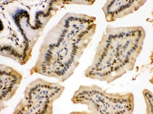
IHC (Immunohistochemistry)
(Figure 4. IHC analysis of EIF6 using anti-EIF6 antibody (AAA11655).EIF6 was detected in paraffin-embedded section of Human Intestinal Cancer Tissue. Heat mediated antigen retrieval was performed in citrate buffer (pH6, epitope retrieval solution) for 20 mins. The tissue section was blocked with 10% goat serum. The tissue section was then incubated with 1ug/ml rabbit anti-EIF6 Antibody (AAA11655) overnight at 4 degree C. Biotinylated goat anti-rabbit IgG was used as secondary antibody and incubated for 30 minutes at 37 degree C. The tissue section was developed using Strepavidin-Biotin-Complex (SABC) with DAB as the chromogen.)
IHC (Immunohistochemistry)
(Figure 3. IHC analysis of EIF6 using anti-EIF6 antibody (AAA11655).EIF6 was detected in paraffin-embedded section of Rat Testis Tissue. Heat mediated antigen retrieval was performed in citrate buffer (pH6, epitope retrieval solution) for 20 mins. The tissue section was blocked with 10% goat serum. The tissue section was then incubated with 1ug/ml rabbit anti-EIF6 Antibody (AAA11655) overnight at 4 degree C. Biotinylated goat anti-rabbit IgG was used as secondary antibody and incubated for 30 minutes at 37 degree C. The tissue section was developed using Strepavidin-Biotin-Complex (SABC) with DAB as the chromogen.)
IHC (Immunohistochemistry)
(Figure 2. IHC analysis of EIF6 using anti-EIF6 antibody (AAA11655).EIF6 was detected in paraffin-embedded section of Mouse Intestine Tissue. Heat mediated antigen retrieval was performed in citrate buffer (pH6, epitope retrieval solution) for 20 mins. The tissue section was blocked with 10% goat serum. The tissue section was then incubated with 1ug/ml rabbit anti-EIF6 Antibody (AAA11655) overnight at 4 degree C. Biotinylated goat anti-rabbit IgG was used as secondary antibody and incubated for 30 minutes at 37 degree C. The tissue section was developed using Strepavidin-Biotin-Complex (SABC) with DAB as the chromogen.)
WB (Western Blot)
(Figure 1. Western blot analysis of EIF6 using anti-EIF6 antibody (AAA11655).Electrophoresis was performed on a 5-20% SDS-PAGE gel at 70V (Stacking gel) / 90V (Resolving gel) for 2-3 hours. The sample well of each lane was loaded with 50ug of sample under reducing conditions.Lane 1: Rat Cardiac Muscle Tissue Lysate,Lane 2: Rat Liver Tissue Lysate,Lane 3: Mouse Liver Tissue Lysate,Lane 4: Human Placenta Tissue Lysate,Lane 5: COLO320 Whole Cell Lysate,Lane 6: HELA Whole Cell Lysate.After Electrophoresis, proteins were transferred to a Nitrocellulose membrane at 150mA for 50-90 minutes. Blocked the membrane with 5% Non-fat Milk/ TBS for 1.5 hour at RT. The membrane was incubated with rabbit anti-EIF6 antigen affinity purified polyclonal antibody at 0.5ug/mL overnight at 4 degree C, then washed with TBS-0.1%Tween 3 times with 5 minutes each and probed with a goat anti-rabbit IgG-HRP secondary antibody at a dilution of 1:10000 for 1.5 hour at RT. The signal is developed using an Enhanced Chemiluminescent detection (ECL) kit with Tanon 5200 system. A specific band was detected for EIF6 at approximately 27KD. The expected band size for EIF6 is at 27KD.)
Background: EIF6 (Eukaryotic Translation Initiation Factor 6), also called EIF3A or ITGB4BP, is a human gene. By fluorescence in situ hybridization, Sanvito et al. (1998) mapped the ITGB4BP gene to 20q11.2. Ceci et al. (2003) demonstrated that the ribosomal 60S subunit is activated by release of EIF6. In the cytoplasm, EIF6 is bound to free 60S but not to 80S subunits. Furthermore, EIF6 interacts in the cytoplasm with RACK1, a receptor for activated protein kinase C. Gandin et al. (2008) demonstrated that mammalian eIF6 is required for efficient initiation of translation in vivo. Eif6-null mouse embryos were lethal at preimplantation. Heterozygous mice had 50% reduction of eIF6 levels in all tissues, and showed reduced mass of hepatic and adipose tissues due to a lower number of cells and to impaired G1/S cell cycle progression.
NCBI and Uniprot Product Information
Similar Products


ANP Antibody

ANP Antibody


ANP Antibody


