Human Fc Fragment of IgG Low Affinity IIIb Receptor ELISA Kit | FcgammaR3B elisa kit
Human Fc Fragment of IgG Low Affinity IIIb Receptor ELISA Kit
Reactivity
Human
Synonyms
Fc Fragment of IgG Low Affinity IIIb Receptor; N/A; Human Fc Fragment of IgG Low Affinity IIIb Receptor ELISA Kit; FcgammaR3B/CD16b/FCG3/FCGR3B/FcR-10/FCRIIIB/IGFR3/CD16/CD16b/CD16B/CD16b antigen/Fc fragment of IgG; low affinity IIIb; receptor (CD16b)/Fc fragment of IgG; receptor for (CD16)/FCG3/Fc-gamma receptor IIIb (CD 16)/Fc-gamma RIII/Fc-gamma RIIIb/Fc-gamma RIII-beta/FCGR3/FcR-10/fcR-10/fcRIII/FcRIII/FcRIIIb/fcRIIIb/IGFR3/IgG Fc receptor III-1/low affinity immunoglobulin gamma Fc region receptor III-B; FcgammaR3B elisa kit
Reactivity
Human
Specificity
This assay has high sensitivity and excellent specificity for detection of FcgammaR3A. No significant cross-reactivity or interference between FcgammaR3A and analogues was observed.
Assay Type
Sandwich ELISA, Double Antibody
Samples
Serum, Plasma, Cell Culture Supernatant, cell or tissue lysate, Other liquid samples
Detection Range
15.625-1000pg/ml
Sensitivity
9.375pg/ml
Intra-assay Precision
Intra-assay Precision (Precision within an assay): 3 samples with low, middle and high level Fcgamma R3A were tested 20 times on one plate, respectively. Intra-Assay: CV<8%
Inter-assay Precision
Inter-assay Precision (Precision between assays): 3 samples with low, middle and high level Fc gammaR3A were tested on 3 different plates, 8 replicates in each plate. CV (%) = SD/meanX100. Inter-Assay: CV<10%
Preparation and Storage
Store entire kit at 2-8C for short-term. For longer-term, please store the microplate & standard at -20C, while the remaining reagents can be stored at 2-8C
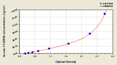
Standard Curve (Sample)
Related Product Information for FcgammaR3B elisa kit
Principle of the Assay: This kit was based on sandwich enzyme-linked immune-sorbent assay technology. Anti-Fcgamma R3A antibody was pre-coated onto 96-well plates. And the biotin conjugated anti-FcgammaR3A antibody was used as detection antibodies. The standards, test samples and biotin conjugated detection antibody were added to the wells subsequently, and washed with wash buffer. HRP-Streptavidin was added and unbound conjugates were washed away with wash buffer. TMB substrates were used to visualize HRP enzymatic reaction. TMB was catalyzed by HRP to produce a blue color product that changed into yellow after adding acidic stop solution. The density of yellow is proportional to the FcgammaR3A amount of sample captured in plate. Read the O.D. absorbance at 450nm in a microplate reader, and then the concentration of FcgammaR3A can be calculated.