


Goat MDH/MDH2 Polyclonal Antibody | anti-MDH2 antibody
MDH/MDH2 Goat anti-Human Polyclonal (Internal Region) Antibody
Peptide-ELISA: 1:8000
WB: 0.1-0.3ug/ml
Peptide ELISA: antibody detection limit dilution 1:8000. Western blot: Approx 35kDa band observed in lysates of cell lines HeLa, HepG2 and NIH3T3 (calculated MW of 35.5kDa according to Human NP_005909.2 and 35.6kDa according to Mouse NP_032643.2). Recommended concentration: 0.1-0.3ug/ml. Approx 35kDa band observed in Human, Mouse, Rat and Pig Heart lysates (calculated MW of 35.5kDa according to Human NP_005909.2 and 35.6kDa according to Mouse NP_032643.2, Rat NP_112413.2 and Pig NP_001231082.1). Recommended concentration: 0.01-0.03ug/ml.
IHC (Immunohistochemistry)
(MDH/MDH2 Antibody-Human Heart: Formalin-Fixed, Paraffin-Embedded (FFPE))
IHC (Immunohistchemistry)
(MDH/MDH2 Antibody-Human Liver: Formalin-Fixed, Paraffin-Embedded (FFPE))
IHC (Immunohistochemistry)
(MDH/MDH2 Antibody-Human Skeletal Muscle: Formalin-Fixed, Paraffin-Embedded (FFPE))
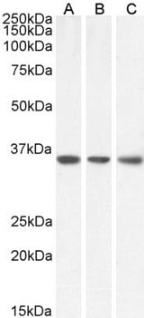
WB (Western Blot)
(MDH/MDH2 Antibody-Antibody (0.1ug/ml) staining of HeLa (A), HepG2 (B) and NIH3T3 (C) lysate (35ug protein in RIPA buffer). Primary incubation was 1 hour. Detected by chemiluminescence.)
WB (Western Blot)
(MDH/MDH2 Antibody-Antibody (0.01ug/ml) staining of Human (A), Mouse (B), Rat (C) and Pig (D) Heart lysate (35ug protein in RIPA buffer). Primary incubation was 1 hour. Detected by chemiluminescence.)
WB (Western Blot)
(MDH/MDH2 Antibody-Goat anti-MDH2 Antibody (0.1ug/ml) staining of HeLa (A), HepG2 (B) and NIH3T3 (C) lysate (35ug protein in RIPA buffer). Primary incubation was 1 hour. Detected by chemiluminescencence.)
WB (Western Blot)
(MDH/MDH2 Antibody-Goat anti-MDH2 Antibody (0.01ug/ml) staining of Human (A), Mouse (B), Rat (C) and Pig (D) Heart lysate (35ug protein in RIPA buffer). Primary incubation was 1 hour. Detected by chemiluminescencence.)


