Rabbit anti-Human Phosphoinositide 3 Kinase, p110 beta Polyclonal Antibody | anti-PI3Kbeta antibody
Phosphoinositide 3 Kinase, p110 beta (DKFZp779K1237, MGC133043, p110beta, Phosphatidylinositol 3 Kinase Catalytic beta Polypeptide, Phosphatidylinositol-4,5-bisphosphate 3-kinase Catalytic Subunit beta Isoform, Phosphoinositide 3 Kinase Catalytic beta Pol
ELISA: 0.5ng/well
WB: 0.05-0.1ug/ml
Applications are based on unconjugated antibody.
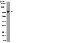
WB (Western Blot)
(Western Blot Analysis: Representative lot data. Lysate from HEK-293 cells was resolved by electrophoresis, transferred to PVDF and probed with anti-PI3 Kinase, p110beta (0.05 ug/mL). Proteins were visualized using donkey anti-rabbit secondary antibody conjugated to HRP and chemiluminescence detection. Arrow indicates PI3 Kinase, p110beta (~110kD).)
IP (Immunoprecipitation)
(Immunoprecipitation: 10ug of was used to immunoprecipitate from 500ug of Jurkat whole cell lysate. The antibody was collected on Protein A beads and eluted with sample buffer. 5uL of Jurkat whole cell lysate was then resolved via SDS-PAGE, transferred to PVDF and probed with 0.5ug . Proteins were visualized using anti-rabbit-HRP conjugate and an ECL system.)
ICC (Immunocytochemistry)
(Immunocytochemistry: PI3-Kinase, p110b staining of human skeletal muscle. Tissue pre-treated with Citrate pH 6.0 antigen-retrieval. diluted to 1:100, IHC-Select Detection with HRP-DAB. Immunoreactivity is seeing as a staining pattern of cross banding striated muscle fibers.)
ICC (Immunocytochemistry)
(Immunocytochemistry: PI3-Kinase, p110b staining in colorectal carcinoma. Tissue pre-treated with Citrate pH 6.0 antigen-retrieval. diluted to 1:100, IHC-Select Detection with HRP-DAB. Immunoreactivity is seeing as a plasma membrane staining pattern.)
ICC (Immunocytochemistry)
(Confocal Immunocytochemistry Analysis: HeLa cells were fixed, permeablized, and stained with (Cy3, red), DAPI (blue, nuclei), and Phalloidin-AlexaFluor488 (actin, green). Figure on the left has the DAPI filter off.)
ICC (Immunocytochemistry)
(Immunocytochemistry: PI3-Kinase, p110b staining of human kidney 2 mm array spot. Tissue pre-treated with Citrate pH 6.0 antigen-retrieval. diluted to 1:100, IHC-Select Detection with HRP-DAB. Immunoreactivity is see as a staining to include the thin and distal microtubules.)