


Rabbit FUS Polyclonal Antibody | anti-FUS antibody
FUS Polyclonal Antibody
IHC: 1:50 - 1:100
IF: 1:50 - 1:100
IF (Immunofluorescence)
(Immunofluorescence analysis of NIH/3T3 cells using FUS antibody (AAA28170) at dilution of 1:100. Blue: DAPI for nuclear staining.)
IF (Immunofluorescence)
(Immunofluorescence analysis of C6 cells using FUS antibody (AAA28170) at dilution of1:100. Blue: DAPI for nuclear staining.)
IHC (Immunohistchemistry)
(Immunohistochemistry of paraffin-embedded mouse spleen using FUS antibody (AAA28170) at dilution of 1:100 (40x lens).)
IHC (Immunohistochemistry)
(Immunohistochemistry of paraffin-embedded mouse brain using FUS antibody (AAA28170) at dilution of 1:100 (40x lens).)
IHC (Immunohistochemistry)
(Immunohistochemistry of paraffin-embedded human breast cancer using FUS antibody (AAA28170) at dilution of 1:100 (40x lens).)
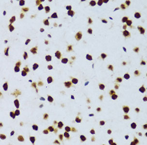
IHC (Immunohistochemistry)
(Immunohistochemistry of paraffin-embedded rat brain using FUS antibody (AAA28170) at dilution of 1:100 (40x lens).)
WB (Western Blot)
(Western blot analysis of extracts from normal (control) and FUS knockout (KO) 293T cells, using FUS antibody (AAA28170) at 1:3000 dilution.Secondary antibody: HRP Goat Anti-Rabbit IgG (H+L) at 1:10000 dilution.Lysates/proteins: 25ug per lane.Blocking buffer: 3% non fat dry milk in TBST.Detection: ECL Basic Kit.Exposure time: 1s.)
WB (Western Blot)
(Western blot analysis of extracts ofvarious cell lines, using FUS antibody (AAA28170) at 1:1000 dilution.Secondary antibody: HRP Goat Anti-Rabbit IgG (H+L) at 1:10000 dilution.Lysates/proteins: 25ug per lane.Blocking buffer: 3% nonfat dry milk in TBST.Detection: ECL Basic Kit.Exposure time: 1s.)
NCBI and Uniprot Product Information
Observed MW: 70kDa


