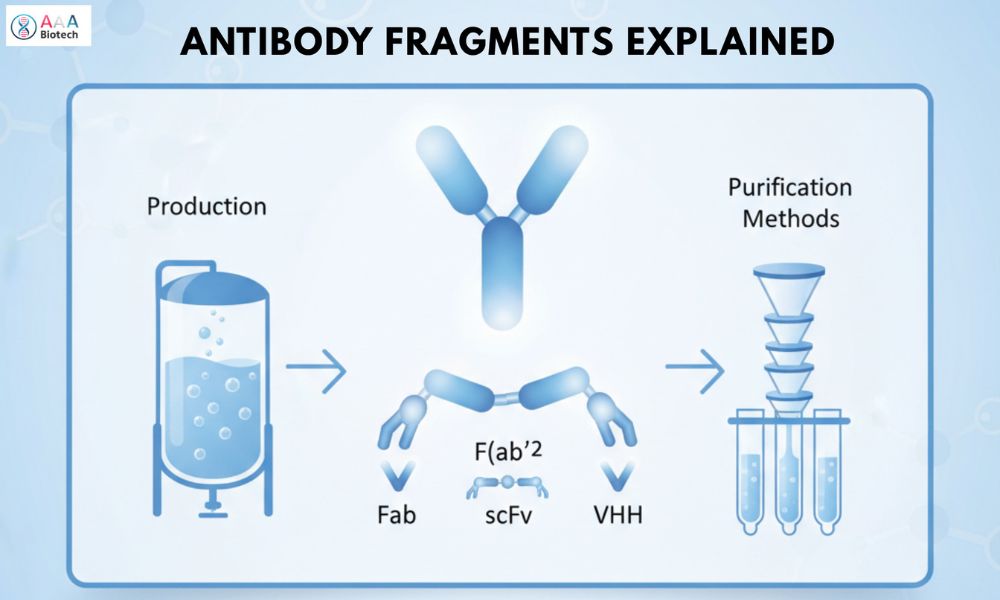
Antibody Fragments Explained: Types, Production, and Purification Methods
 Cynthia
12 min read
Cynthia
12 min read
In this Article
All of the products listed in AAA Biotech’s catalog are strictly for research-use only (RUO).
Key Takeaways
- Antibody fragments are smaller, engineered versions of full antibodies that retain target specificity.Antibody fragments are smaller, engineered versions of full antibodies that retain target specificity.
- Fab, scFv, and VHH (Nanobody) are the main fragment types.
- Fragmentation can be enzymatic or recombinant, with recombinant methods preferred for their superior consistency.
- pH and enzyme concentration critically affect efficiency in enzymatic cleavage.
- Fragment size affects stability, tissue penetration, and half-life.
- E. coli, yeast, and mammalian cells serve as the primary expression systems.
- “Protein L”-affinity chromatography enables selective purification.
- Engineering can extend fragment stability and systemic persistence.
The clock is ticking in modern medicine, and every day, researchers face critical challenges in delivering therapeutic agents through dense tumor tissue, crossing the blood-brain barrier, or neutralizing fast-acting toxins before they cause irreparable damage.
The IgG antibody (the body’s traditional hero) is often too large, too slow, and too prone to triggering unwanted immune responses to win these battles.
“This challenge has created an urgent, multi-billion-dollar race to master the miniature: antibody fragments.”
These Y-shaped, highly precise tools help in eliminating unnecessary molecular bulk and achieve tissue penetration and modularity that full-sized biologics simply cannot match, unlocking a new frontier in diagnostics and therapeutics.
With this expert guide, we aim to help medical scholars and researchers understand the emerging field of antibody fragments, their types, production, and purification methods that help deliver reliable research results.
Defining The Precision Toolkit
What is an Antibody Fragment?
In simple terms, an antibody fragment is a reduced-size derivative of a full-length immunoglobulin, engineered to retain the full antigen-binding specificity of the parent molecule while shedding the bulk of the constant domains.
These mini-biologics are characterized by their smaller size, low immunogenicity, and vastly superior tissue-penetrative capabilities.
The core design philosophy of modern biotherapeutics: less is often more, provided the specific function is retained.
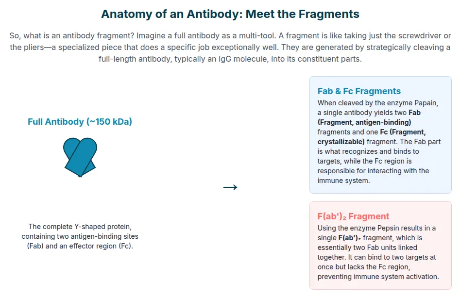
What are the Fab and Fc Fragments of Antibodies?
The logic behind fragment therapeutics rests on the structural segregation achieved through fragmentation:
01. The Fab Fragment (Fragment antigen-binding): This is the business end of the antibody, the functional unit responsible for recognizing and binding the target. A single Fab fragment consists of two structural pairings:
NOTE: The Variable Light (VL) and Constant Light (CL) domains of the light chain are paired with the Variable Heavy (VH) and first Constant Heavy (CH1) domains of the heavy chain.
These four domains are held securely by native inter-chain disulfide bonds and non-covalent interactions.
02. The Fc Fragment (Fragment crystallizable): This region, derived solely from the constant domains, is the functional tail of the antibody. In a full-length IgG, the Fc fragment is critical for mediating immune functions like alerting immune cells via Antibody-Dependent Cell-mediated Cytotoxicity (ADCC) or triggering the Complement system (CDC) and for ensuring long systemic presence by interacting with the neonatal Fc receptor (FcRn).
The intentional removal of the Fc region in fragments is highly desirable for applications where immune activation is dangerous (e.g., diagnostics, neutralizing toxins) and thus hopefully avoided.
The Magnificent Miniatures: Size, Structure, and Trade-offs
The engineering of antibody fragments revolves around two primary binding formats: the classic Fab format and the variable-domain-only (Fv) format. Understanding the types of antibody fragments is crucial, as their specific design dictates their utility.
The Classics: Fab and F(ab')₂
- Fab Fragment: The monovalent Fab fragment typically weighs
in around 50–55 kDa. Its structure, held firm by disulfide bonds, gives it
moderate stability and excellent binding capability. It is a workhorse in
research, being the largest core fragment.
- F(ab')₂ Fragment: This fragment is slightly larger, approximately ~100 kDa. Crucially, it is bivalently linked, retaining two binding sites connected by disulfide bonds in the hinge region. This bivalency confers higher functional avidity - a stronger, more persistent binding effect without the liability of the Fc domain.
The Engineered Workhorse: scFv
The Single-Chain Fv (scFv fragment) is the modular backbone of next-generation therapies. It represents the absolute minimum structure necessary to retain binding: the VH and VL domains only.
The challenge?
These two domains dissociate easily. The solution, pioneered through genetic engineering, was the incorporation of a flexible peptide linker.
- Size and Stability: At 25–30 kDa, the scFv is half the size of a Fab fragment, lending it better penetration. However, this format is notoriously prone to aggregation and stability issues due to its reduced domain interactions. Overcoming this requires extensive engineering, such as modifying hydrophobic patches or introducing engineered disulfide bonds into the framework regions, a topic frequently highlighted in discussions amongst bioprocess engineers.
The Nanobody Niche: VHH (Single-Domain Antibodies)
The VHH domain, often trademarked as a Nanobody, offers the highest stability and smallest size among all practical fragments. Derived from the unique heavy-chain-only antibodies of camelids (like llamas), the VHH consists solely of the heavy-chain variable domain.
- Extreme Robustness: With a molecular weight of only 12–15
kDa, about one-tenth the size of a conventional antibody, VHHs are exceptionally
resistant to high temperatures, extreme pH, and proteases.
- Targeting Advantage: Their single-domain structure often
features long CDR3 loops that jut out like a probe, allowing them to bind to
cryptic epitopes - hidden pockets or canyons on target antigens, that are
physically inaccessible to larger antibodies.
| Fragment Type | Structure | Molecular Weight (Approx.) | Key Advantage | Major Limitation |
|---|---|---|---|---|
| Fab | VH, CH1, VL, CL (Disulfide linked) | 50–55 kDa | Moderate stability; High affinity | Short half-life; Moderate penetration |
| scFv | VH and VL (Peptide linker) | 25–30 kDa | Modular for engineering; High flexibility | Low stability; Aggregation-prone |
| VHH (Nanobody) | Single Variable Heavy Domain | 12–15 kDa | Extreme stability; Highest penetration | Very short systemic half-life |
Table 01: Key Differences of Fab, scFv, and VHH Fragments
The Bioprocess Journey: Fragment Production
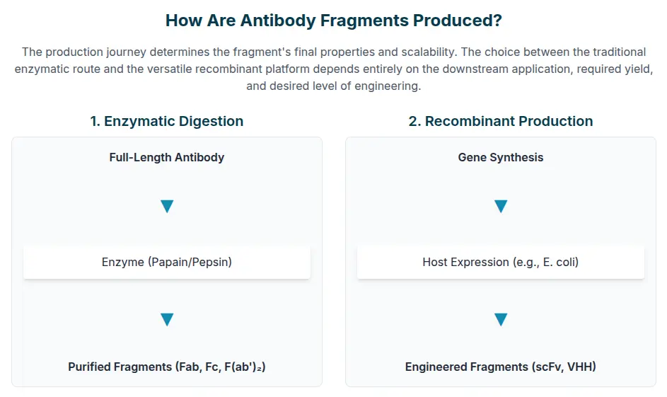
How are Antibody Fragments Produced?
Antibody fragment production relies on two distinct methods:
01. Technologically sophisticated pathways: Enzymatic method
02. The scalable and genetically controlled: Recombinant method
The Old School: Enzymatic Fragmentation
The initial method involved controlled proteolysis using enzymes like Papain (yielding Fab and Fc) or Pepsin (yielding the F(ab')₂ fragment). The advantage here is speed - fragments can be generated rapidly from commercially available full-length IgG.
- This method represents a classic trial-and-error pain point, and research on
fragmentation optimization highlights that the precise cleavage pattern varies
unpredictably for each new antibody clone.
- Researchers must systematically screen multiple parameters — pH (which has
the greatest impact on efficiency), enzyme concentration, and reaction
time.
- These screenings consume large and costly quantities of starting
material.
- The high cost in both time and resources often makes the modern recombinant antibody fragments approach the preferred standard.
Engineering the Future: Recombinant Production
Recombinant antibody fragments are produced by genetically engineering a host cell to express the fragment's specific DNA sequence. Host selection is a critical, consequential decision that balances cost, speed, and product quality:
1. Prokaryotic Systems (E. coli): High Yield, High Risk
Escherichia coli (E. coli) is the economic powerhouse of
fragment production, offering superior growth kinetics, low-cost media, and the
ability to achieve high cell-density fermentation, leading to massive potential
yields.
Yet, E. coli faces a major biophysical obstacle: it cannot perform the complex folding or disulfide bond formation required for complex fragments like Fab. This often results in the over-expressed protein aggregating into dense, inactive masses called Inclusion Bodies (I.B.) inside the cell.
Recovering the active fragment requires stripping the protein with harsh denaturing agents (like urea) followed by arduous and often low-yield protein refolding procedures.
2. Eukaryotic Systems: The Quality Trade-off
For fragments requiring precise native folding and complex post-translational
modifications, eukaryotic systems are often necessary:
- Lower Eukaryotes (Pichia pastoris): This yeast
system provides a better balance than E. coli, utilizing secretion
pathways and allowing proper disulfide bond formation, often yielding
better-folded fragments.
- Mammalian Cells (e.g., CHO): These systems represent the
gold standard for therapeutic-grade quality, providing the native environment
necessary for correct folding and modification. They are typically mandatory for
complex, multi-chain structures and bispecific fragments. The trade-off is a
significantly higher cost and lower expression titer compared to microbial
systems.
| Expression System | Typical Yield | Key Advantage | Major Challenge | Ideal Fragment Types |
|---|---|---|---|---|
| E. coli (Bacterial) | Highest | Lowest cost; Fastest scale-up | Inclusion bodies; Mandatory refolding | scFv, VHH |
| Pichia pastoris (Yeast) | Intermediate | Proper disulfide bond formation | Potential for hyper-glycosylation | Simple Fab, scFv |
| Mammalian (e.g., CHO) | Lowest | Highest product quality; Native folding | Highest cost; Slowest growth rate | Complex Fab, Bispecifics, Fc-fusion |
Table 02: Comparison of Recombinant Expression Systems
The Purity Mandate: Downstream Purification
The goal of antibody fragment purification is absolute product homogeneity. This typically involves a multi-step chromatography process focusing on rapid capture followed by extensive polishing to eliminate host cell proteins (HCPs), aggregates, and inactive protein variants.
The Critical Capture Phase: Protein L Affinity
For full-length IgG, Protein A affinity chromatography binds the Fc region. Since therapeutic fragments deliberately lack this region, a specialized ligand is mandatory.
Protein L is the definitive standard affinity matrix for capturing Fab and scFv fragment formats. This is because Protein L selectively and robustly binds to the kappa light chain variable region (Vκ), a feature present in most engineered fragments.
This rapid, high-selectivity capture allows for immediate isolation of the target fragment from the complex crude lysate. However, the cost of Protein L matrices is noted as a significant financial limitation, especially for large-scale purification runs. Elution typically occurs using harsh low pH buffers, such as 0.1 mol/L Na-citrate at pH 2.3.
The Polishing Phases: Aggregation Management
Following capture, polishing is non-negotiable, particularly for the highly aggregation-prone scFv format. Aggregates such as dimers, trimers, or larger complexes must be removed because they can significantly reduce therapeutic efficacy and may induce a severe immunogenic response.
- Ion Exchange Chromatography (IEX): This step separates
fragments based on their surface charge (isoelectric point, pI). IEX is crucial
for removing charge-related process impurities and ensuring charge-based
homogeneity.
- Size Exclusion Chromatography (SEC): Also known as Gel Filtration, this technique separates fragments based on their hydrodynamic size. SEC is the mandatory final step for fragments, as it effectively resolves and eliminates aggregate species (dimers, trimers, etc.), ensuring that only the functionally correct monomeric product is carried forward.
Future Frontiers: The Antibody Development Platform
Overcoming the Pharmacokinetic (PK) Challenge
The ultimate paradox of the fragment is that its small size, which grants superior tissue penetration, also leads to its rapid demise. Fragments under 50 kDa, including scFv and VHH, are cleared from the body through renal filtration within minutes to a few hours after administration. This poor PK profile makes simple, unmodified fragments unsuitable for most systemic therapeutic uses.
To solve this, significant molecular engineering is required to extend the serum half-life, a process that leverages the body's natural recycling machinery: the neonatal Fc receptor (FcRn).
- Albumin Fusion/Binding: The most effective strategy is to
fuse the fragment to a domain that binds Human Serum Albumin (HSA), the most
abundant and long-lived plasma protein. This 'hitch-hiking' technique has been
clinically validated. For instance, an engineered Fab construct (Fab-dsFv) fused
to an anti-albumin Fv domain achieved an extended serum half-life of 7.9
days in cynomolgus monkeys, comparable to PEGylated Fab' fragments.
- Fc Fusion: While counterintuitive to the fragment's core premise, single-domain fragments like VHH can be fused to a modified Fc domain to create Fc-fusion proteins. This directly engages the FcRn pathway, increasing systemic half-life and, optionally, restoring some desirable Fc-mediated functions.
Engineering for Potency: The Modular Antibody Development Platform
The high stability of VHH and the modularity of the scFv fragment have made them the core components of the next generation of targeted treatments. This ease of genetic manipulation forms the basis of the modern Antibody Development Platform:
- Bispecific T-Cell Engagers (BiTEs): These groundbreaking
therapeutics are typically constructed from two scFv fragment units linked in
tandem. One scFv binds to a T-cell-specific marker (usually CD3), and the other
targets a tumor-associated antigen. This effectively creates a physical,
localized bridge between the T-cell and the cancer cell, inducing highly potent
tumor killing.
- CAR-T Cell Therapies: In Chimeric Antigen Receptor (CAR) T-cell therapies, the scFv fragment serves as the crucial external targeting domain. Its small, flexible nature allows it to efficiently recognize and bind the target antigen on the cancer cell surface, initiating the T-cell activation cascade.
Despite the enormous promise, researchers must proceed with caution. The research indicates that increased complexity comes with increased biophysical risk. Highly complex formats - such as bispecific and tandem scFv fragments - often exhibit a decreased overall developability potential compared to traditional full-length antibodies, showing a significantly larger number of flags related to stability and aggregation. This highlights the necessity of continuous, rigorous biophysical characterization throughout the entire pipeline.
Conclusion: The Era of the Ultra-Specialized Biologic
The shift from the conventional IgG antibody to ultra-specialized antibody fragments is not merely a trend; it is a fundamental engineering response to the limitations of size and complexity in drug delivery.
From the classic, disulfide-stabilized Fab fragment to the flexible, modular scFv fragment and the robust VHH Nanobody, these miniature molecules are enabling unprecedented access to cryptic tumor targets and fast-acting diagnostics.
The future of the antibody development platform is dependent on our collective ability to tame the fragment paradox - to successfully navigate the high-risk production of recombinant antibody fragments in systems like E. coli, perfect the specialized antibody fragment purification methods required to yield high-purity product, and ultimately engineer solutions like HSA fusion that grant these powerful molecules the necessary systemic persistence to save lives.
The fight against complex disease requires precision, and in the world of biologics, the greatest power often comes in the smallest package.
Faq's
What is an antibody fragment?
An antibody fragment is a smaller derivative of a full immunoglobulin that keeps the antigen-binding specificity of the parent antibody while having improved tissue penetration, lower immunogenicity, and faster systemic clearance.
How are antibody fragments produced?
They are produced through two main methods: enzymatic digestion of full-length antibodies (using enzymes like papain or pepsin) or recombinant expression in host cells such as E. coli, yeast, or mammalian systems using gene engineering.
What are the Fab and Fc fragments of antibodies?
The Fab fragment binds antigens and performs most of the recognition work, while the Fc fragment activates immune response mechanisms like ADCC or complement activation. Removal of Fc prevents immune activation, useful for diagnostics or toxin neutralization.
What is the size of an antibody fragment?
Antibody fragments vary in size: Fab (~50–55 kDa), scFv (~25–30 kDa), and VHH (Nanobody, ~12–15 kDa), depending on structure and intended functionality.
Can antibody fragments be used for therapy?
Yes, fragments are integral to modern therapies such as bispecific antibodies and CAR-T treatments, offering rapid tissue access and tunable binding properties.
Why are recombinant methods preferred over enzymatic methods?
Recombinant methods provide higher consistency, scalability, and precise control over expression and structure compared to unpredictable enzymatic cleavage patterns.
What are the main challenges in antibody fragment production?
Common challenges include protein aggregation, misfolding, instability, and short half-life, which require sophisticated expression systems and engineering solutions.
How is stability improved in antibody fragments?
Stability can be increased by adding disulfide bonds, modifying hydrophobic patches, using peptide linkers, or fusing fragments to stable proteins like albumin or Fc domains.
Lead Clinical Research Coordinator (LCRC)
Cynthia Lee is the President of AAA Biotech and specializes in understanding highly validated and characterized monoclonal/polyclonal antibodies, recombinant proteins, and ELISA kits.