Best ELISA Type for Antibody Detection: Indirect vs Competitive
 Cynthia
12 min read
Cynthia
12 min read
In this Article
- Decoding the ELISA Core: A Refresher on Immunoassay Functionality
- Defining the Technology and Principle
- The Contenders: Types of ELISA Test for Serology
- Performance Face-Off: Strategic Selection Matrix
- Overcoming Technical Hurdles: Restrictions of ELISA Tests
- The Future of High-Sensitivity Antibody Detection
- Final Expert Recommendation
- Conclusion
All of the products listed in AAA Biotech’s catalog are strictly for research-use only (RUO).

Key Takeaways
- Indirect ELISA is widely used for antibody detection due to signal amplification and flexibility.
- Competitive ELISA offers strong specificity in complex samples and early seroconversion scenarios.
- Sandwich ELISA is excellent for antigen detection, but not ideal for antibody screening.
- Direct ELISA is fast but often less sensitive for antibody detection.
- Secondary antibodies enable broad applicability across many primary antibodies.
- Matrix effects and cross-reactivity are major ELISA challenges to mitigate.
- A well-validated antibody pair and optimized blocking are essential.
- Choose a format based on target, sample, and desired sensitivity and throughput.
In critical applications, from infectious disease screening to monitoring the efficacy of cutting-edge vaccines, the accuracy and speed of serological diagnostics are non-negotiable. A delay or error in identifying a specific immune response, the presence and concentration of antibodies can have profound clinical and research consequences. The Enzyme-Linked Immunosorbent Assay (ELISA) is the foundational technology for this task, yet selecting the correct assay architecture is the primary determinant of success.
The fundamental question facing laboratory professionals is this, which type of ELISA is best for antibody detection?
The answer depends entirely on your target analyte, sample complexity, and sensitivity requirements. This guide will provide an expert analysis, and help facilitate the strategic choice between the two dominant serological formats: Indirect ELISA and Competitive ELISA.
Decoding the ELISA Core: A Refresher on Immunoassay Functionality
The reliability of any antibody detection ELISA hinges on the foundational principles of the assay. Understanding what an ELISA test is and how an ELISA test works is typically the best starting point for determining the best method for a given project.
Defining the Technology and Principle
ELISA, also referenced as Enzyme Immunoassay (EIA), is a quantitative, plate-based technique designed for the detection and measurement of soluble substances, including proteins, hormones, and the specific antibodies that provide evidence of immune status.
The methodology is based on the highly selective binding of an antigen to an antibody. In the ELISA test for antibodies, the target antigen is immobilized on a solid surface, typically a 96- or 384-well polystyrene microplate. Detection is achieved by linking one of the binding components to a reporter enzyme. The resulting enzymatic reaction with a specific substrate produces a measurable product, usually a color change, whose intensity correlates with the amount of analyte present.
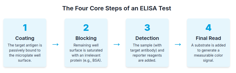
Method of ELISA Test: The Four Core Steps
The method of the ELISA test is standardized into four primary phases, which require rigorous washing cycles between each step, usually with a buffered solution (such as PBS containing a detergent like Tween-20), to eliminate unbound material and reduce background noise.
- Coating: The target macromolecule in antibody detection
assays (the pure or semi-pure antigen), is passively bound to the microplate
well surface. This immobilization provides the solid phase for the subsequent
binding reactions.
- Blocking: To prevent non-specific binding of later
reagents, the remaining unoccupied surface area of the well is saturated using
an irrelevant protein, such as Bovine Serum Albumin (BSA).
- Detection: The sample containing the analyte (target
antibody) is introduced, followed by the specific binding of reporter
reagents.
- Final Read: A substrate is added to generate a signal
measurable by a microplate reader.
Key Detection Reagents
The final, measurable signal relies on the highly active reporter enzyme conjugated to a detection antibody. The most common reporter enzymes are Horseradish Peroxidase (HRP) and Alkaline Phosphatase (AP). The selection of the substrate determines the final color and absorbance requirement:
Table 01: ELISA Detection Substrates and Readouts
| Enzyme | Substrate | Absorbance Peak | Color Reaction Notes |
|---|---|---|---|
| HRP | TMB (3,3’,5,5’-tetramethylbenzidine) | 450 nm | Most common. Yields the strongest signal after stop solution addition (blue to yellow transition). |
| HRP | OPD (o-phenylenediamine dihydrochloride) | 492 nm | Changes from yellow to orange upon the addition of the stop solution. |
| AP | pNPP (p-Nitrophenyl Phosphate) | 405 nm | Results in a stable yellow color. Slower reaction time compared to HRP substrates. |
Quantitative Rigor
For research and clinical use, the quantitative output is crucial. Accurate concentration measurements are derived by plotting the assay results against a standard curve generated from serial dilutions of a known reference standard. This standard curve typically plots concentration logarithmically against absorbance linearly.
The Contenders: Types of ELISA Test for Serology
While there are four primary types of ELISA test (Direct, Indirect, Sandwich,
Competitive), only two are strategically suited for quantifying specific
antibodies in biological fluids (serology): Indirect ELISA and Competitive
ELISA.
Sandwich ELISA, for instance, is designed for antigen capture between two antibodies and is therefore unsuitable for detecting antibodies in serum.
The optimal selection depends on how the detection system interacts with the sample analyte of interest.
Indirect ELISA: The Power of Amplification and Flexibility
The Indirect ELISA is widely favored for general serological screening because its design provides powerful signal amplification, making it one of the most sensitive assay formats.
So, how does ELISA detect antibodies in the Indirect format?
The core mechanism involves sequential binding, leveraging the flexibility of non-labeled primary antibodies, followed by a universal, labeled secondaryantibody.
Step-by-Step Protocol
- Antigen Capture: The specific target antigen is immobilized
onto the microplate well surface.
- Primary Antibody Binding: The sample (containing the
primary antibody analyte) is added. If present, the analyte binds specifically
to the captured antigen.
- Secondary Antibody Binding (The Amplification Step): An
enzyme-conjugated secondary antibody is introduced. This secondary reagent
targets the host species and isotype of the primary antibody. These are the
critically important components used in ELISA for detection.
- Signal Generation: Substrate is added, and the resulting
signal directly correlates with the amount of primary antibody present.
Advantages of Indirect ELISA
The superiority of the Indirect format stems primarily from the use of the
secondary antibody:
- High Sensitivity via Amplification: The secondary antibody,
often polyclonal, can recognize and bind to multiple epitopes on the captured
primary antibody molecule. This allows multiple enzyme labels to bind to a
single analyte molecule, creating a robust signal cascade that yields
significantly higher sensitivity than direct methods.
- Flexibility and Economy: This is the most flexible format.
A single labeled secondary antibody reagent can be used to detect hundreds of
different unlabeled primary antibodies, provided they were raised in the same
host species. This greatly reduces inventory and procedural costs for research
laboratories.
- Ideal for Titer Measurement: It is the most suitable
technique for accurately determining the total concentration or titer of
antibodies in a sample, especially in response to vaccination or infection.
Disadvantages and Cross-Reactivity Risk
The main drawback of the Indirect ELISA is the added layer of complexity and
time required for the secondary incubation step. Crucially, the introduction of
a secondary detection antibody carries an inherent risk of cross-reactivity with
components in the sample matrix or the blocking agents. This can lead to
non-specific binding and elevated background noise, potentially obscuring a
weak, specific signal.
Competitive ELISA: The Champion of Small Analytes and Specific Epitopes
Competitive ELISA offers a highly specific and robust method for detecting antibodies, particularly valuable when samples are complex or the target is an early, low-titer antibody response.
So, how does ELISA detect antibodies in the Competitive format?
The Competitive format operates on the principle of signal inversion, where the unknown antibody in the sample competes with a reference reagent for limited binding sites on an immobilized target. For antibody detection, the plate is coated with the target antigen.
Competition Mechanism and Interpretation
- Simultaneous Binding: The sample (containing the unknown
antibody analyte) is added concurrently with a known, pre-labeled reference
antibody (or labeled antigen). Both compete fiercely for the limited number of
binding sites on the immobilized antigen.
- Inverse Signal Correlation: The resulting signal is
inversely proportional to the amount of target antibody in the sample.
- Positive Result (Antibody Present): A High concentration of
the sample antibody blocks the labeled reference antibody from binding. This
results in a low or absent signal (no color
change).
- Negative Result (Antibody Absent): Low concentration of
sample antibody allows the labeled reference antibody to bind freely. This
results in a high signal (strong color change).
Advantages of Competitive ELISA
The distinct mechanism of competitive binding provides several key advantages
over non-competitive formats:
- Superior Robustness in Complex Matrices: Competitive ELISA
requires significantly less sample purification because its reliance on a
specific competition event reduces interference from general matrix components.
This makes it exceptionally reliable for testing crude biological fluids like
serum, plasma, or extracts.
- Ideal for Early Seroconversion Studies: This assay has
demonstrated superior capability in detecting antibodies shortly after infection
(early seroconversion) when titers are still very low. For diagnostics requiring
early confirmation of immune status, its targeted specificity is invaluable.
- Effective for Small Molecules: It is the gold standard for
detecting small analytes that possess only a single epitope, such as haptens or
various small antigens, which cannot be captured in a sandwich format.
- Low Variability: The controlled, simultaneous competition
step often results in lower intra-assay and inter-assay variability.
Disadvantages of Competitive ELISA
Competitive ELISA generally exhibits lower overall sensitivity compared to the
signal-amplified Indirect method and cannot be used reliably with extremely
dilute samples. Furthermore, the lack of flexibility is a practical restriction:
a unique, labeled competitive reagent often must be developed for each target
assay, increasing upfront development cost.
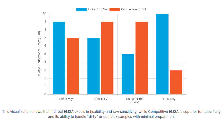
Performance Face-Off: Strategic Selection Matrix
The decision regarding the optimal antibody test also depends on ELISA kits. Here’s the direct comparison between these two ELISA formats:
Table 02: Indirect vs. Competitive ELISA Performance Matrix for Antibody Detection
| Metric | Indirect ELISA | Competitive ELISA | Strategic Implication |
|---|---|---|---|
| Sensitivity | High (Achieves signal amplification) | Moderate to High (No inherent amplification mechanism) | Indirect is favored when maximizing signal detection is paramount. |
| Specificity | Good, but sensitive to secondary Ab cross-reactivity | High (Specific epitope targeting via competition) | Competitive excels in complex samples by limiting non-specific binding. |
| Target Analytes | Antibodies, proteins (high molecular weight) | Small molecules, haptens, specific antibodies (low molecular weight) | - |
| Sample Prep | Moderate (Requires more purification for low background) | Low (Highly tolerant of crude samples) | - |
| Flexibility | Highest (One labeled secondary for many primaries) | Low (Requires target-specific labeled reference reagent) | - |
| Data Interpretation | Direct Correlation | Inverse Correlation | - |
| Best Use Case | Total Titer Determination, R&D Screening | Early Seroconversion, Complex Clinical Matrices | - |
Overcoming Technical Hurdles: Restrictions of ELISA Tests
All ELISA methods are subject to technical limitations that require rigorous
optimization. Recognizing these restrictions is critical for assay validation.
Matrix Effects and Background Noise
The components inherent in biological fluids, such as heterophilic antibodies, complement factors, or high levels of irrelevant proteins, can interfere with the specific antigen-antibody interaction. This "matrix effect" can skew quantitative results or increase background noise.
Mitigation Strategies:
- Sample Dilution: Highly complex samples (serum, plasma,
extracts) must be diluted significantly, often by 50% or more, using specialized
dilution buffers to minimize matrix interference.
- Optimization: Thorough optimization of coating, blocking,
and washing steps, utilizing specialized buffers containing detergents and
blocking agents, is essential to ensure high signal-to-noise ratios.
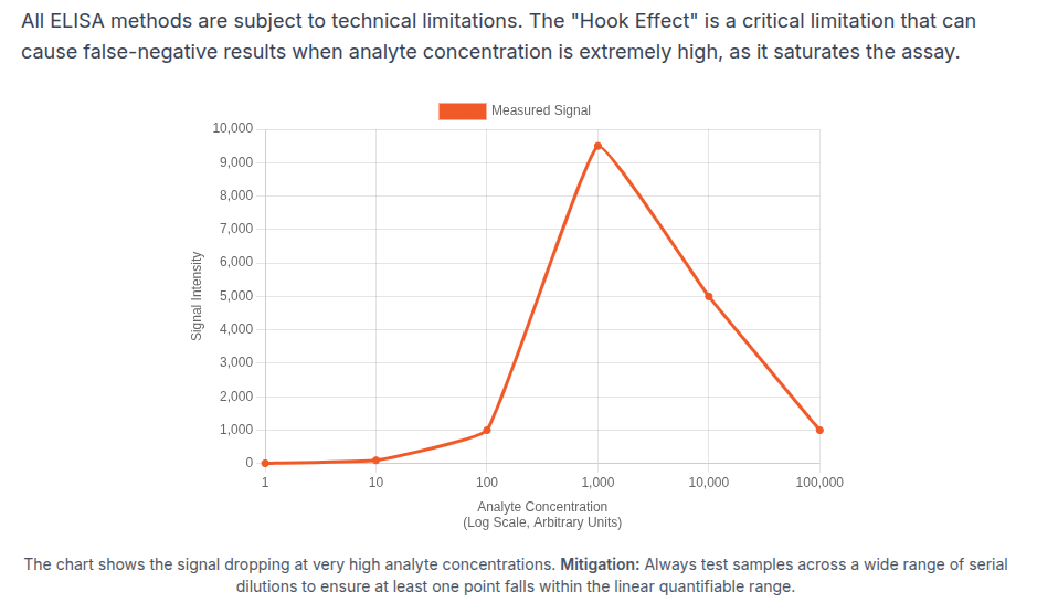
The Hook Effect (High-Dose Non-Linearity)
The Hook Effect is a severe limitation, often resulting in a false-negative or falsely low reading when the analyte concentration far exceeds the assay’s linear range. This occurs when excess analyte saturates both the capture and detection reagents, preventing the formation of stable complexes.
Mitigation Strategies:
- Serial Dilution: The most effective control is to test
every complex sample across a wide range of serial dilutions. This ensures that
at least one dilution point falls within the linear, quantifiable range of the
assay.
- Two-Step Protocols: Employing a multi-step protocol, which separates the binding phases, can physically minimize the competitive reactions responsible for the Hook Effect.
The Future of High-Sensitivity Antibody Detection
Technological advancements are rapidly addressing the inherent limitations of traditional ELISA methods, particularly the constraints on sensitivity, speed, and multiplexing capability.
- Nanotechnology Integration: The convergence of immunology
and materials science has introduced artificial enzyme mimics and Quantum Dots
(QDs). QDs, with their high stability and adjustable light emissions,
dramatically enhance detection sensitivity and facilitate running multiple,
simultaneous tests on a single sample (multiplexing).
- Automation and Microfluidics: Advances in automation,
incorporating robotic microfluidic handling and sophisticated sensor systems,
increase throughput while minimizing human error. These systems allow
researchers to analyze a broad spectrum of proteins and agents with greater
precision, essential for fields like neurodegenerative research.
Final Expert Recommendation
Choosing the best antibody detection ELISA requires aligning
the assay format with the specific requirements of the biological analyte of
interest:
- Choose Indirect ELISA when flexibility is prioritized,
large-scale screening of many primary antibodies is required, and maximum signal
amplification is needed to determine the total antibody concentration or titer
in a sample with a relatively clean matrix.
- Choose Competitive ELISA when the analytical goal is
targeted specificity, rapid diagnostics (such as early seroconversion), or when
dealing with highly complex biological fluids that are difficult or impractical
to purify extensively. The assay’s robustness and ability to handle complex
matrices often outweigh the raw sensitivity advantage of the Indirect format in
these critical applications.
Conclusion

Selecting the right ELISA format hinges on the specific antibody biology, the
sample matrix, and the required sensitivity. Indirect ELISA offers robust signal
amplification and broad flexibility, making it ideal for antibody detection
across diverse primary antibodies and high-throughput screening. Competitive
ELISA excels when target specificity matters, especially in complex samples or
early seroconversion scenarios, even though it may sacrifice some sensitivity.
For AAA Biotech customers, the best approach is to match the assay architecture to the analytical goal: quantify total antibody titer with indirect formats or pinpoint early, highly specific antibody responses with competitive formats.
In developing your own, validation of antibody pairs, optimization of blocking and washing, and confirmation of performance with appropriate controls are essential.
Faq's
Which ELISA type is best for antibody detection?
Indirect ELISA is typically preferred for antibody detection due to signal amplification and flexibility, though competitive ELISA suits complex matrices and early seroconversion.
What is the main difference between Indirect and Competitive ELISA?
The main difference is the binding principle. Indirect ELISA uses a labeled secondary antibody for signal amplification and a direct signal correlation. Competitive ELISA relies on a competition event between the sample antibody and a labeled reference, resulting in an inverse signal.
Which type of ELISA is considered the most sensitive?
Indirect ELISA is generally considered the most sensitive for antibody detection. This high sensitivity is achieved because the labeled secondary antibody can bind to multiple sites on the primary antibody, creating a powerful signal amplification cascade.
Why is Competitive ELISA better for complex samples like serum?
Competitive ELISA is superior for complex samples because its reliance on a very specific, limited-site competition event significantly reduces interference from the sample matrix. It requires less sample purification than the Indirect format.
What is the "Hook Effect" in ELISA, and how is it solved?
The "Hook Effect" is when an extremely high concentration of the analyte causes a falsely low or negative reading. It is mitigated by testing every complex sample across a wide range of serial dilutions, ensuring one reading falls within the assay’s quantifiable linear range.
Lead Clinical Research Coordinator (LCRC)
Cynthia Lee is the President of AAA Biotech and specializes in understanding highly validated and characterized monoclonal/polyclonal antibodies, recombinant proteins, and ELISA kits.
Related Posts