Secondary Antibodies Roles in Immunoassays: Structure, Selection, & Applications
 Cynthia
12 min read
Cynthia
12 min read
In this Article
All of the products listed in AAA Biotech’s catalog are strictly for research-use only (RUO).

Key Takeaways
- Core role & benefit of secondary antibodies
- Key structural elements
- Crucial selection rules
- Types and labels
- Applications
- Future enhancements
Primary antibodies – first line of detection in immunoassays,
and bind specifically to a target antigen (a protein, peptide, or other
biomolecule).
What are secondary antibodies?
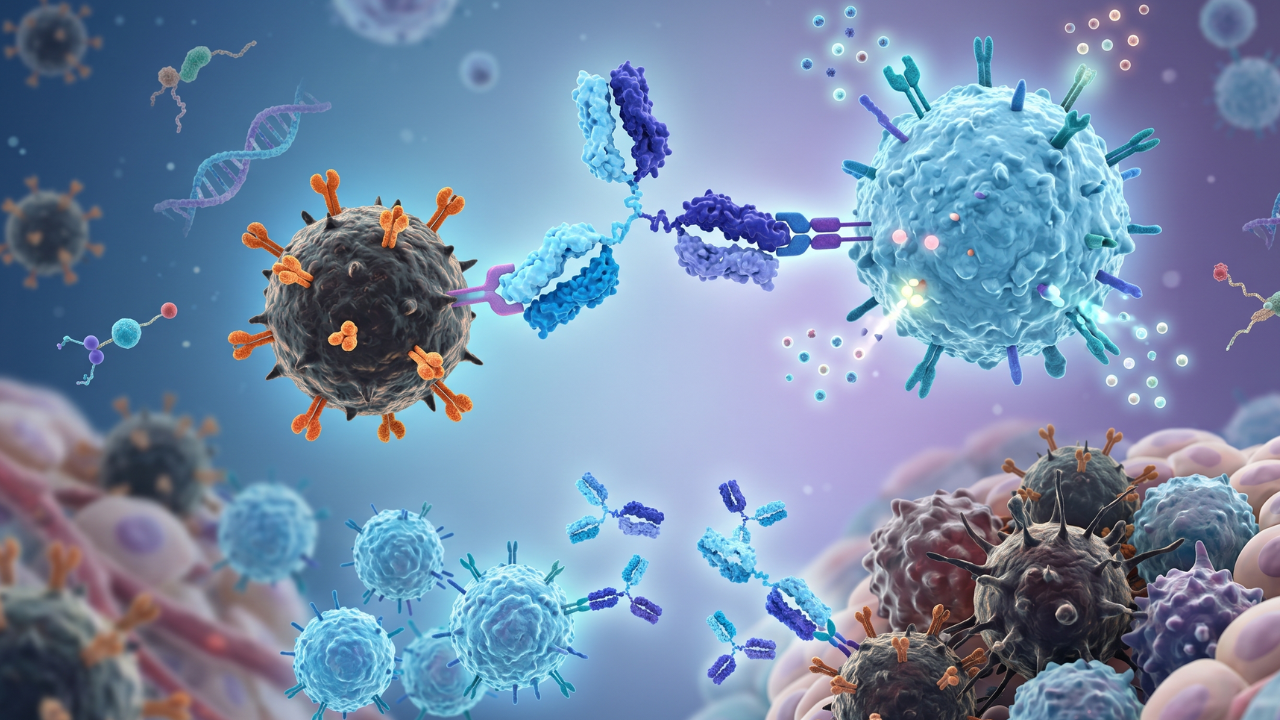
Secondary antibodies are labeled with markers (like an enzyme, dye, or fluorescent tag). When a secondary antibody binds with a primary antibody, the marker reacts to produce a color, light or fluorescence for signal amplification in immunodetection.
For simplification,
Let’s assume that the target antigen is a lock. The primary antibody is the key to that lock and the secondary antibody is a flashlight attached to the key.
However, achieving consistent and reliable results is a significant challenge.
According to Journal of Cell Biology, 68% of researchers report variability issues undermining data reproducibility.
Key Structural Properties
Secondary antibodies share the same basic Y-shaped structure of all antibodies. However, the key structural properties depend on the production and modification to serve a specific role in immunoassays.
Basic Y-Shaped Immunoglobulin Structure
Four Polypeptide Chains
A typical secondary antibody is consisting of two identical heavy chains and two
Label/Tag
identical light chains. Disulfide bonds hold these chains together.
Variable Regions (V regions)
These regions are at the tips of the “Y” arms. There is one region on each heavy chain and one on each light chain. Each V region has its own specific amino acid sequence. Combined variable regions of one heavy and one light chain form an antigen-binding site that binds specifically to the constant region of a primary antibody.
Constant Regions
Constant regions are just below the variable regions forming the ‘stalk’ and the lower part of the ‘arms’. These regions determine the effector functions of the antibody.
Fab (Fragment antigen-binding) Regions
These regions are antigen-binding portions of an antibody molecule. The two arms of the ‘Y’, each containing one light chain and one heavy chain, bind to the target. Fab regions in a secondary antibody bind to the primary antibody.
Fc (Fragment crystallizable) Region
While the Fc Region (“stem” of the “Y”) is of importance for primary antibody effector functions, Fc (Fragment crystallizable) Region in secondary antibodies dictates the class (IgG, IgM, etc.) of the secondary antibody. Sometimes other reagents can also bind to this region.
Hinge Region
This flexible segment connects the Fab arms and the Fc region and also supports adaptability for binding.
Detection Labels: Mechanisms and Applications
Secondary antibodies have specialized detectable tags/labels attached to their
constant regions. These tags/labels generate measurable signals. The choice of
the tag depends on the sensitivity, equipment needs, and potential limitations.
In research laboratories, the following four types of detectable tags/labels are
the widest used:
- HRP (Horseradish Peroxidase)
- Alkaline Phosphatase (AP)
- Fluorophores
- Biotin
Label/Tag Detection Method Common Applications
| Label/Tag | Detection Method | Common Applications | Key Limitations |
|---|---|---|---|
| HRP (Horseradish Peroxidase) | Colorimetric (e.g., TMB/DAB) or Chemiluminescence |
|
|
| Alkaline Phosphatase (AP) | Colorimetric (e.g., BCIP/NBT) |
|
|
| Fluorophores | Fluorescence (light emission at specific wavelengths) |
|
|
| Biotin | Streptavidin–conjugates (enzyme/fluorophore) |
|
|
Species/Isotype Selection Rules
The choice of secondary antibody dictates the success and specificity of any immunoassay that they are a component of. A secondary antibody must bind exclusively to the intended primary antibody. The following crucial factors related to primary antibody rules govern this specificity:
- Its Host Species
- Its Isotype or Subclass
Its Host Species
The primary antibody and the secondary antibody must not be generated in the same animal species. For example, a secondary antibody for a mouse primary antibody must be raised in a non-mouse species.
Its Isotype or Subclass
Primary antibodies vary by subclass (e.g., IgG1, IgG2a) or isotype (e.g., IgG, IgM). “Pan-specific” secondary antibodies bind to a broad range of primary antibodies. Multiplexing requires isotype-specific secondary antibodies.
Types of Secondary Antibodies
Secondary antibodies are categorized in the following ways:
- Structural Classification
- Classification by Conjugates
- Additional Classifications
Structural Classification
Classifying secondary antibodies on the basis of their structural forms is critical especially in applications that require reduction in background noise or target particular regions of the primary antibody.
Whole IgG
Whole IgG secondary antibodies have intact antibody structure forming a Y-shaped molecule with two Fab (Fragment antigen-binding) regions and one Fc (Fragment crystallizable) region. It effectively binds to the primary antibody.
However, Fc region binding to Fc receptors on lymph nodes or spleen or other cells or tissues can potentially lead to high background in assays like immunohistochemistry.
F(ab')₂ Fragments
These are the products of pepsin digestion of IgG antibodies. Most of the Fc fragment is removed and the two Fab arms and the hinge are retained. This bivalent fragment has the ability to bind to the primary antibody. The lack of Fc region means no Fc receptor binding and reduced background noise. F(ab')₂ Fragments are beneficial in immunohistochemistry and immunofluorescence on tissues with high Fc receptor expression.
Fab Fragments
Fab Fragments are produced through digestion of whole IgG with papain. As a result, a single whole IgG molecule yields two separate monovalent Fab fragments and one Fc fragment. Each Fab fragment has the ability to bind but lacks the hinge and Fc region. The small size allows better tissue penetration. As Fab fragments keep cross-reactivity to a minimum, they are particularly ideal for multiple labeling experiments involving primary antibodies from the same species. They can also block endogenous immunoglobulins on cells or tissues.
Fc-Specific Antibodies
Fc-specific antibodies specifically bind to the primary antibody’s Fc region. These secondary antibodies are useful in applications where the Fc region of the primary antibody is exposed or when detecting specific fragments.
Fragment-Specific Antibodies
These secondary antibodies are engineered to bind specific fragments of the primary antibody, such as F(ab) or F(ab')₂. They are useful for binding fragmented primary antibodies or primary antibodies with exposed regions that are exposed for detection. Since they often target fragments that lack an Fc region, these secondary antibodies can also help reduce background in tissues with high Fc receptor expression.
Classification by Conjugates
The label or conjugate type on the secondary antibody is crucial to:
- Detect and amplify signal
- Determine the visualization mechanism
Enzyme-Conjugated Secondary Antibodies
Horseradish Peroxidase (HRP)
These secondary antibodies are conjugated to HRP and used for chemiluminescent or colorimetric (e.g., TMB, DAB) detection. (Refer to Table 1 for more details on applications and limitations.)
Alkaline Phosphatase (AP)
These secondary antibodies are conjugated to AP and used for colorimetric detection. (Refer to Table 1 for more details on applications and limitations.)
Fluorophore-Conjugated Secondary Antibodies
Labeled/tagged with fluorescent dyes such as tetramethylrhodamine isothiocyanate (TRITC), fluorescein isothiocyanate (FITC), Alexa Fluor dyes or other fluorescent dyes, these secondary antibodies are used in live-cell imaging, flow cytometry and fluorescence microscopy. These secondary antibodies require specialized filters or excitation sources.
Biotin-Conjugated Secondary Antibodies
These secondary antibodies are labeled with biotin which is a small molecule that binds with high affinity to streptavidin or avidin and provides ultra-sensitive signal amplification. (Refer to Table 1 for more details on applications and limitations.)
Other Conjugates
These secondary antibodies include gold particles and radioisotopes.
- Gold particles are used in electron microscopy for high-resolution imaging.
- Radioisotopes are used in radioimmunoassays for quantitative detection.
Additional Classifications
Secondary antibodies that are classified on the basis of some other attributes, with said attributes being crucial for their selection and specificity.
Based on Source
Polyclonal Secondary Antibodies
These secondary antibodies are the product of immunization of an animal with a
whole immunoglobulin. Polyclonal secondary antibodies can recognize multiple
epitopes on the primary antibody.
The high sensitivity of these antibodies, due to their ability to have many of themselves binding to a single primary antibody, make them suitable for detecting low abundance targets. However, non-specific binding may result in a higher background.
Monoclonal Secondary Antibodies
These secondary antibodies are produced from a single clone of B cells and can
recognize a single epitope on the primary antibody. ,. antibodies provide superior specificity for highly specific target detection.
Recombinant Secondary Antibodies
These secondary antibodies include nano-secondaries and multi-rAB. The
combination of high sensitivity and superior specificity offers minimal
cross-reactivity, low background and high detectable brightness. This is ideal
for super-resolution microscopy.
Based on Host Species
Common host species for raising secondary antibodies include:
- Goat
- Rabbit
- Donkey
- Sheep
- Mouse
Based on Isotype Specificity
Secondary antibodies can be pan-specific or isotype-specific. Pan-specific
antibodies bind many primary antibody types. Isotype-specific antibodies bind
only one type. Isotype-specific antibodies are crucial for multiplexing, as they
allow for the detection and differentiation of multiple targets in the same
experiment.
Applications of Secondary Antibodies in Immunoassays
Secondary antibodies are reagents used in a wide range of immunoassay techniques
to provide signal detection, amplification and versatility. Indirect detection
methods use secondary antibodies across various biological research fields.
Western Blotting (WB)
Western blotting is a laboratory technique used to detect specific proteins within a sample. These proteins are separated by gel electrophoresis and transferred onto a membrane. Secondary antibodies bind to the primary antibody bound to the target protein and enable its detection and visualization through an attached label. This allows:
Signal Amplification
Multiple secondary antibodies binding to a single primary antibody significantly
boosts the signal - this is helpful for detecting low-abundance target proteins.
Versatility
A single conjugated secondary antibody, capable of binding with any primary
antibody from a specific host species, eliminates the need for labelling each
primary antibody directly.
Multiplexing
Fluorescently labeled secondary antibodies allow the simultaneous detection of
multiple proteins on the same blot.
ELISA (Enzyme-Linked Immunosorbent Assay)
ELISA, or enzyme-linked immunosorbent assay is used to detect and quantify substances like antibodies or antigens in a sample. Indirect ELISA and some sandwich ELISA use secondary antibodies for signal detection and amplification.
Indirect ELISA
This immunoassay method detects and measures antibodies in a sample. If the
sample contains specific primary antibodies, they bind to a known antigen coated
onto a plate. The secondary antibody binds to the primary antibody. The enzyme
linked to the secondary antibody reacts with a substrate to produce a detectable
signal. This signal helps researchers determine the presence and quantity of the
target antibodies.
Sandwich ELISA
Sandwich ELISA is used to detect and quantify antigens in a sample. The antigen
is sandwiched between two layers of antibodies. The bottom layer is a capture
antibody coated on a plate that binds to the antigen. The top layer is a
detection antibody (detection primary) that binds to a different epitope on the
antigen. A conjugated secondary antibody binds to the detection antibody to
generate a signal.
Immunohistochemistry (IHC) and Immunofluorescence (IF)
Immunohistochemistry (IHC) and immunofluorescence (IF) are techniques to visualize antigens without destroying the cellular or tissue morphology.
Indirect Detection
In this method of antigen location visualization, a conjugated secondary
antibody binds to an unlabeled primary antibody bound to the target antigen.
Signal Amplification
Multiple conjugated secondary antibodies binding to a single primary antibody
lead to concentration of detectable labels at the target site, significantly
amplifying the signal. This helps in visualizing low-expression antigens within
cells and tissues.
Multiplexing
Multiplexing uses different secondary antibodies with each antibody conjugated
to a unique fluorophore. These secondary antibodies bind to primary antibodies
with different isotypes or primary antibodies from distinct host species. The
result is simultaneous visualization of multiple antigens within a cell or
tissue.
Reduced Background
Immunohistochemistry (IHC) and Immunofluorescence (IF) use secondary antibodies
with no Fc region to reduce background when working with tissues with high
endogenous Fc receptor expression. This prevents the non-specific binding that
can lead to high background signal.
Flow Cytometry
This technique quickly analyzes cell populations by detecting specific proteins expressed by these cells.
Cell Surface and Intracellular Staining
First, unlabeled primary antibodies are incubated with cells. These antibodies
target and bind to unique antigens on or within the cells.
Fluorophore-conjugated secondary antibodies bind to primary antibodies already
bound to the antigens.
The fluorophores are excited and subsequently emit light as stained cells pass through a laser beam in a flow cytometer. This light is detected and measured for precise quantification and phenotypic analysis of different cell populations based on their specific protein expression.
Multiplexing
In Flow Cytometry, each antibody in a panel of different primary antibodies
targets a unique antigen. A corresponding secondary antibody detects each
primary antibody. Each secondary antibody is conjugated to a different
fluorophore, emitting light at a distinct wavelength. Multiple emitted
fluorescent signals are detected simultaneously as cells pass through the flow
cytometer's lasers. As a result, researchers can analyze multiple cellular
markers at once for complex phenotypic analysis.
Immunoprecipitation (IP) and Chromatin Immunoprecipitation (ChIP)
Immunoprecipitation (IP) and Chromatin Immunoprecipitation (ChIP) use antibodies to isolate specific proteins (IP) or protein-DNA complexes (ChIP) from a sample.
Detection of Pulled-Down Proteins
After a primary antibody captures and immobilizes the target protein or
protein-DNA complex, Western blotting is performed on the immunoprecipitated
material. A secondary antibody binds to the primary antibody bound to the target
protein to confirm the presence of the protein by amplifying the signal.
Minimizing Interference
The heavy chain of the primary antibody can interfere in IP/Western Blot due to
similar size as the target protein. This interference can obscure the signal.
Using light chain-specific secondary antibodies can optimize detection after
immunoprecipitation.
Future Directions
The secondary antibodies that are utilized by researchers are continuously
evolving to achieve greater precision, sensitivity and reproducibility in
immunoassays.
Advancements in Recombinant Secondary Antibodies
Recombinant antibodies are produced using genetic engineering to
offer:
- Batch-to-batch consistency
- Well defined specificity
- Ability to engineer novel formats
Advancements in recombinant antibodies directly address the current challenges
in research by reducing variability and enhancing reproducibility.
Development of Nanobodies and Single-Domain Antibodies
Nanobodies and single-domain antibodies are small in size and highly stable. Their ability to be easily conjugated, superior tissue penetration and reduced steric hindrance make them ideal for:
- Live-cell imaging
- Super-resolution microscopy
- Highly multiplexed assays
Enhanced Conjugation Technologies
New methods being developed to attach enzymes, fluorescent dyes and other labels can lead to brighter, more stable, signals and higher levels of multiplexing.
Integration with Automation and AI
The integration of automation and AI for analysis is transforming immunoassay research. These technologies can enable higher throughput and more sophisticated data interpretation.
Faq's
Is IgG a secondary antibody?
No, IgG is a class of antibodies. A secondary antibody binds to a primary antibody. Many secondary antibodies are of the IgG class, but IgG itself is not inherently "secondary."
How do I select the correct secondary antibody for my immunoassay?
Make sure that the secondary antibody and primary antibody are raised from different host species and isotype. Match the secondary antibody's specificity to your primary antibody's host species and isotype.
Why are secondary antibodies preferred over directly labeled primary antibodies?
Multiple secondary antibodies binding to a single primary antibody significantly amplifies the signal and allows the detection of even low-abundance antigens. A single type of labeled secondary antibody can bind to various primary antibodies, provided the secondaries are designed to recognize and bind primaries from that specific host species and of that particular isotype. This saves time and cost. So, secondaries offer three benefits: amplification, versatility, and cost-effectiveness.
What types of detection labels are commonly attached to secondary antibodies?
The following are the types of detection labels commonly attached to secondary antibodies:
Enzymes (Horseradish Peroxidase (HRP) and Alkaline Phosphatase (AP))
Fluorophores (Fluorescent dyes such as FITC, Alexa Fluor dyes)
Biotin
Some less common detection labels include gold particles (for electron microscopy) and radioisotopes (for radioimmunoassays).
Lead Clinical Research Coordinator (LCRC)
Cynthia Lee is the President of AAA Biotech and specializes in understanding highly validated and characterized monoclonal/polyclonal antibodies, recombinant proteins, and ELISA kits.
Related Posts