


Mouse CD14 Monoclonal Antibody | anti-CD14 antibody
CD14 Monoclonal Antibody
Preservative: 0.03% Proclin 300
Constituents: 50% Glycerol, 0.01M PBS, pH 7.4
IHC: 1:50-1:200
IF: 1:50-1:200
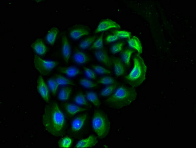
IF (Immunofluorescence)
(Immunofluorescence staining of Hela cells with AAA27028 at 1:90, counter-stained with DAPI. The cells were blocked in 10% normal Goat Serum and then incubated with the primary antibody overnight at 4 degree C. The secondary antibody was Alexa Fluor 488-congugated AffiniPure Goat Anti-Mouse IgG(H+L).)
IF (Immunofluorescence)
(Immunofluorescence staining of A549 cells with AAA27028 at 1:90, counter-stained with DAPI. The cells were blocked in 10% normal Goat Serum and then incubated with the primary antibody overnight at 4 degree C. The secondary antibody was Alexa Fluor 488-congugated AffiniPure Goat Anti-Mouse IgG(H+L).)
IHC (Immunohistochemistry)
(IHC image of AAA27028 diluted at 1:100 and staining in paraffin-embedded human adrenal gland tissue performed on a Leica BondTM system. After dewaxing and hydration, antigen retrieval was mediated by high pressure in a citrate buffer (pH 6.0). Section was blocked with 10% normal goat serum 30min at RT. Then primary antibody (1% BSA) was incubated at 4 degree C overnight. The primary is detected by a biotinylated secondary antibody and visualized using an HRP conjugated SP system.)
IHC (Immunohistochemistry)
(IHC image of AAA27028 diluted at 1:100 and staining in paraffin-embedded human stomach tissue performed on a Leica BondTM system. After dewaxing and hydration, antigen retrieval was mediated by high pressure in a citrate buffer (pH 6.0). Section was blocked with 10% normal goat serum 30min at RT. Then primary antibody (1% BSA) was incubated at 4 degree C overnight. The primary is detected by a biotinylated secondary antibody and visualized using an HRP conjugated SP system.)
IHC (Immunohistchemistry)
(IHC image of AAA27028 diluted at 1:100 and staining in paraffin-embedded human breast cancer performed on a Leica BondTM system. After dewaxing and hydration, antigen retrieval was mediated by high pressure in a citrate buffer (pH 6.0). Section was blocked with 10% normal goat serum 30min at RT. Then primary antibody (1% BSA) was incubated at 4 degree C overnight. The primary is detected by a biotinylated secondary antibody and visualized using an HRP conjugated SP system.)
IHC (Immunohistochemistry)
(IHC image of AAA27028 diluted at 1:100 and staining in paraffin-embedded human colon cancer performed on a Leica BondTM system. After dewaxing and hydration, antigen retrieval was mediated by high pressure in a citrate buffer (pH 6.0). Section was blocked with 10% normal goat serum 30min at RT. Then primary antibody (1% BSA) was incubated at 4 degree C overnight. The primary is detected by a biotinylated secondary antibody and visualized using an HRP conjugated SP system.)
IHC (Immunohistochemistry)
(IHC image of AAA27028 diluted at 1:100 and staining in paraffin-embedded human lung cancer performed on a Leica BondTM system. After dewaxing and hydration, antigen retrieval was mediated by high pressure in a citrate buffer (pH 6.0). Section was blocked with 10% normal goat serum 30min at RT. Then primary antibody (1% BSA) was incubated at 4 degree C overnight. The primary is detected by a biotinylated secondary antibody and visualized using an HRP conjugated SP system.)
IHC (Immunohistochemistry)
(IHC image of AAA27028 diluted at 1:100 and staining in paraffin-embedded human tonsil tissue performed on a Leica BondTM system. After dewaxing and hydration, antigen retrieval was mediated by high pressure in a citrate buffer (pH 6.0). Section was blocked with 10% normal goat serum 30min at RT. Then primary antibody (1% BSA) was incubated at 4 degree C overnight. The primary is detected by a biotinylated secondary antibody and visualized using an HRP conjugated SP system.)
WB (Western Blot)
(Western BlotPositive WB detected in: Hela whole cell lysate, A549 whole cell lysate, HepG2 whole cell lysateAll lanes: CD14 antibody at 1:1800SecondaryGoat polyclonal to Mouse IgG at 1/10000 dilutionPredicted band size: 41 kDaObserved band size: 55 kDa)
WB (Western Blot)
(Western BlotPositive WB detected in: Rabbit spleen tissue, Rabbit small intestine tissueAll lanes: CD14 antibody at 1:2500SecondaryGoat polyclonal to Mouse IgG at 1/10000 dilutionPredicted band size: 41 kDaObserved band size: 55 kDa)


