


 Application Data
(Published Customer Image:Rat anti Mouse Gr-1 antibody, clone RB6-8C5 used for the identification of neutrophils by immunofluorescence.Image caption:S. typhimurium-Infected Macrophages Containing Phagocytosed Neutrophils and T Cells Confocal fluorescence microscopy of 50-mum-thick liver sections from 1-wk-infected Slc11a1 wild-type mice. (A-C) S.Typhimurium (O-antigen, arrows) are red, macrophages (F4-80 and MOMA-2) are blue, DNA (DAPI) is gray, phalloidin is green, and neutrophils (Gr-1/Ly-6G/RB6-8C5) are pink (arrowheads). (A) Collapsed image from a 40-mum Z-stack. Scale bar is 20 mum. (B and C) Sections from (A) that are 4 mum apart. The video from which (A-C) were derived (Video S2) is available online. (D-G) T cells within multinucleate macrophages.Macrophages (F4-80 and MOMA-2) are blue (D, G, and H), T cells (CD3zeta) are red (D, G, arrowheads), DAPI is gray (E, G), actin-bound phalloidin is green (F, G). (G) Is a composite of (D, E, and F). Scale bars are 16 mum. (H) An image from a different mouse stained and labeled as described for (D-G). Scale bar is 8 mum. A video showing a T cell inside of a macrophage is available online (Video S3).From: Nix RN, Altschuler SE, Henson PM, Detweiler CS (2007) Hemophagocytic Macrophages Harbor Salmonella enterica during Persistent Infection.PLoS Pathog 3(12): e193.)
Application Data
(Published Customer Image:Rat anti Mouse Gr-1 antibody, clone RB6-8C5 used for the identification of neutrophils by immunofluorescence.Image caption:S. typhimurium-Infected Macrophages Containing Phagocytosed Neutrophils and T Cells Confocal fluorescence microscopy of 50-mum-thick liver sections from 1-wk-infected Slc11a1 wild-type mice. (A-C) S.Typhimurium (O-antigen, arrows) are red, macrophages (F4-80 and MOMA-2) are blue, DNA (DAPI) is gray, phalloidin is green, and neutrophils (Gr-1/Ly-6G/RB6-8C5) are pink (arrowheads). (A) Collapsed image from a 40-mum Z-stack. Scale bar is 20 mum. (B and C) Sections from (A) that are 4 mum apart. The video from which (A-C) were derived (Video S2) is available online. (D-G) T cells within multinucleate macrophages.Macrophages (F4-80 and MOMA-2) are blue (D, G, and H), T cells (CD3zeta) are red (D, G, arrowheads), DAPI is gray (E, G), actin-bound phalloidin is green (F, G). (G) Is a composite of (D, E, and F). Scale bars are 16 mum. (H) An image from a different mouse stained and labeled as described for (D-G). Scale bar is 8 mum. A video showing a T cell inside of a macrophage is available online (Video S3).From: Nix RN, Altschuler SE, Henson PM, Detweiler CS (2007) Hemophagocytic Macrophages Harbor Salmonella enterica during Persistent Infection.PLoS Pathog 3(12): e193.)
Rat anti-Mouse Gr-1 Monoclonal Antibody | anti-Gr-1 antibody
Rat Anti Mouse Gr-1: FITC
This product should be stored undiluted. Storage in frost-free freezers is not recommended. Avoid repeated freezing and thawing as this may denature the antibody.
Should this product contain a precipitate we recommend microcentrifugation before use.
Application Data
(Published Customer Image:Rat anti Mouse Gr-1 antibody, clone RB6-8C5 used for the identification of neutrophils by immunofluorescence.Image caption:S. typhimurium-Infected Macrophages Containing Phagocytosed Neutrophils and T Cells Confocal fluorescence microscopy of 50-mum-thick liver sections from 1-wk-infected Slc11a1 wild-type mice. (A-C) S.Typhimurium (O-antigen, arrows) are red, macrophages (F4-80 and MOMA-2) are blue, DNA (DAPI) is gray, phalloidin is green, and neutrophils (Gr-1/Ly-6G/RB6-8C5) are pink (arrowheads). (A) Collapsed image from a 40-mum Z-stack. Scale bar is 20 mum. (B and C) Sections from (A) that are 4 mum apart. The video from which (A-C) were derived (Video S2) is available online. (D-G) T cells within multinucleate macrophages.Macrophages (F4-80 and MOMA-2) are blue (D, G, and H), T cells (CD3zeta) are red (D, G, arrowheads), DAPI is gray (E, G), actin-bound phalloidin is green (F, G). (G) Is a composite of (D, E, and F). Scale bars are 16 mum. (H) An image from a different mouse stained and labeled as described for (D-G). Scale bar is 8 mum. A video showing a T cell inside of a macrophage is available online (Video S3).From: Nix RN, Altschuler SE, Henson PM, Detweiler CS (2007) Hemophagocytic Macrophages Harbor Salmonella enterica during Persistent Infection.PLoS Pathog 3(12): e193.)
Application Data
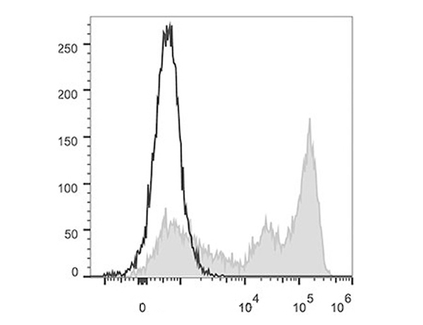
(Published customer image:Rat anti Mouse Gr-1 antibody, clone RB6-8C5 used for the evaluation of Gr-1 expression on circulating monocytes from mouse blood by flow cytometry.Image caption:Effect of CD206+ M2 macrophage depletion on collagen deposition and cell infiltration within the infarct 2 weeks post MI in MAFIA mice. A. Representative Sirius Red staining on histological sections from MAFIA mice 2 weeks post MI. B. Quantification of collagen staining as a percentage of LV from MAFIA mice 2 weeks post MI (Scale bar: 1 mm, n = 6 animals per group, >=8 images per animal). C. Representative hematoxilin and eosin staining on 20x infarct or remote histological section from MAFIA mice 2 weeks post MI showing an increase in inflammatory infiltrates in animals treated with GW2580. D. Quantification of nuclei number per mm2 in MAFIA remote and infarct zone in MAFIA mice 2 weeks post MI. (n = 4-6 animals per group). E. Representative images of immunofluorescent staining of CD206+ M2 macrophages, Gr1+ M1 macrophages and Ly6G+ neutrophils within the infarct zone from MAFIA mice 2 weeks post MI. Quantification of Ly6G+ neutrophil, Gr1+ M1 macrophage and CD206+ M2 macrophage infiltration within the infarct zone from MAFIA mice 2 weeks post MI (n = 4 animals per group, 10 images/animal). * p)
Application Data
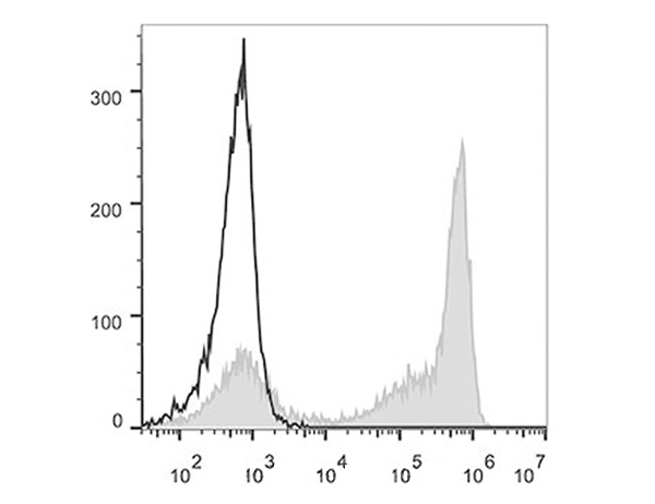
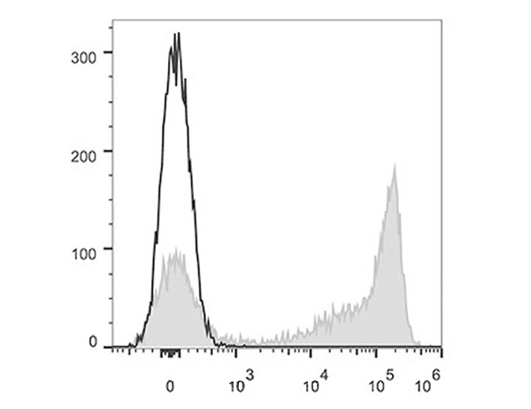
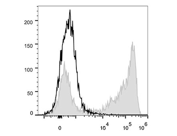
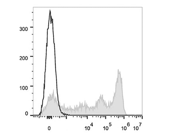
(Published customer image:Rat anti Mouse Gr-1 antibody, clone RB6-8C5 used for the evaluation of Gr-1 expression on circulating monocytes from mouse blood by flow cytometry.Image caption:Depletion of the circulating monocyte and Gr1lo and F4/80hi populations following 1 week GW2580 treatment. A. Identification of total monocyte population in mouse blood using MAFIA-GFP. B Following one week of GW2580 treatment, no difference in total circulating monocytes was observed. FACS quantification of Gr1hi (M1) and Gr1lo (M2; C,D) and F4/80hi (E,F) in total monocytes following 1 week GW2580 treatment (n = 6 and n = 4 animals per group, respectively).From: Leblond A-L, Klinkert K, Martin K, Turner EC, Kumar AH, Browne T, et al. (2015) Systemic and Cardiac Depletion of M2 Macrophage through CSF-1R Signaling Inhibition Alters Cardiac Function Post Myocardial Infarction.PLoS ONE 10(9): e0137515.)
Application Data
(Figure A : RPE conjugated Rat anti Mouse CD11b and Alexa Fluor 647 conjugated Rat IgG2b isotype control . Figure B. RPE conjugated Rat anti Mouse CD11b and Alexa Fluor 647 conjugated Rat anti Mouse Gr-1 . All experiments performed on murine bone marrow in the presence of Murine SeroBlock (BUF041A).)
Application Data
(Figure A : Pacific Blue conjugated Rat anti Mouse CD11b and FITC conjugated Rat IgG2b isotype control . Figure B. Pacific Blue conjugated Rat anti Mouse CD11b and FITC conjugated Rat anti Mouse GR-1 . All experiments performed on red cell lysed mouse bone marrow gated on mononuclear cells in the presence of Mouse Seroblock (BUF041A). Data acquired on the ZE5 cell analyzer.)
Application Data
(Figure A. Pacific Blue conjugated Rat anti Mouse CD11b and Alexa Fluor700 conjugated Rat IgG2b isotype control . Figure B. Pacific Blue conjugated Rat anti Mouse CD11b and Alexa Fluor700 conjugated Rat anti Mouse GR-1 . All experiments performed on red cell lysed mouse bone marrow gated on mononuclear cells in the presence of Mouse Seroblock (BUF041A). Data acquired on the ZE5 cell analyzer.)
Application Data
(Figure A. RPE conjugated Rat anti Mouse CD11b and Pacific Blue conjugated Rat IgG2b isotype control . Figure B. RPE conjugated Rat anti Mouse CD11b and Pacific Blue conjugated Rat anti Mouse GR-1 . All experiments performed on red cell lysed mouse bone marrow gated on mononuclear cells in the presence of Mouse Seroblock (BUF041A). Data acquired on the ZE5 cell analyzer.)
The Gr-1 antigen is primarily a marker of myeloid differentiation. In the bone marrow the level of Gr-1 expression is low on immature myeloblasts and increases as the myeloid cells mature to granulocytes. Gr-1 is also expressed on macrophages and transiently on differentiating monocytes.
Rat anti Mouse Gr-1 antibody, clone RB6-8C5 has been used successfully for the depletion of mature neutrophils in vivo (Czuprynski et al 1994, Daley et al. 2008).
NCBI and Uniprot Product Information
Customer Reviews
Loading reviews...
Share Your Experience
Similar Products


Product Notes
The Gr-1 ly6g (Catalog #AAA12257) is an Antibody produced from Rat and is intended for research purposes only. The product is available for immediate purchase. The Rat Anti Mouse Gr-1: FITC reacts with Mouse and may cross-react with other species as described in the data sheet. AAA Biotech's Gr-1 can be used in a range of immunoassay formats including, but not limited to, FCM/FACS (Flow Cytometry). Researchers should empirically determine the suitability of the Gr-1 ly6g for an application not listed in the data sheet. Researchers commonly develop new applications and it is an integral, important part of the investigative research process. It is sometimes possible for the material contained within the vial of "Gr-1, Monoclonal Antibody" to become dispersed throughout the inside of the vial, particularly around the seal of said vial, during shipment and storage. We always suggest centrifuging these vials to consolidate all of the liquid away from the lid and to the bottom of the vial prior to opening. Please be advised that certain products may require dry ice for shipping and that, if this is the case, an additional dry ice fee may also be required.Precautions
All products in the AAA Biotech catalog are strictly for research-use only, and are absolutely not suitable for use in any sort of medical, therapeutic, prophylactic, in-vivo, or diagnostic capacity. By purchasing a product from AAA Biotech, you are explicitly certifying that said products will be properly tested and used in line with industry standard. AAA Biotech and its authorized distribution partners reserve the right to refuse to fulfill any order if we have any indication that a purchaser may be intending to use a product outside of our accepted criteria.Disclaimer
Though we do strive to guarantee the information represented in this datasheet, AAA Biotech cannot be held responsible for any oversights or imprecisions. AAA Biotech reserves the right to adjust any aspect of this datasheet at any time and without notice. It is the responsibility of the customer to inform AAA Biotech of any product performance issues observed or experienced within 30 days of receipt of said product. To see additional details on this or any of our other policies, please see our Terms & Conditions page.Item has been added to Shopping Cart
If you are ready to order, navigate to Shopping Cart and get ready to checkout.