 IF (Immunofluorescence)
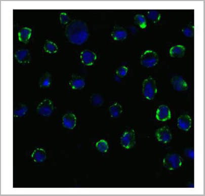
(Figure 11 Immunofluorescence Validation of ACE2 In Caco2 CellsImmunofluorescent analysis of 4% paraformaldehyde-fixed Caco2 cells labeling ACE2 with AAA10945 at 20 ug/mL, followed by goat anti-rabbit IgG secondary antibody at 1/500 dilution (green) and DAPI staining (blue). Imageshowing membrane staining on Caco2 cells.)
IF (Immunofluorescence)
(Figure 11 Immunofluorescence Validation of ACE2 In Caco2 CellsImmunofluorescent analysis of 4% paraformaldehyde-fixed Caco2 cells labeling ACE2 with AAA10945 at 20 ug/mL, followed by goat anti-rabbit IgG secondary antibody at 1/500 dilution (green) and DAPI staining (blue). Imageshowing membrane staining on Caco2 cells.)
Rabbit ACE2 Polyclonal Antibody | anti-ACE2 antibody
ACE2 Antibody
IHC: 2 ug/mL
IF: 10 ug/mL
Antibody validated: Western Blot in human, mouse and rat samples; Immunohistochemistry in human samples; Immunofluorescence in human, mouse andrat samples. All other applications and species not yet tested.
IF (Immunofluorescence)
(Figure 11 Immunofluorescence Validation of ACE2 In Caco2 CellsImmunofluorescent analysis of 4% paraformaldehyde-fixed Caco2 cells labeling ACE2 with AAA10945 at 20 ug/mL, followed by goat anti-rabbit IgG secondary antibody at 1/500 dilution (green) and DAPI staining (blue). Imageshowing membrane staining on Caco2 cells.)
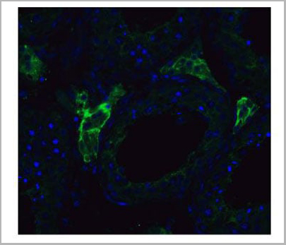
IF (Immunofluorescence)
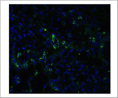
(Figure 9 Immunofluorescence Validation of ACE2 in Rat Lung Tissue Immunofluorescent analysis of 4% paraformaldehyde-fixed rat lung tissue labeling ACE-2 with AAA10945 at 20 ug/mL, followed by goat anti-rabbit IgG secondary antibody at 1/500 dilution (green) and DAPI staining (blue).)
IF (Immunofluorescence)
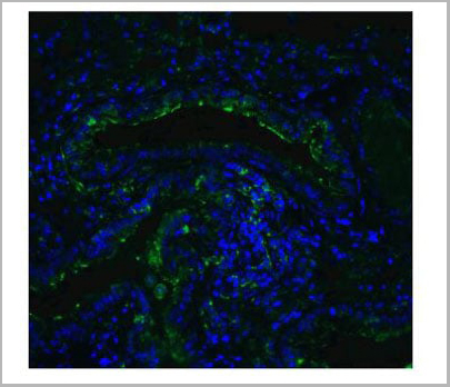
(Figure 8 Immunofluorescence Validation of ACE2 in Mouse Lung Tissue Immunofluorescent analysis of 4% paraformaldehyde-fixed mouse lung tissue labeling ACE-2 with AAA10945 at 20ug/mL, followed by goat anti-rabbit IgG secondary antibody at 1/500 dilution (green) and DAPI staining (blue).)
IF (Immunofluorescence)
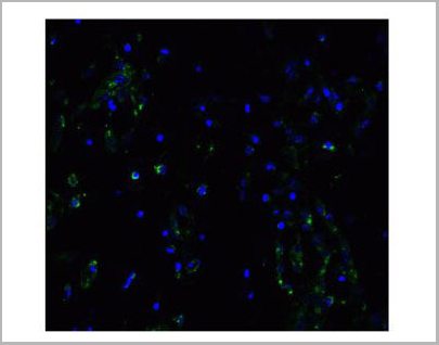
(Figure 7 Immunofluorescence Validation of ACE2 in Human Lung Tissue Immunofluorescent analysis of 4% paraformaldehyde-fixed human lung tissue labeling ACE-2 with AAA10945 at 20ug/mL, followed by goat anti-rabbit IgG secondary antibody at 1/500 dilution (green) and DAPI staining (blue).)
IF (Immunofluorescence)
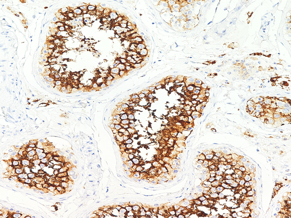
(Figure 6 Immunofluorescence Validation of ACE2 in Human Testis Tissue Immunofluorescent analysis of 4% paraformaldehyde-fixed human testis tissue labeling ACE-2 with AAA10945 at 20ug/mL, followed by goat anti-rabbit IgG secondary antibody at 1/500 dilution (green) and DAPI staining (blue).)
IF (Immunofluorescence)
(Figure 6 Immunofluorescence Validation of ACE2 in Human Testis Tissue Immunofluorescent analysis of 4% paraformaldehyde-fixed human testis tissue labeling ACE-2 with AAA10945 at 20ug/mL, followed by goat anti-rabbit IgG secondary antibody at 1/500 dilution (green) and DAPI staining (blue).)
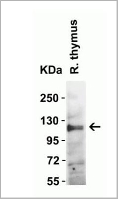
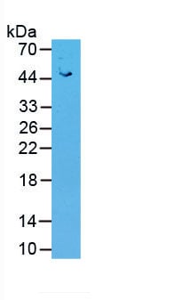
WB (Western Blot)
(Figure 4 Western Blot Validation in Rat Thymus Tissue Loading: 15 ug of lysates per lane. Antibodies: ACE2, AAA10945(2 ug/mL), 1h incubation at RT in 5% NFDM/TBST. Secondary: Goat anti-rabbit IgG HRP conjugate at 1:10000 dilution.)
WB (Western Blot)
(Figure 4 Western Blot Validation in Rat Thymus Tissue Loading: 15 ug of lysates per lane. Antibodies: ACE2, AAA10945(2 ug/mL), 1h incubation at RT in 5% NFDM/TBST. Secondary: Goat anti-rabbit IgG HRP conjugate at 1:10000 dilution.)
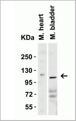
WB (Western Blot)
(Figure 3 Western Blot Validation in Mouse Tissues Loading: 15 ug of lysates per lane. Antibodies: ACE2, AAA10945(2 ug/mL), 1h incubation at RT in 5% NFDM/TBST. Secondary: Goat anti-rabbit IgG HRP conjugate at 1:10000 dilution.)
WB (Western Blot)
(Figure 3 Western Blot Validation in Mouse Tissues Loading: 15 ug of lysates per lane. Antibodies: ACE2, AAA10945(2 ug/mL), 1h incubation at RT in 5% NFDM/TBST. Secondary: Goat anti-rabbit IgG HRP conjugate at 1:10000 dilution.)
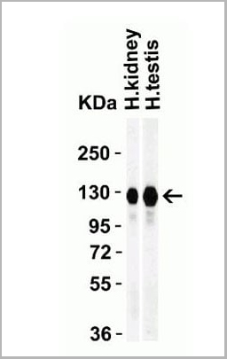
WB (Western Blot)
(Figure 2 Western Blot Validation in Human Tissues Loading: 15 ug of lysates per lane. Antibodies: ACE2, AAA10945 (1 ug/mL), 1h incubation at RT in 5% NFDM/TBST. Secondary: Goat anti-rabbit IgG HRP conjugate at 1:10000 dilution.)
WB (Western Blot)
(Figure 2 Western Blot Validation in Human Tissues Loading: 15 ug of lysates per lane. Antibodies: ACE2, AAA10945 (1 ug/mL), 1h incubation at RT in 5% NFDM/TBST. Secondary: Goat anti-rabbit IgG HRP conjugate at 1:10000 dilution.)
Application Data
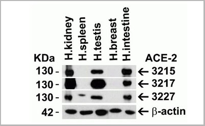
(Figure 1 Independent Antibody Validation (IAV) via Protein Expression Profile in Human Tissues Loading: 15 ug of lysates per lane. Antibodies: ACE2, (2 ug/mL), ACE2, (2 ug/mL), ACE2, AAA10945 (2 ug/mL) and beta-actin (1 ug)
Application Data
(Figure 1 Independent Antibody Validation (IAV) via Protein Expression Profile in Human Tissues Loading: 15 ug of lysates per lane. Antibodies: ACE2, (2 ug/mL), ACE2, (2 ug/mL), ACE2, AAA10945 (2 ug/mL) and beta-actin (1 ug)
IF (Immunofluorescence)
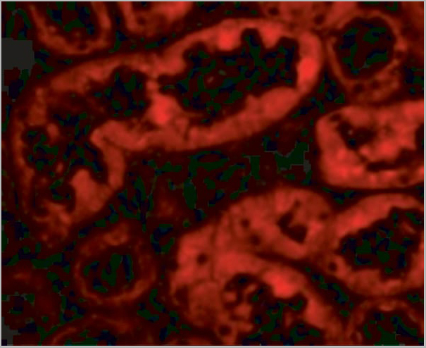
(Figure 10 Immunofluorescence Validation of ACE2 in Human Kidney Cells Immunofluorescent analysis of 4% paraformaldehyde-fixed human kidney cells labeling ACE2 with AAA10945 at 10ug/mL, followed by goat anti-rabbit IgG secondary antibody at 1/500 dilution (red).)
IF (Immunofluorescence)
(Figure 10 Immunofluorescence Validation of ACE2 in Human Kidney Cells Immunofluorescent analysis of 4% paraformaldehyde-fixed human kidney cells labeling ACE2 with AAA10945 at 10ug/mL, followed by goat anti-rabbit IgG secondary antibody at 1/500 dilution (red).)
IHC (Immunohistochemistry)
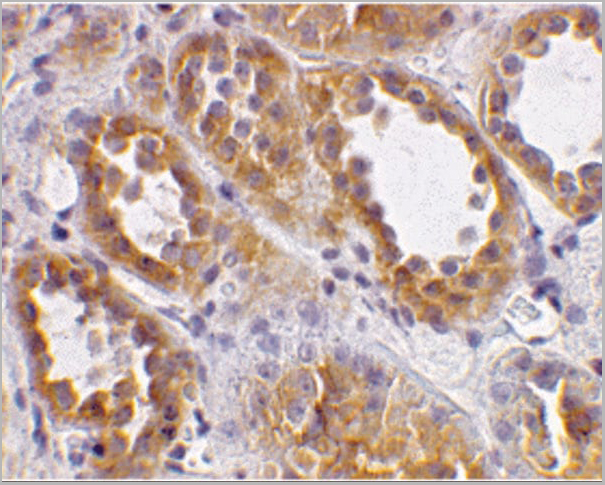
(Figure 5 Immunohistochemistry Validation of ACE2 inHuman Kidney Tissue Immunohistochemical analysis of paraffin-embedded human kidney tissue using anti-ACE2 antibody (3227) at 2ug/ml. Tissue was fixed with formaldehyde and blockedwith 10% serum for 1 h at RT; antigen retrieval was byheat mediation with a citrate buffer (pH6). Samples wereincubated with primary antibody overnight at 4°C. A goatanti-rabbit IgG H&L (HRP) at 1/250 was used as secondary.Counter stained with Hematoxylin.)
IHC (Immunohistochemistry)
(Figure 5 Immunohistochemistry Validation of ACE2 inHuman Kidney Tissue Immunohistochemical analysis of paraffin-embedded human kidney tissue using anti-ACE2 antibody (3227) at 2ug/ml. Tissue was fixed with formaldehyde and blockedwith 10% serum for 1 h at RT; antigen retrieval was byheat mediation with a citrate buffer (pH6). Samples wereincubated with primary antibody overnight at 4°C. A goatanti-rabbit IgG H&L (HRP) at 1/250 was used as secondary.Counter stained with Hematoxylin.)
NCBI and Uniprot Product Information
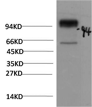
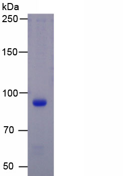
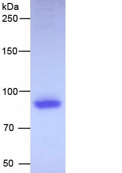
Observed MW: 130 kD (7 N-linked glycostation)
Customer Reviews
Loading reviews...
Share Your Experience
Similar Products


Product Notes
The ACE2 ace2 (Catalog #AAA10945) is an Antibody produced from Rabbit and is intended for research purposes only. The product is available for immediate purchase. The ACE2 Antibody reacts with Human, Mouse, Rat and may cross-react with other species as described in the data sheet. AAA Biotech's ACE2 can be used in a range of immunoassay formats including, but not limited to, ELISA, WB (Western Blot), IHC (Immunohistochemistry), IF (Immunofluorescence). WB: 1-2 ug/mL IHC: 2 ug/mL IF: 10 ug/mL Antibody validated: Western Blot in human, mouse and rat samples; Immunohistochemistry in human samples; Immunofluorescence in human, mouse andrat samples. All other applications and species not yet tested. Researchers should empirically determine the suitability of the ACE2 ace2 for an application not listed in the data sheet. Researchers commonly develop new applications and it is an integral, important part of the investigative research process. It is sometimes possible for the material contained within the vial of "ACE2, Polyclonal Antibody" to become dispersed throughout the inside of the vial, particularly around the seal of said vial, during shipment and storage. We always suggest centrifuging these vials to consolidate all of the liquid away from the lid and to the bottom of the vial prior to opening. Please be advised that certain products may require dry ice for shipping and that, if this is the case, an additional dry ice fee may also be required.Precautions
All products in the AAA Biotech catalog are strictly for research-use only, and are absolutely not suitable for use in any sort of medical, therapeutic, prophylactic, in-vivo, or diagnostic capacity. By purchasing a product from AAA Biotech, you are explicitly certifying that said products will be properly tested and used in line with industry standard. AAA Biotech and its authorized distribution partners reserve the right to refuse to fulfill any order if we have any indication that a purchaser may be intending to use a product outside of our accepted criteria.Disclaimer
Though we do strive to guarantee the information represented in this datasheet, AAA Biotech cannot be held responsible for any oversights or imprecisions. AAA Biotech reserves the right to adjust any aspect of this datasheet at any time and without notice. It is the responsibility of the customer to inform AAA Biotech of any product performance issues observed or experienced within 30 days of receipt of said product. To see additional details on this or any of our other policies, please see our Terms & Conditions page.Item has been added to Shopping Cart
If you are ready to order, navigate to Shopping Cart and get ready to checkout.