


Rabbit Catenin-gamma Polyclonal Antibody | anti-CTNNG antibody
Catenin-gamma Antibody
Phosphate buffered saline, pH 7.4, 150mM NaCl, 0.02% sodium azide and 50% glycerol.
IHC: 1:50-1:200
IF/ICC: 1:100-1:500
WB (Western Blot)
(Western blot analysis of Catenin-gamma expression in mouse tissue extract)
IHC (Immunohistochemistry)
(Catenin-gamma Antibody for IHC in human colon tissue.)
IHC (Immunohistochemistry)
(Catenin-gamma Antibody for IHC in human colon tissue.)
IHC (Immunohistchemistry)
(AAA31082 at 1/200 staining human breast cancer tissue sections by IHC-P. The tissue was formaldehyde fixed and a heat mediated antigen retrieval step in citrate buffer was performed. The tissue was then blocked and incubated with the antibody for 1.5 hours at 22 degree C. An HRP conjugated goat anti-rabbit antibody was used as the secondary.)
IHC (Immunohistochemistry)
(AAA31082 at 1/50 staining human colon cancer tissue sections by IHC-P. The tissue was formaldehyde fixed and a heat mediated antigen retrieval step in citrate buffer was performed. The tissue was then blocked and incubated with the antibody for 1.5 hours at 22 degree C. An HRP conjugated goat anti-rabbit antibody was used as the secondary.)
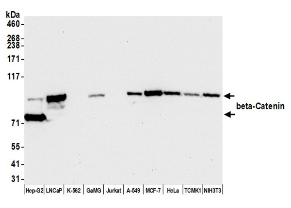
WB (Western Blot)
(Western blot analysis of extracts from rat brain, mouse spleen, using Catenin-gamma Antibody.)
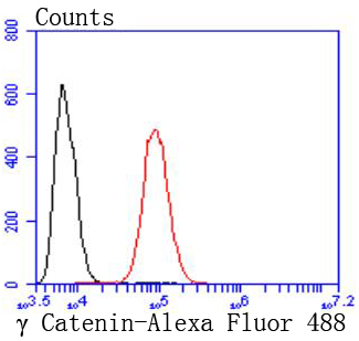
IF (Immunofluorescence)
(AAA31082 staining HeLa by IF/ICC. The sample were fixed with PFA and permeabilized in 0.1% Triton X-100, then blocked in 10% serum for 45 minutes at 25 degree C. The primary antibody was diluted at 1/200 and incubated with the sample for 1 hour at 37 degree C. An Alexa Fluor 594 conjugated goat anti-rabbit IgG (H+L) Ab, diluted at 1/600, was used as the secondary antibody.)
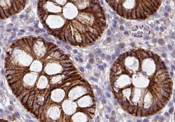
IHC (Immunohistochemistry)
(AAA31082 at 1/200 staining human colon cancer tissue sections by IHC-P. The tissue was formaldehyde fixed and a heat mediated antigen retrieval step in citrate buffer was performed. The tissue was then blocked and incubated with the antibody for 1.5 hours at 22 degree C. An HRP conjugated goat anti-rabbit antibody was used as the secondary.)
IHC (Immunohistochemistry)
(AAA31082 at 1/200 staining human breast cancer tissue sections by IHC-P. The tissue was formaldehyde fixed and a heat mediated antigen retrieval step in citrate buffer was performed. The tissue was then blocked and incubated with the antibody for 1.5 hours at 22 degree C. An HRP conjugated goat anti-rabbit antibody was used as the secondary.)
Subunit Structure: Homodimer. Component of an E-cadherin/catenin adhesion complex composed of at least E-cadherin/CDH1 and gamma-catenin/JUP, and possibly alpha-catenin/CTNNA1; the complex is located to adherens junctions. The stable association of CTNNA1 is controversial as CTNNA1 was shown not to bind to F-actin when assembled in the complex. Interacts with MUC1. Interacts with CAV1 (By similarity). Interacts with PTPRJ. Interacts with DSG1. Interacts with DSC1 and DSC2. Interacts with PKP2.
Post-translational Modifications: May be phosphorylated by FER.
Similarity: The entire ARM repeats region mediates binding to CDH1/E-cadherin. The N-terminus and first three ARM repeats are sufficient for binding to DSG1. The N-terminus and first ARM repeat are sufficient for association with CTNNA1. DSC1 association requires both ends of the ARM repeat region. Belongs to the beta-catenin family.
NCBI and Uniprot Product Information
Predicted: 82 kDa