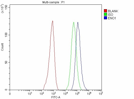
 FCM (Flow Cytometry)
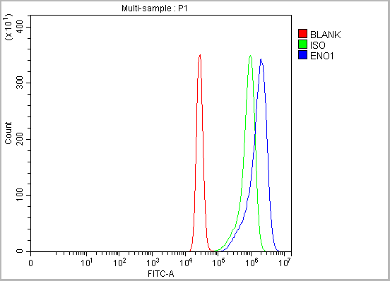
(Figure 10. Flow Cytometry analysis of K562 cells using anti-ENO1 antibody (AAA19420).Overlay histogram showing K562 cells stained with AAA19420 (Blue line). The cells were blocked with 10% normal goat serum. And then incubated with rabbit anti-ENO1 Antibody (AAA19420, 1 ug/1x10^6 cells) for 30 min at 20 degree C. DyLight488 conjugated goat anti-rabbit IgG was used as secondary antibody for 30 minutes at 20 degree C. Isotype control antibody (Green line) was rabbit IgG (1 ug/1x10^6) used under the same conditions. Unlabelled sample (Red line) was also used as a control.)
FCM (Flow Cytometry)
(Figure 10. Flow Cytometry analysis of K562 cells using anti-ENO1 antibody (AAA19420).Overlay histogram showing K562 cells stained with AAA19420 (Blue line). The cells were blocked with 10% normal goat serum. And then incubated with rabbit anti-ENO1 Antibody (AAA19420, 1 ug/1x10^6 cells) for 30 min at 20 degree C. DyLight488 conjugated goat anti-rabbit IgG was used as secondary antibody for 30 minutes at 20 degree C. Isotype control antibody (Green line) was rabbit IgG (1 ug/1x10^6) used under the same conditions. Unlabelled sample (Red line) was also used as a control.)
Rabbit anti-Human ENO1 Polyclonal Antibody | anti-ENO1 antibody
Anti-ENO1 Antibody Picoband
Each vial contains 4 mg Trehalose, 0.9 mg NaCl, 0.2 mg Na2HPO4.
IHC-P: 2-5 ug/ml, Human
FC/FACS: 1-3 ug/1x10^6 cells, Human
Direct ELISA: 0.1-0.5 ug/ml, Human
Tested Species: In-house tested species with positive results.
Enhanced Chemiluminescent Kit with anti-Rabbit IgG for Western blot, and HRP Conjugated anti-Rabbit IgG Super Vision Assay Kit for IHC(P).
FCM (Flow Cytometry)
(Figure 10. Flow Cytometry analysis of K562 cells using anti-ENO1 antibody (AAA19420).Overlay histogram showing K562 cells stained with AAA19420 (Blue line). The cells were blocked with 10% normal goat serum. And then incubated with rabbit anti-ENO1 Antibody (AAA19420, 1 ug/1x10^6 cells) for 30 min at 20 degree C. DyLight488 conjugated goat anti-rabbit IgG was used as secondary antibody for 30 minutes at 20 degree C. Isotype control antibody (Green line) was rabbit IgG (1 ug/1x10^6) used under the same conditions. Unlabelled sample (Red line) was also used as a control.)
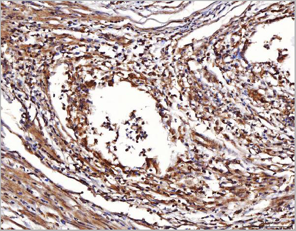
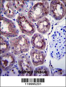
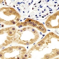
IHC (Immunohistchemistry)
(Figure 9. IHC analysis of ENO1 using anti-ENO1 antibody (AAA19420).ENO1 was detected in a paraffin-embedded section of human renal cell carcinoma tissue. Heat mediated antigen retrieval was performed in EDTA buffer (pH 8.0, epitope retrieval solution). The tissue section was blocked with 10% goat serum. The tissue section was then incubated with 2 ug/ml rabbit anti-ENO1 Antibody (AAA19420) overnight at 4 degree C. Peroxidase Conjugated Goat Anti-rabbit IgG was used as secondary antibody and incubated for 30 minutes at 37 degree C. The tissue section was developed using HRP Conjugated Rabbit IgG Super Vision Assay Kit with DAB as the chromogen.)
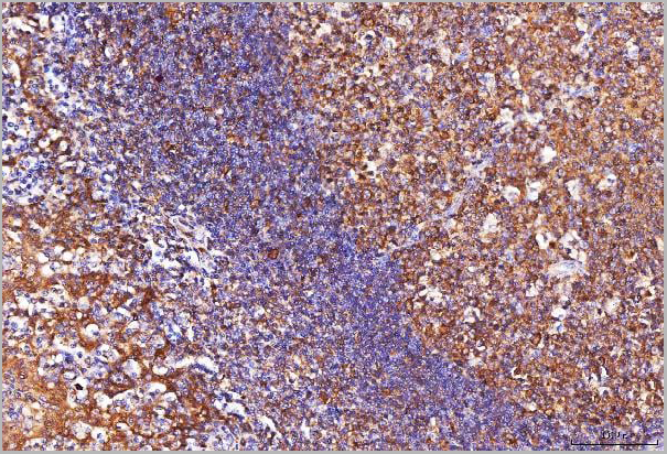
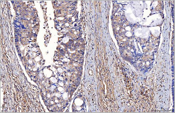
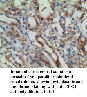
IHC (Immunohistochemistry)
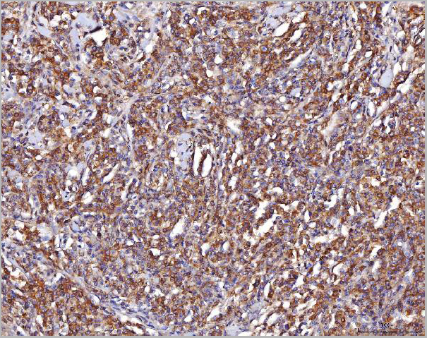
(Figure 8. IHC analysis of ENO1 using anti-ENO1 antibody (AAA19420).ENO1 was detected in a paraffin-embedded section of human lymphoma tissue. Heat mediated antigen retrieval was performed in EDTA buffer (pH 8.0, epitope retrieval solution). The tissue section was blocked with 10% goat serum. The tissue section was then incubated with 2 ug/ml rabbit anti-ENO1 Antibody (AAA19420) overnight at 4 degree C. Peroxidase Conjugated Goat Anti-rabbit IgG was used as secondary antibody and incubated for 30 minutes at 37 degree C. The tissue section was developed using HRP Conjugated Rabbit IgG Super Vision Assay Kit with DAB as the chromogen.)
IHC (Immunohistochemistry)
(Figure 8. IHC analysis of ENO1 using anti-ENO1 antibody (AAA19420).ENO1 was detected in a paraffin-embedded section of human lymphoma tissue. Heat mediated antigen retrieval was performed in EDTA buffer (pH 8.0, epitope retrieval solution). The tissue section was blocked with 10% goat serum. The tissue section was then incubated with 2 ug/ml rabbit anti-ENO1 Antibody (AAA19420) overnight at 4 degree C. Peroxidase Conjugated Goat Anti-rabbit IgG was used as secondary antibody and incubated for 30 minutes at 37 degree C. The tissue section was developed using HRP Conjugated Rabbit IgG Super Vision Assay Kit with DAB as the chromogen.)
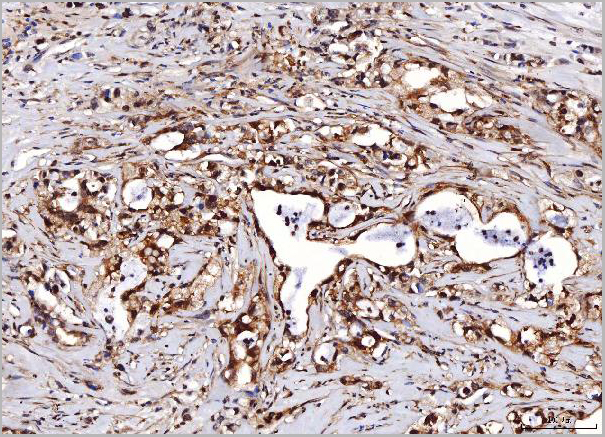
IHC (Immunohistochemistry)
(Figure 7. IHC analysis of ENO1 using anti-ENO1 antibody (AAA19420).ENO1 was detected in a paraffin-embedded section of human lung cancer tissue. Heat mediated antigen retrieval was performed in EDTA buffer (pH 8.0, epitope retrieval solution). The tissue section was blocked with 10% goat serum. The tissue section was then incubated with 2 ug/ml rabbit anti-ENO1 Antibody (AAA19420) overnight at 4 degree C. Peroxidase Conjugated Goat Anti-rabbit IgG was used as secondary antibody and incubated for 30 minutes at 37 degree C. The tissue section was developed using HRP Conjugated Rabbit IgG Super Vision Assay Kit with DAB as the chromogen.)
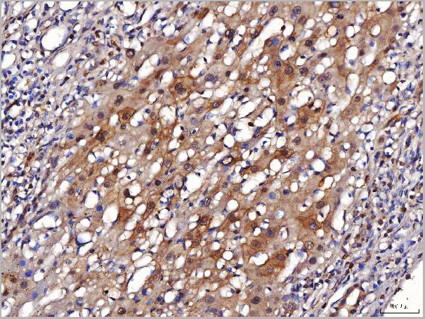
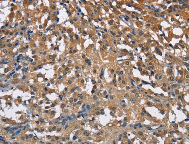
IHC (Immunohistchemistry)
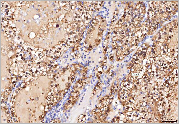
(Figure 6. IHC analysis of ENO1 using anti-ENO1 antibody (AAA19420).ENO1 was detected in a paraffin-embedded section of human liver cancer tissue. Heat mediated antigen retrieval was performed in EDTA buffer (pH 8.0, epitope retrieval solution). The tissue section was blocked with 10% goat serum. The tissue section was then incubated with 2 ug/ml rabbit anti-ENO1 Antibody (AAA19420) overnight at 4 degree C. Peroxidase Conjugated Goat Anti-rabbit IgG was used as secondary antibody and incubated for 30 minutes at 37 degree C. The tissue section was developed using HRP Conjugated Rabbit IgG Super Vision Assay Kit with DAB as the chromogen.)
IHC (Immunohistchemistry)
(Figure 6. IHC analysis of ENO1 using anti-ENO1 antibody (AAA19420).ENO1 was detected in a paraffin-embedded section of human liver cancer tissue. Heat mediated antigen retrieval was performed in EDTA buffer (pH 8.0, epitope retrieval solution). The tissue section was blocked with 10% goat serum. The tissue section was then incubated with 2 ug/ml rabbit anti-ENO1 Antibody (AAA19420) overnight at 4 degree C. Peroxidase Conjugated Goat Anti-rabbit IgG was used as secondary antibody and incubated for 30 minutes at 37 degree C. The tissue section was developed using HRP Conjugated Rabbit IgG Super Vision Assay Kit with DAB as the chromogen.)
IHC (Immunohistochemistry)
(Figure 5. IHC analysis of ENO1 using anti-ENO1 antibody (AAA19420).ENO1 was detected in a paraffin-embedded section of human gastric carcinoma tissue. Heat mediated antigen retrieval was performed in EDTA buffer (pH 8.0, epitope retrieval solution). The tissue section was blocked with 10% goat serum. The tissue section was then incubated with 2 ug/ml rabbit anti-ENO1 Antibody (AAA19420) overnight at 4 degree C. Peroxidase Conjugated Goat Anti-rabbit IgG was used as secondary antibody and incubated for 30 minutes at 37 degree C. The tissue section was developed using HRP Conjugated Rabbit IgG Super Vision Assay Kit with DAB as the chromogen.)
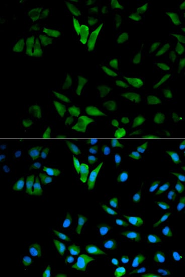
IHC (Immunohistochemistry)
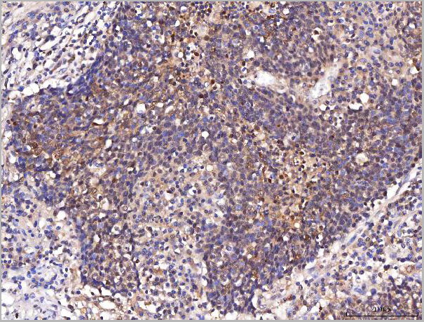
(Figure 4. IHC analysis of ENO1 using anti-ENO1 antibody (AAA19420).ENO1 was detected in a paraffin-embedded section of human chronic tonsillitis tissue. Heat mediated antigen retrieval was performed in EDTA buffer (pH 8.0, epitope retrieval solution). The tissue section was blocked with 10% goat serum. The tissue section was then incubated with 2 ug/ml rabbit anti-ENO1 Antibody (AAA19420) overnight at 4 degree C. Peroxidase Conjugated Goat Anti-rabbit IgG was used as secondary antibody and incubated for 30 minutes at 37 degree C. The tissue section was developed using HRP Conjugated Rabbit IgG Super Vision Assay Kit with DAB as the chromogen.)
IHC (Immunohistochemistry)
(Figure 3. IHC analysis of ENO1 using anti-ENO1 antibody (AAA19420).ENO1 was detected in a paraffin-embedded section of human cervical cancer tissue. Heat mediated antigen retrieval was performed in EDTA buffer (pH 8.0, epitope retrieval solution). The tissue section was blocked with 10% goat serum. The tissue section was then incubated with 2 ug/ml rabbit anti-ENO1 Antibody (AAA19420) overnight at 4 degree C. Peroxidase Conjugated Goat Anti-rabbit IgG was used as secondary antibody and incubated for 30 minutes at 37 degree C. The tissue section was developed using HRP Conjugated Rabbit IgG Super Vision Assay Kit with DAB as the chromogen.)
IHC (Immunohistochemistry)
(Figure 2. IHC analysis of ENO1 using anti-ENO1 antibody (AAA19420).ENO1 was detected in a paraffin-embedded section of human adenocarcinoma of the right colon tissue. Heat mediated antigen retrieval was performed in EDTA buffer (pH 8.0, epitope retrieval solution). The tissue section was blocked with 10% goat serum. The tissue section was then incubated with 2 ug/ml rabbit anti-ENO1 Antibody (AAA19420) overnight at 4 degree C. Peroxidase Conjugated Goat Anti-rabbit IgG was used as secondary antibody and incubated for 30 minutes at 37 degree C. The tissue section was developed using HRP Conjugated Rabbit IgG Super Vision Assay Kit with DAB as the chromogen.)
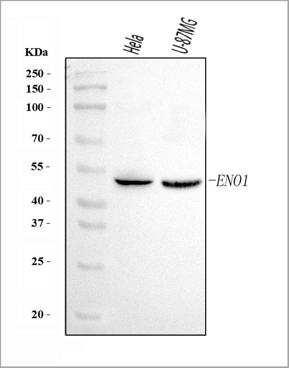
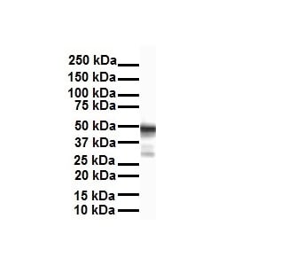
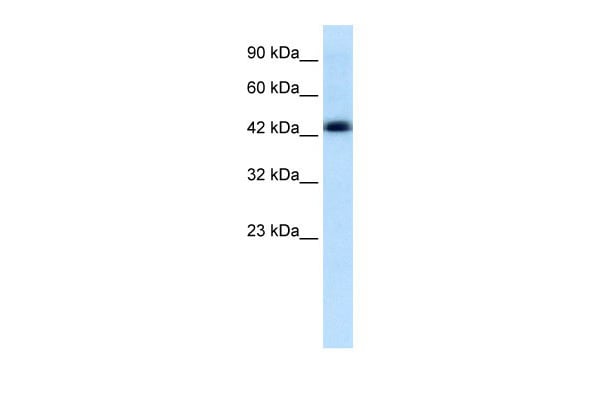
WB (Western Blot)
(Figure 1. Western blot analysis of ENO1 using anti-ENO1 antibody (AAA19420).Electrophoresis was performed on a 5-20% SDS-PAGE gel at 70V (Stacking gel)/90V (Resolving gel) for 2-3 hours. The sample well of each lane was loaded with 30 ug of sample under reducing conditions.Lane 1: human Hela whole cell lysates,Lane 2: human U-87MG whole cell lysates.After electrophoresis, proteins were transferred to a nitrocellulose membrane at 150 mA for 50-90 minutes. Blocked the membrane with 5% non-fat milk/TBS for 1.5 hour at RT. The membrane was incubated with rabbit anti-ENO1 antigen affinity purified polyclonal antibody (#AAA19420) at 0.5 ug/mL overnight at 4 degree C, then washed with TBS-0.1%Tween 3 times with 5 minutes each and probed with a goat anti-rabbit IgG-HRP secondary antibody at a dilution of 1:5000 for 1.5 hour at RT. The signal is developed using an Enhanced Chemiluminescent detection (ECL) kit with Tanon 5200 system. A specific band was detected for ENO1 at approximately 50 kDa. The expected band size for ENO1 is at 50 kDa.)
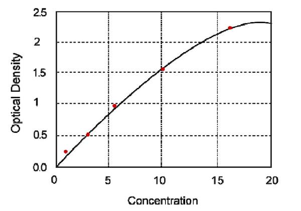
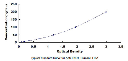
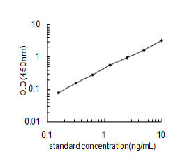
Principle of the Assay: The Picokine™ Human CD79B Pre-Coated ELISA (Enzyme-Linked Immunosorbent Assay) kit is a solid-phase immunoassay specially designed to measure Human CD79B with a 96-well strip plate that is pre-coated with antibody specific for CD79B. The detection antibody is a biotinylated antibody specific for CD79B. The capture antibody is a monoclonal antibody from mouse and the detection antibody is a biotinylated polyclonal antibody from goat. The kit contains recombinant Human CD79B with immunogen: Expression system for standard: CHO; Immunogen sequence: A29-D159. The kit is analytically validated with ready-to-use reagents. To measure Human CD79B, add standards and samples to the wells, then add the biotinylated detection antibody. Wash the wells with PBS or TBS buffer, and add Avidin-Biotin-Peroxidase Complex (ABC-HRP). Wash away the unbounded ABC-HRP with PBS or TBS buffer and add TMB. TMB is an HRP substrate and will be catalyzed to produce a blue color product, which changes into yellow after adding the acidic stop solution. The absorbance of the yellow product at 450nm is linearly proportional to Human CD79B in the sample. Read the absorbance of the yellow product in each well using a plate reader, and benchmark the sample wells' readings against the standard curve to determine the concentration of Human CD79B in the sample.
NCBI and Uniprot Product Information
Customer Reviews
Loading reviews...
Share Your Experience
Similar Products


Product Notes
The ENO1 eno1 (Catalog #AAA19420) is an Antibody produced from Rabbit and is intended for research purposes only. The product is available for immediate purchase. The Anti-ENO1 Antibody Picoband reacts with Human and may cross-react with other species as described in the data sheet. AAA Biotech's ENO1 can be used in a range of immunoassay formats including, but not limited to, ELISA, FCM/FACS (Flow Cytometry), IHC (Immunohistochemistry), WB (Western Blot). WB: 0.25-0.5 ug/ml, Human IHC-P: 2-5 ug/ml, Human FC/FACS: 1-3 ug/1x10^6 cells, Human Direct ELISA: 0.1-0.5 ug/ml, Human Tested Species: In-house tested species with positive results. Enhanced Chemiluminescent Kit with anti-Rabbit IgG for Western blot, and HRP Conjugated anti-Rabbit IgG Super Vision Assay Kit for IHC(P). Researchers should empirically determine the suitability of the ENO1 eno1 for an application not listed in the data sheet. Researchers commonly develop new applications and it is an integral, important part of the investigative research process. It is sometimes possible for the material contained within the vial of "ENO1, Polyclonal Antibody" to become dispersed throughout the inside of the vial, particularly around the seal of said vial, during shipment and storage. We always suggest centrifuging these vials to consolidate all of the liquid away from the lid and to the bottom of the vial prior to opening. Please be advised that certain products may require dry ice for shipping and that, if this is the case, an additional dry ice fee may also be required.Precautions
All products in the AAA Biotech catalog are strictly for research-use only, and are absolutely not suitable for use in any sort of medical, therapeutic, prophylactic, in-vivo, or diagnostic capacity. By purchasing a product from AAA Biotech, you are explicitly certifying that said products will be properly tested and used in line with industry standard. AAA Biotech and its authorized distribution partners reserve the right to refuse to fulfill any order if we have any indication that a purchaser may be intending to use a product outside of our accepted criteria.Disclaimer
Though we do strive to guarantee the information represented in this datasheet, AAA Biotech cannot be held responsible for any oversights or imprecisions. AAA Biotech reserves the right to adjust any aspect of this datasheet at any time and without notice. It is the responsibility of the customer to inform AAA Biotech of any product performance issues observed or experienced within 30 days of receipt of said product. To see additional details on this or any of our other policies, please see our Terms & Conditions page.Item has been added to Shopping Cart
If you are ready to order, navigate to Shopping Cart and get ready to checkout.