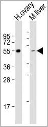

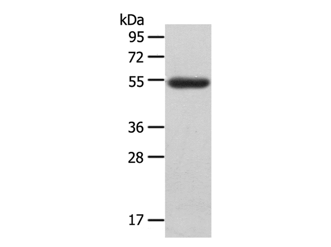
 WB (Western Blot)
(All lanes : Anti-LAG3 Antibody (Center) at 1:1000 dilutionLane 1: human ovary lysateLane 2: mouse liver lysate Lysates/proteins at 20 ug per lane.Secondary Goat Anti-Rabbit IgG, (H+L), Peroxidase conjugated at 1/10000 dilution.Predicted band size : 57 kDaBlocking/Dilution buffer: 5% NFDM/TBST.)
WB (Western Blot)
(All lanes : Anti-LAG3 Antibody (Center) at 1:1000 dilutionLane 1: human ovary lysateLane 2: mouse liver lysate Lysates/proteins at 20 ug per lane.Secondary Goat Anti-Rabbit IgG, (H+L), Peroxidase conjugated at 1/10000 dilution.Predicted band size : 57 kDaBlocking/Dilution buffer: 5% NFDM/TBST.)
Rabbit anti-Human, Mouse LAG3 Polyclonal Antibody | anti-LAG3 antibody
LAG3 Antibody (Center)
FC~~1:25
WB~~1:1000
WB (Western Blot)
(All lanes : Anti-LAG3 Antibody (Center) at 1:1000 dilutionLane 1: human ovary lysateLane 2: mouse liver lysate Lysates/proteins at 20 ug per lane.Secondary Goat Anti-Rabbit IgG, (H+L), Peroxidase conjugated at 1/10000 dilution.Predicted band size : 57 kDaBlocking/Dilution buffer: 5% NFDM/TBST.)
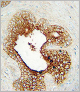
IHC (Immunohistochemistry)
(LAG3 Antibody (Center) immunohistochemistry analysis in formalin fixed and paraffin embedded human prostate carcinoma followed by peroxidase conjugation of the secondary antibody and DAB staining. This data demonstrates the use of the LAG3 Antibody (Center) for immunohistochemistry. Clinical relevance has not been evaluated.)
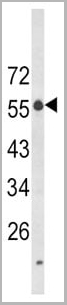
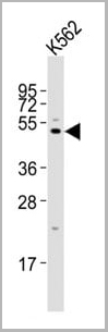
WB (Western Blot)
(Western blot analysis of LAG3 Antibody (Center) in K562 cell line lysates (35ug/lane). LAG3 (arrow) was detected using the purified Pab.)
WB (Western Blot)
(Western blot analysis of LAG3 Antibody (Center) in K562 cell line lysates (35ug/lane). LAG3 (arrow) was detected using the purified Pab.)
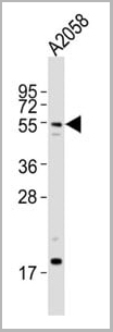
WB (Western Blot)
(Anti-LAG3 Antibody (Center)at 1:2000 dilution (please inquire) whole cell lysates Lysates/proteins at 20 ug per lane. Secondary Goat Anti-Rabbit IgG, (H+L), Peroxidase conjugated at 1/10000 dilution Predicted band size : 57 kDa Blocking/Dilution buffer: 5% NFDM/TBST.)
WB (Western Blot)
(Anti-LAG3 Antibody (Center)at 1:2000 dilution (please inquire) whole cell lysates Lysates/proteins at 20 ug per lane. Secondary Goat Anti-Rabbit IgG, (H+L), Peroxidase conjugated at 1/10000 dilution Predicted band size : 57 kDa Blocking/Dilution buffer: 5% NFDM/TBST.)
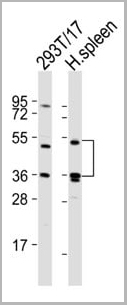
WB (Western Blot)
(All lanes : Anti-LAG3 Antibody (Center) at 1:2000 dilution Lane 1: 293T/17 whole cell lysates Lane 2: human spleen lysates Lysates/proteins at 20 ug per lane. Secondary Goat Anti-Rabbit IgG, (H+L), Peroxidase conjugated at 1/10000 dilution Predicted band size : 57 kDa Blocking/Dilution buffer: 5% NFDM/TBST)
WB (Western Blot)
(Anti-LAG3 Antibody (Center)at 1:2000 dilution + K562 whole cell lysates Lysates/proteins at 20 ug per lane. Secondary Goat Anti-Rabbit IgG, (H+L), Peroxidase conjugated at 1/10000 dilution Predicted band size : 57 kDa Blocking/Dilution buffer: 5% NFDM/TBST.)
WB (Western Blot)
(Anti-LAG3 Antibody (Center)at 1:2000 dilution + K562 whole cell lysates Lysates/proteins at 20 ug per lane. Secondary Goat Anti-Rabbit IgG, (H+L), Peroxidase conjugated at 1/10000 dilution Predicted band size : 57 kDa Blocking/Dilution buffer: 5% NFDM/TBST.)
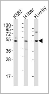
WB (Western Blot)
(All lanes : Anti-LAG3 Antibody (Center) at 1:2000 dilution Lane 1: K562 whole cell lysates Lane 2: human liver lysates Lane 3: human ovary lysates Lysates/proteins at 20 ug per lane. Secondary Goat Anti-Rabbit IgG, (H+L), Peroxidase conjugated at 1/10000 dilution Predicted band size : 57 kDa Blocking/Dilution buffer: 5% NFDM/TBST.)
WB (Western Blot)
(Anti-LAG3 Antibody (Center) at 1:2000 dilution + mouse heart lysate Lysates/proteins at 20 ug per lane. Secondary Goat Anti-Rabbit IgG, (H+L), Peroxidase conjugated at 1/10000 dilution. Predicted band size : 57 kDa Blocking/Dilution buffer: 5% NFDM/TBST.)
WB (Western Blot)
(Anti-LAG3 Antibody (Center) at 1:2000 dilution + mouse heart lysate Lysates/proteins at 20 ug per lane. Secondary Goat Anti-Rabbit IgG, (H+L), Peroxidase conjugated at 1/10000 dilution. Predicted band size : 57 kDa Blocking/Dilution buffer: 5% NFDM/TBST.)
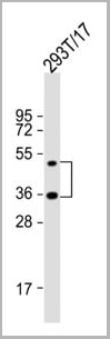
WB (Western Blot)
(Anti-LAG3 Antibody (Center) at 1:2000 dilution + 293T/17 whole cell lysate Lysates/proteins at 20 ug per lane. Secondary Goat Anti-Rabbit IgG, (H+L), Peroxidase conjugated at 1/10000 dilution. Predicted band size : 57 kDa Blocking/Dilution buffer: 5% NFDM/TBST.)
WB (Western Blot)
(Anti-LAG3 Antibody (Center) at 1:2000 dilution + 293T/17 whole cell lysate Lysates/proteins at 20 ug per lane. Secondary Goat Anti-Rabbit IgG, (H+L), Peroxidase conjugated at 1/10000 dilution. Predicted band size : 57 kDa Blocking/Dilution buffer: 5% NFDM/TBST.)
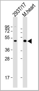
WB (Western Blot)
(All lanes : Anti-LAG3 Antibody (Center) at 1:2000 dilution Lane 1: 293T/17 whole cell lysate Lane 2: mouse heart lysate Lysates/proteins at 20 ug per lane. Secondary Goat Anti-Rabbit IgG, (H+L), Peroxidase conjugated at 1/10000 dilution. Predicted band size : 57 kDa Blocking/Dilution buffer: 5% NFDM/TBST.)
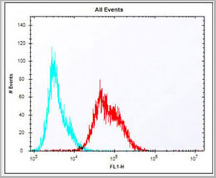
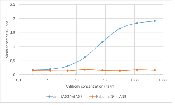
FCM (Flow Cytometry)
(Overlay histogram showing Jurkat cells stained with AAA28766 (red line). The cells were fixed with 2% paraformaldehyde (10 min) and then permeabilized with 90% methanol for 10 min. The cells were then icubated in 2% bovine serum albumin to block non-specific protein-protein interactions followed by the antibody (AAA28766, 1:25 dilution) for 60 min at 37ºC. The secondary antibody used was Alexa Fluor® 488 goat anti-rabbit lgG (H+L) (1583138) at 1/400 dilution for 40 min at 37ºC. Isotype control antibody (blue line) was rabbit IgG1 (1ug/1x10^6 cells) used under the same conditions. Acquisition of >10, 000 events was performed.)
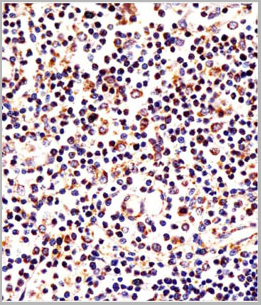
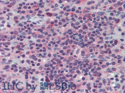
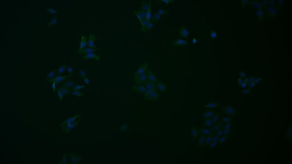
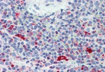
IHC (Immunohistochemistry)
(AAA28766 staining LAG3 in human thymus tissue sections by Immunohistochemistry (IHC-P - paraformaldehyde-fixed, paraffin-embedded sections). Tissue was fixed with formaldehyde and blocked with 3% BSA for 0. 5 hour at room temperature; antigen retrieval was by heat mediation with a citrate buffer (pH6). Samples were incubated with primary antibody (1/25) for 1 hours at 37°C. A undiluted biotinylated goat polyvalent antibody was used as the secondary antibody.)
NCBI and Uniprot Product Information
Customer Reviews
Loading reviews...
Share Your Experience
Similar Products


Product Notes
The LAG3 lag3 (Catalog #AAA28766) is an Antibody produced from Rabbit and is intended for research purposes only. The product is available for immediate purchase. The immunogen sequence is 103-132. The LAG3 Antibody (Center) reacts with Human, Mouse and may cross-react with other species as described in the data sheet. AAA Biotech's LAG3 can be used in a range of immunoassay formats including, but not limited to, WB (Western Blot), ELISA, IHC (Immunohistochemistry), FCM/FACS (Flow Cytometry). IHC-P~~1:10~50 FC~~1:25 WB~~1:1000. Researchers should empirically determine the suitability of the LAG3 lag3 for an application not listed in the data sheet. Researchers commonly develop new applications and it is an integral, important part of the investigative research process. It is sometimes possible for the material contained within the vial of "LAG3, Polyclonal Antibody" to become dispersed throughout the inside of the vial, particularly around the seal of said vial, during shipment and storage. We always suggest centrifuging these vials to consolidate all of the liquid away from the lid and to the bottom of the vial prior to opening. Please be advised that certain products may require dry ice for shipping and that, if this is the case, an additional dry ice fee may also be required.Precautions
All products in the AAA Biotech catalog are strictly for research-use only, and are absolutely not suitable for use in any sort of medical, therapeutic, prophylactic, in-vivo, or diagnostic capacity. By purchasing a product from AAA Biotech, you are explicitly certifying that said products will be properly tested and used in line with industry standard. AAA Biotech and its authorized distribution partners reserve the right to refuse to fulfill any order if we have any indication that a purchaser may be intending to use a product outside of our accepted criteria.Disclaimer
Though we do strive to guarantee the information represented in this datasheet, AAA Biotech cannot be held responsible for any oversights or imprecisions. AAA Biotech reserves the right to adjust any aspect of this datasheet at any time and without notice. It is the responsibility of the customer to inform AAA Biotech of any product performance issues observed or experienced within 30 days of receipt of said product. To see additional details on this or any of our other policies, please see our Terms & Conditions page.Item has been added to Shopping Cart
If you are ready to order, navigate to Shopping Cart and get ready to checkout.