Rabbit anti-Human OPN-a/b Polyclonal Antibody | anti-OPN-a/b antibody
OPN-a/b, NT (SPP1, BNSP, OPN, Osteopontin, Bone sialoprotein 1, Nephropontin, Secreted phosphoprotein 1, Urinary stone protein, Uropontin) (MaxLight 750)
IF (Immunofluorescence)
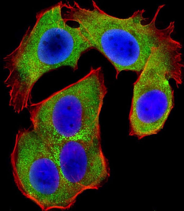
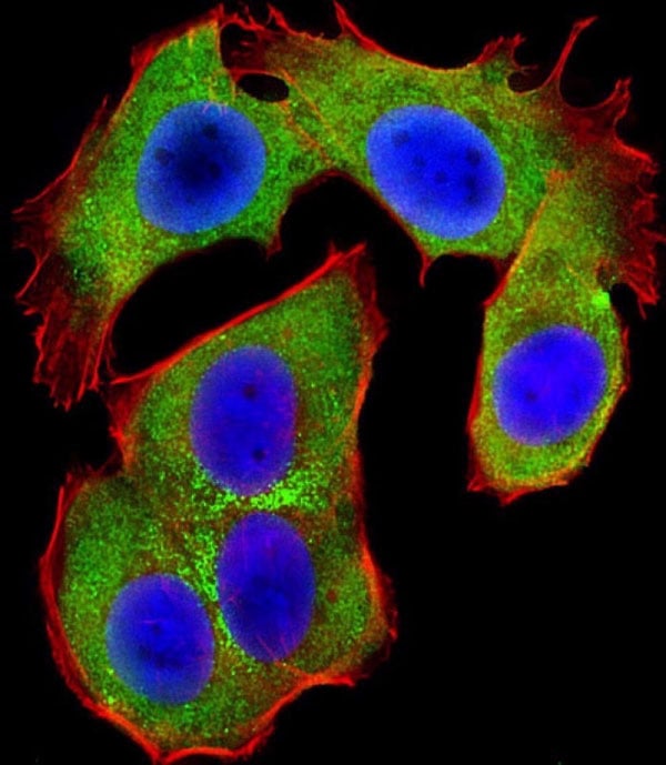
(Immunofluorescent analysis of 4% paraformaldehyde-fixed, 0.1% Triton X-100 permeabilized MCF-7 (human breast cancer cell line) cells labeling Pdx1 with at 1:25 dilution, followed by DyLight 488-conjugated IgG goat anti-rabbit secondary antibody at 1:200 dilution (green). Immunofluorescence image showing cytoplasm staining on MCF-7 cell line. Cytoplasmic actin is detected with DyLight 554 Phalloidin (PD18466410) at 1:100 dilution (red). The nuclear counter stain is DAPI (blue).)
IHC (Immunohistochemistry)
(Immunohistochemistry analysis in formalin fixed and paraffin embedded human kidney tissue using followed by peroxidase conjugation of the secondary antibody and DAB staining.This data demonstrates the use of for immunohistochemistry.)
Application Data
(All lanes: at 1:1000 dilution Lane 1: human liver lysates Lane 2: MDA-MB-453 whole cell lysates Lane 3: MOLT-4 whole cell lysates Lysates/proteins at 20ug/lane. Secondary IgG, (H+L) goat anti-rabbit, Peroxidase conjugated at 1:10000 dilution. Predicted band size : 35kD Blocking/Dilution buffer: 5% NFDM/TBST.)
Application Data
(039288 at 1:2000 dilution + Hela whole cell lysate Lysates/proteins at 20ug/lane. Secondary IgG, (H+L) goat anti-rabbit Peroxidase conjugated at 1:10000 dilution. Predicted band size: 35kD Blocking/Dilution buffer: 5% NFDM/TBST.)
Application Data
(All lanes: at 1:2000 dilution Lane 1: Hela whole cell lysate Lane 2: HL-60 whole cell lysate Lane 3: Jurkat whole cell lysate Lane 4: MOLT-4 whole cell lysate Lysates/proteins at 20ug/lane. Secondary IgG (H+L) goat anti-rabbit, Peroxidase conjugated at 1:10000 dilution. Predicted band size: 35kD Blocking/Dilution buffer: 5% NFDM/TBST.)


