


Rabbit SHANK3 Polyclonal Antibody | anti-SHANK3 antibody
Anti-SHANK3 Antibody
IHC-P: Concentration: 2-5ug/ml; Tested Species: Human, Rat
FC: Concentration: 1-3ug/1x106 cells; Tested Species: Human, Mouse, Rat
Direct ELISA: Concentration:0.1-0.5ug/ml; Tested Species: Human
Tested Species : In house tesed species with positive results.
Other applications have not been tested.
Optimal dilutions should be determined by end users.
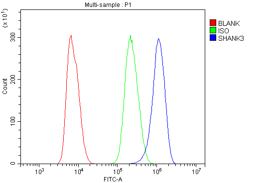
FCM (Flow Cytometry)
(Figure 6. Flow Cytometry analysis of NRK cells using anti-SHANK3 antibody (AAA19237).Overlay histogram showing NRK cells stained with AAA19237 (Blue line). The cells were blocked with 10% normal goat serum. And then incubated with rabbit anti-SHANK3 Antibody (AAA19237,1μg/1x106 cells) for 30 min at 20 degree C. DyLight®488 conjugated goat anti-rabbit IgG (5-10μg/1x106 cells) was used as secondary antibody for 30 minutes at 20 degree C. Isotype control antibody (Green line) was rabbit IgG (1μg/1x106) used under the same conditions. Unlabelled sample (Red line) was also used as a control.)
FCM (Flow Cytometry)
(Figure 5. Flow Cytometry analysis of HEPA1-6 cells using anti-SHANK3 antibody (AAA19237).Overlay histogram showing HEPA1-6 cells stained with AAA19237 (Blue line). The cells were blocked with 10% normal goat serum. And then incubated with rabbit anti-SHANK3 Antibody (AAA19237,1μg/1x106 cells) for 30 min at 20 degree C. DyLight®488 conjugated goat anti-rabbit IgG (5-10μg/1x106 cells) was used as secondary antibody for 30 minutes at 20 degree C. Isotype control antibody (Green line) was rabbit IgG (1μg/1x106) used under the same conditions. Unlabelled sample (Red line) was also used as a control.)
FCM (Flow Cytometry)
(Figure 4. Flow Cytometry analysis of Raji cells using anti-SHANK3 antibody (AAA19237).Overlay histogram showing Raji cells stained with AAA19237 (Blue line). The cells were blocked with 10% normal goat serum. And then incubated with rabbit anti-SHANK3 Antibody (AAA19237,1μg/1x106 cells) for 30 min at 20 degree C. DyLight®488 conjugated goat anti-rabbit IgG (5-10μg/1x106 cells) was used as secondary antibody for 30 minutes at 20 degree C. Isotype control antibody (Green line) was rabbit IgG (1μg/1x106) used under the same conditions. Unlabelled sample (Red line) was also used as a control.)
IHC (Immunohistochemistry)
(Figure 3. IHC analysis of SHANK3 using anti SHANK3 antibody (AAA19237).SHANK3 was detected in paraffin-embedded section of rat brain tissue. Heat mediated antigen retrieval was performed in EDTA buffer (pH8. 0, epitope retrieval solution). The tissue section was blocked with 10% goat serum. The tissue section was then incubated with 2μg/ml rabbit anti-SHANK3 Antibody (AAA19237) overnight at 4 degree C. Biotinylated goat anti-rabbit IgG was used as secondary antibody and incubated for 30 minutes at 37 degree C. The tissue section was developed using Strepavidin-Biotin-Complex (SABC) (Catalog #) with DAB as the chromogen.)
IHC (Immunohistochemistry)
(Figure 2. IHC analysis of SHANK3 using anti SHANK3 antibody (AAA19237).SHANK3 was detected in paraffin-embedded section of human meningoma tissue. Heat mediated antigen retrieval was performed in EDTA buffer (pH8. 0, epitope retrieval solution). The tissue section was blocked with 10% goat serum. The tissue section was then incubated with 2μg/ml rabbit anti-SHANK3 Antibody (AAA19237) overnight at 4 degree C. Biotinylated goat anti-rabbit IgG was used as secondary antibody and incubated for 30 minutes at 37 degree C. The tissue section was developed using Strepavidin-Biotin-Complex (SABC) (Catalog # with DAB as the chromogen.)
WB (Western Blot)
(Figure 1. Western blot analysis of SHANK3 using anti-SHANK3 antibody (AAA19237).Electrophoresis was performed on a 5-20% SDS-PAGE gel at 70V (Stacking gel) / 90V (Resolving gel) for 2-3 hours. The sample well of each lane was loaded with 50ug of sample under reducing conditions.Lane 1: rat brain tissue lysatesLane 2: mouse brain tissue lysates.After Electrophoresis, proteins were transferred to a Nitrocellulose membrane at 150mA for 50-90 minutes. Blocked the membrane with 5% Non-fat Milk/ TBS for 1. 5 hour at RT. The membrane was incubated with rabbit anti-SHANK3 antigen affinity purified polyclonal antibody (Catalog # AAA19237) at 0. 25 μg/mL overnight at 4 degree C, then washed with TBS-0. 1%Tween 3 times with 5 minutes each and probed with a goat anti-rabbit IgG-HRP secondary antibody at a dilution of 1:10000 for 1. 5 hour at RT. The signal is developed using an Enhanced Chemiluminescent detection (ECL) kit (Catalog # with Tanon 5200 system. A specific band was detected for SHANK3 at approximately 180-190KD. The expected band size for SHANK3 is at 185KD.)
2. Boeckers TM, Bockmann J, Kreutz MR, Gundelfinger ED (2002). "ProSAP/Shank proteins - a family of higher order organizing molecules of the postsynaptic density with an emerging role in human neurological disease". J. Neurochem. 81 (5): 903-10.


