


Rabbit TORC1/CRTC1 Polyclonal Antibody | anti-CRTC1 antibody
Anti-TORC1/CRTC1 Antibody
IHC-P: 2-5ug/ml|Human, Mouse, Rat|
ICC/IF: 5ug/ml|Human|
FC/FACS/FCM: 1-3ug/1x106 cells|Human, Rat|
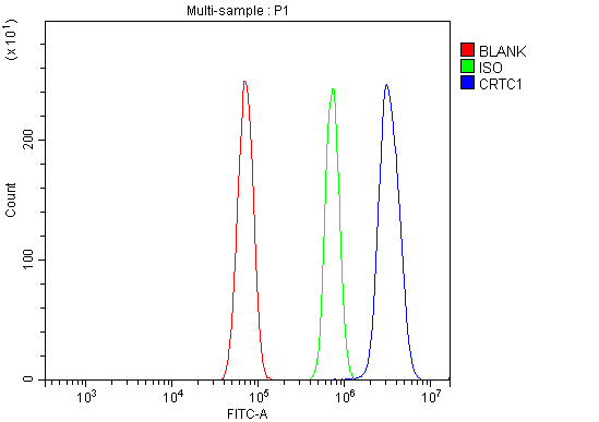
FCM (Flow Cytometry)
(Figure 8. Flow Cytometry analysis of RH35 cells using anti-TORC1/CRTC1 antibody (AAA19251).Overlay histogram showing RH35 cells stained with AAA19251 (Blue line). The cells were blocked with 10% normal goat serum. And then incubated with rabbit anti-TORC1/CRTC1 Antibody (AAA19251, 1μg/1x106 cells) for 30 min at 20 degree C. DyLight®488 conjugated goat anti-rabbit IgG (5-10μg/1x106 cells) was used as secondary antibody for 30 minutes at 20 degree C. Isotype control antibody (Green line) was rabbit IgG (1μg/1x106) used under the same conditions. Unlabelled sample (Red line) was also used as a control.)
FCM (Flow Cytometry)
(Figure 7. Flow Cytometry analysis of A431 cells using anti-TORC1/CRTC1 antibody (AAA19251).Overlay histogram showing A431 cells stained with AAA19251 (Blue line). The cells were blocked with 10% normal goat serum. And then incubated with rabbit anti-TORC1/CRTC1 Antibody (AAA19251, 1μg/1x106 cells) for 30 min at 20 degree C. DyLight®488 conjugated goat anti-rabbit IgG (5-10μg/1x106 cells) was used as secondary antibody for 30 minutes at 20 degree C. Isotype control antibody (Green line) was rabbit IgG (1μg/1x106) used under the same conditions. Unlabelled sample (Red line) was also used as a control.)
IF (Immunofluorescence)
(Figure 6. IF analysis of TORC1/CRTC1 using anti- TORC1/CRTC1 antibody (AAA19251).TORC1/CRTC1 was detected in immunocytochemical section of MCF-7 cells. Enzyme antigen retrieval was performed using IHC enzyme antigen retrieval reagent for 15 mins. The cells were blocked with 10% goat serum. And then incubated with 5μg/mL rabbit anti- TORC1/CRTC1 Antibody (AAA19251) overnight at 4 degree C. DyLight®488 Conjugated Goat Anti-Rabbit IgG was used as secondary antibody at 1:100 dilution and incubated for 30 minutes at 37 degree C. The section was counterstained with DAPI. Visualize using a fluorescence microscope and filter sets appropriate for the label used.)
IHC (Immunohistochemistry)
(Figure 5. IHC analysis of TORC1/CRTC1 using anti-TORC1/CRTC1 antibody (AAA19251).TORC1/CRTC1 was detected in paraffin-embedded section of human lung cancer tissue. Heat mediated antigen retrieval was performed in EDTA buffer (pH8. 0, epitope retrieval solution). The tissue section was blocked with 10% goat serum. The tissue section was then incubated with 2μg/ml rabbit anti-TORC1/CRTC1 Antibody (AAA19251) overnight at 4 degree C. Biotinylated goat anti-rabbit IgG was used as secondary antibody and incubated for 30 minutes at 37 degree C. The tissue section was developed using Strepavidin-Biotin-Complex (SABC) (Catalog # with DAB as the chromogen.)
IHC (Immunohistochemistry)
(Figure 4. IHC analysis of TORC1/CRTC1 using anti-TORC1/CRTC1 antibody (AAA19251).TORC1/CRTC1 was detected in paraffin-embedded section of mouse brain tissue. Heat mediated antigen retrieval was performed in EDTA buffer (pH8. 0, epitope retrieval solution). The tissue section was blocked with 10% goat serum. The tissue section was then incubated with 2μg/ml rabbit anti-TORC1/CRTC1 Antibody (AAA19251) overnight at 4 degree C. Biotinylated goat anti-rabbit IgG was used as secondary antibody and incubated for 30 minutes at 37 degree C. The tissue section was developed using Strepavidin-Biotin-Complex (SABC) (Catalog # with DAB as the chromogen.)
IHC (Immunohistochemistry)
(Figure 3. IHC analysis of TORC1/CRTC1 using anti-TORC1/CRTC1 antibody (AAA19251).TORC1/CRTC1 was detected in paraffin-embedded section of mouse brain tissue. Heat mediated antigen retrieval was performed in EDTA buffer (pH8. 0, epitope retrieval solution). The tissue section was blocked with 10% goat serum. The tissue section was then incubated with 2μg/ml rabbit anti-TORC1/CRTC1 Antibody (AAA19251) overnight at 4 degree C. Biotinylated goat anti-rabbit IgG was used as secondary antibody and incubated for 30 minutes at 37 degree C. The tissue section was developed using Strepavidin-Biotin-Complex (SABC) (Catalog # with DAB as the chromogen.)
IHC (Immunohistochemistry)
(Figure 2. IHC analysis of TORC1/CRTC1 using anti-TORC1/CRTC1 antibody (AAA19251).TORC1/CRTC1 was detected in paraffin-embedded section of rat brain tissue. Heat mediated antigen retrieval was performed in EDTA buffer (pH8. 0, epitope retrieval solution). The tissue section was blocked with 10% goat serum. The tissue section was then incubated with 2μg/ml rabbit anti-TORC1/CRTC1 Antibody (AAA19251) overnight at 4 degree C. Biotinylated goat anti-rabbit IgG was used as secondary antibody and incubated for 30 minutes at 37 degree C. The tissue section was developed using Strepavidin-Biotin-Complex (SABC) (Catalog # with DAB as the chromogen.)
WB (Western Blot)
(Figure 1. Western blot analysis of TORC1/CRTC1 using anti-TORC1/CRTC1 antibody (AAA19251).Electrophoresis was performed on a 5-20% SDS-PAGE gel at 70V (Stacking gel) / 90V (Resolving gel) for 2-3 hours. The sample well of each lane was loaded with 30ug of sample under reducing conditions.Lane 1: human Hela whole cell lysatesLane 2: human HEK293 whole cell lysatesLane 3: human PC-3 whole cell lysatesLane 4: human HepG2 whole cell lysatesLane 5: human U20S whole cell lysatesLane 6: rat brain tissue lysatesLane 7: mouse brain tissue lysates.After Electrophoresis, proteins were transferred to a Nitrocellulose membrane at 150mA for 50-90 minutes. Blocked the membrane with 5% Non-fat Milk/ TBS for 1. 5 hour at RT. The membrane was incubated with rabbit anti-TORC1/CRTC1 antigen affinity purified polyclonal antibody (Catalog # AAA19251) at 0. 25 μg/mL overnight at 4 degree C, then washed with TBS-0. 1%Tween 3 times with 5 minutes each and probed with a goat anti-rabbit IgG-HRP secondary antibody at a dilution of 1:5000 for 1. 5 hour at RT. The signal is developed using an Enhanced Chemiluminescent detection (ECL) kit (Catalog # with Tanon 5200 system. A specific band was detected for TORC1/CRTC1 at approximately 78KD. The expected band size for TORC1/CRTC1 is at 78KD.)
2. Mair, W., Morantte, I., Rodrigues, A. P. C., Manning, G., Montminy, M., Shaw, R. J., Dillin, A. Lifespan extension induced by AMPK and calcineurin is mediated by CRTC-1 and CREB. Nature 470: 404-408, 2011.
3. Tonon, G., Modi, S., Wu, L., Kubo, A., Coxon, A. B., Komiya, T., O'Neil, K., Stover, K., El-Naggar, A., Griffin, J. D., Kirsch, I. R., Kaye, F. J. t(11;19)(q21;p13) translocation in mucoepidermoid carcinoma creates a novel fusion product that disrupts a Notch signaling pathway. Nature Genet. 33: 208-213, 2003. Note: Erratum: Nature Genet. 33: 430 only, 2003.