


Rabbit anti-Human PLTP Polyclonal Antibody | anti-PLTP antibody
Anti-PLTP Antibody
IHC-P: 2-5ug/ml|Human|
ICC/IF: 5ug/ml|Human|
FC/FACS/FCM: 1-3ug/1x106 cells|Human|
Direct ELISA: 0.1-0.5ug/ml|Human|
FCM (Flow Cytometry)
(Figure 6. Flow Cytometry analysis of CACO-2 cells using anti-PLTP antibody (AAA19255).Overlay histogram showing CACO-2 cells stained with AAA19255 (Blue line). The cells were blocked with 10% normal goat serum. And then incubated with rabbit anti-PLTP Antibody (AAA19255, 1μg/1x106 cells) for 30 min at 20 degree C. DyLight®488 conjugated goat anti-rabbit IgG (5-10μg/1x106 cells) was used as secondary antibody for 30 minutes at 20 degree C. Isotype control antibody (Green line) was rabbit IgG (1μg/1x106) used under the same conditions. Unlabelled sample (Red line) was also used as a control.)
IF (Immunofluorescence)
(Figure 5. IF analysis of PLTP using anti- PLTP antibody (AAA19255).PLTP was detected in immunocytochemical section of PC-3 cells. Enzyme antigen retrieval was performed using IHC enzyme antigen retrieval reagent for 15 mins. The cells were blocked with 10% goat serum. And then incubated with 5μg/mL rabbit anti- PLTP Antibody (AAA19255) overnight at 4 degree C. DyLight®488 Conjugated Goat Anti-Rabbit IgG was used as secondary antibody at 1:100 dilution and incubated for 30 minutes at 37 degree C. The section was counterstained with DAPI. Visualize using a fluorescence microscope and filter sets appropriate for the label used.)
IHC (Immunohistochemistry)
(Figure 4. IHC analysis of PLTP using anti-PLTP antibody (AAA19255).PLTP was detected in paraffin-embedded section of human rectal cancer tissue. Heat mediated antigen retrieval was performed in EDTA buffer (pH8. 0, epitope retrieval solution). The tissue section was blocked with 10% goat serum. The tissue section was then incubated with 2μg/ml rabbit anti-PLTP Antibody (AAA19255) overnight at 4 degree C. Biotinylated goat anti-rabbit IgG was used as secondary antibody and incubated for 30 minutes at 37 degree C. The tissue section was developed using Strepavidin-Biotin-Complex (SABC) (Catalog # with DAB as the chromogen.)
IHC (Immunohistochemistry)
(Figure 3. IHC analysis of PLTP using anti-PLTP antibody (AAA19255).PLTP was detected in paraffin-embedded section of human ovarian cancer tissue. Heat mediated antigen retrieval was performed in EDTA buffer (pH8. 0, epitope retrieval solution). The tissue section was blocked with 10% goat serum. The tissue section was then incubated with 2μg/ml rabbit anti-PLTP Antibody (AAA19255) overnight at 4 degree C. Biotinylated goat anti-rabbit IgG was used as secondary antibody and incubated for 30 minutes at 37 degree C. The tissue section was developed using Strepavidin-Biotin-Complex (SABC) (Catalog # with DAB as the chromogen.)
IHC (Immunohistochemistry)
(Figure 2. IHC analysis of PLTP using anti-PLTP antibody (AAA19255).PLTP was detected in paraffin-embedded section of human breast cancer tissue. Heat mediated antigen retrieval was performed in EDTA buffer (pH8. 0, epitope retrieval solution). The tissue section was blocked with 10% goat serum. The tissue section was then incubated with 2μg/ml rabbit anti-PLTP Antibody (AAA19255) overnight at 4 degree C. Biotinylated goat anti-rabbit IgG was used as secondary antibody and incubated for 30 minutes at 37 degree C. The tissue section was developed using Strepavidin-Biotin-Complex (SABC) (Catalog # with DAB as the chromogen.)
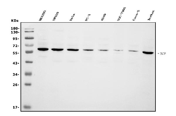
WB (Western Blot)
(Figure 1. Western blot analysis of PLTP using anti-PLTP antibody (AAA19255).Electrophoresis was performed on a 5-20% SDS-PAGE gel at 70V (Stacking gel) / 90V (Resolving gel) for 2-3 hours. The sample well of each lane was loaded with 30ug of sample under reducing conditions.Lane 1: human HEK293 whole cell lysatesLane 2: human SW620 whole cell lysatesLane 3: human Hela whole cell lysatesLane 4: human PC-3 whole cell lysatesLane 5: human A549 whole cell lysatesLane 6: human SGC-7901 whole cell lysatesLane 7: human Caco-2 whole cell lysatesLane 8: human Jurkat whole cell lysates,After Electrophoresis, proteins were transferred to a Nitrocellulose membrane at 150mA for 50-90 minutes. Blocked the membrane with 5% Non-fat Milk/ TBS for 1. 5 hour at RT. The membrane was incubated with rabbit anti-PLTP antigen affinity purified polyclonal antibody (Catalog # AAA19255) at 0. 5 μg/mL overnight at 4 degree C, then washed with TBS-0. 1%Tween 3 times with 5 minutes each and probed with a goat anti-rabbit IgG-HRP secondary antibody at a dilution of 1:5000 for 1. 5 hour at RT. The signal is developed using an Enhanced Chemiluminescent detection (ECL) kit (Catalog # with Tanon 5200 system. A specific band was detected for PLTP at approximately 65KD. The expected band size for PLTP is at 65KD.)
2. Spirin, V., Schmidt, S., Pertsemlidis, A., Cooper, R. S., Cohen, J. C., Sunyaev, S. R. Common single-nucleotide polymorphisms act in concert to affect plasma levels of high-density lipoprotein cholesterol. Am. J. Hum. Genet. 81: 1298-1303, 2007.
3. Teslovich, T. M., Musunuru, K., Smith, A. V., Edmondson, A. C., Stylianou, I. M., Koseki, M., Pirruccello, J. P., Ripatti, S., Chasman, D. I., Willer, C. J., Johansen, C. T., Fouchier, S. W., and 197 others. Biological, clinical and population relevance of 95 loci for blood lipids. Nature 466: 707-713, 2010.


