


Rabbit SV2A Polyclonal Antibody | anti-SV2A antibody
Anti-SV2A Antibody
IHC-P: 2-5ug/ml|Human, Mouse, Rat|
FC/FACS/FCM: 1-3ug/1x106 cells|Human|
FCM (Flow Cytometry)
(Figure 8. Flow Cytometry analysis of U20S cells using anti-SV2A antibody (AAA19279).Overlay histogram showing U20S cells stained with AAA19279 (Blue line). The cells were blocked with 10% normal goat serum. And then incubated with rabbit anti-SV2A Antibody (AAA19279, 1μg/1x106 cells) for 30 min at 20 degree C. DyLight®488 conjugated goat anti-rabbit IgG (5-10μg/1x106 cells) was used as secondary antibody for 30 minutes at 20 degree C. Isotype control antibody (Green line) was rabbit IgG (1μg/1x106) used under the same conditions. Unlabelled sample (Red line) was also used as a control.)
IHC (Immunohistochemistry)
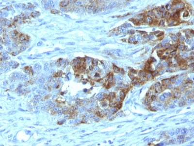
(Figure 7. IHC analysis of SV2A using anti-SV2A antibody (AAA19279).SV2A was detected in paraffin-embedded section of human rectal cancer tissue. Heat mediated antigen retrieval was performed in EDTA buffer (pH8. 0, epitope retrieval solution). The tissue section was blocked with 10% goat serum. The tissue section was then incubated with 2μg/ml rabbit anti-SV2A Antibody (AAA19279) overnight at 4 degree C. Biotinylated goat anti-rabbit IgG was used as secondary antibody and incubated for 30 minutes at 37 degree C. The tissue section was developed using Strepavidin-Biotin-Complex (SABC) (Catalog # with DAB as the chromogen.)
IHC (Immunohistchemistry)
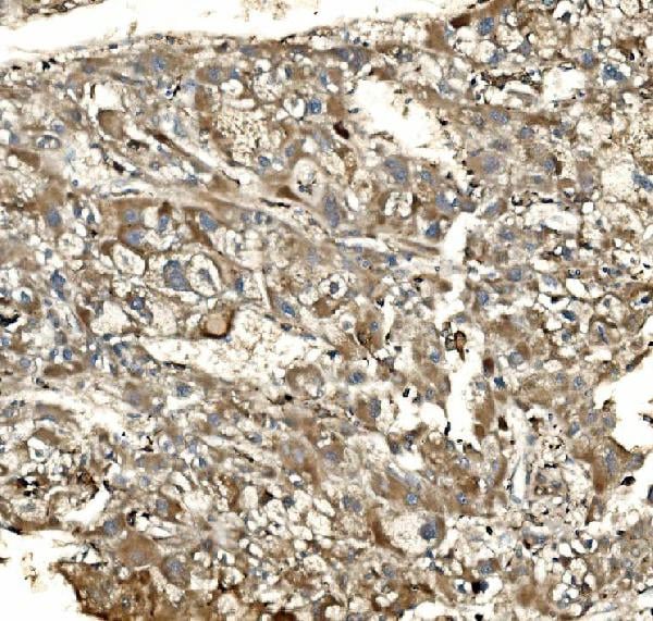
(Figure 6. IHC analysis of SV2A using anti-SV2A antibody (AAA19279).SV2A was detected in paraffin-embedded section of human ovarian serous adenocarcinoma tissue. Heat mediated antigen retrieval was performed in EDTA buffer (pH8. 0, epitope retrieval solution). The tissue section was blocked with 10% goat serum. The tissue section was then incubated with 2μg/ml rabbit anti-SV2A Antibody (AAA19279) overnight at 4 degree C. Biotinylated goat anti-rabbit IgG was used as secondary antibody and incubated for 30 minutes at 37 degree C. The tissue section was developed using Strepavidin-Biotin-Complex (SABC) (Catalog # with DAB as the chromogen.)
IHC (Immunohistochemistry)
(Figure 5. IHC analysis of SV2A using anti-SV2A antibody (AAA19279).SV2A was detected in paraffin-embedded section of human lung cancer tissue. Heat mediated antigen retrieval was performed in EDTA buffer (pH8. 0, epitope retrieval solution). The tissue section was blocked with 10% goat serum. The tissue section was then incubated with 2μg/ml rabbit anti-SV2A Antibody (AAA19279) overnight at 4 degree C. Biotinylated goat anti-rabbit IgG was used as secondary antibody and incubated for 30 minutes at 37 degree C. The tissue section was developed using Strepavidin-Biotin-Complex (SABC) (Catalog # with DAB as the chromogen.)
IHC (Immunohistochemistry)
(Figure 4. IHC analysis of SV2A using anti-SV2A antibody (AAA19279).SV2A was detected in paraffin-embedded section of human liver cancer tissue. Heat mediated antigen retrieval was performed in EDTA buffer (pH8. 0, epitope retrieval solution). The tissue section was blocked with 10% goat serum. The tissue section was then incubated with 2μg/ml rabbit anti-SV2A Antibody (AAA19279) overnight at 4 degree C. Biotinylated goat anti-rabbit IgG was used as secondary antibody and incubated for 30 minutes at 37 degree C. The tissue section was developed using Strepavidin-Biotin-Complex (SABC) (Catalog # with DAB as the chromogen.)
IHC (Immunohistochemistry)
(Figure 3. IHC analysis of SV2A using anti-SV2A antibody (AAA19279).SV2A was detected in paraffin-embedded section of rat brain tissue. Heat mediated antigen retrieval was performed in EDTA buffer (pH8. 0, epitope retrieval solution). The tissue section was blocked with 10% goat serum. The tissue section was then incubated with 2μg/ml rabbit anti-SV2A Antibody (AAA19279) overnight at 4 degree C. Biotinylated goat anti-rabbit IgG was used as secondary antibody and incubated for 30 minutes at 37 degree C. The tissue section was developed using Strepavidin-Biotin-Complex (SABC) (Catalog # with DAB as the chromogen.)
IHC (Immunohistochemistry)
(Figure 2. IHC analysis of SV2A using anti-SV2A antibody (AAA19279).SV2A was detected in paraffin-embedded section of mouse brain tissue. Heat mediated antigen retrieval was performed in EDTA buffer (pH8. 0, epitope retrieval solution). The tissue section was blocked with 10% goat serum. The tissue section was then incubated with 2μg/ml rabbit anti-SV2A Antibody (AAA19279) overnight at 4 degree C. Biotinylated goat anti-rabbit IgG was used as secondary antibody and incubated for 30 minutes at 37 degree C. The tissue section was developed using Strepavidin-Biotin-Complex (SABC) (Catalog # with DAB as the chromogen.)
WB (Western Blot)
(Figure 1. Western blot analysis of SV2A using anti-SV2A antibody (AAA19279).Electrophoresis was performed on a 5-20% SDS-PAGE gel at 70V (Stacking gel) / 90V (Resolving gel) for 2-3 hours. The sample well of each lane was loaded with 30ug of sample under reducing conditions.Lane 1: rat brain tissue lysatesLane 2: mouse brain tissue lysates.After Electrophoresis, proteins were transferred to a Nitrocellulose membrane at 150mA for 50-90 minutes. Blocked the membrane with 5% Non-fat Milk/ TBS for 1. 5 hour at RT. The membrane was incubated with rabbit anti-SV2A antigen affinity purified polyclonal antibody (Catalog # AAA19279) at 0. 5 μg/mL overnight at 4 degree C, then washed with TBS-0. 1%Tween 3 times with 5 minutes each and probed with a goat anti-rabbit IgG-HRP secondary antibody at a dilution of 1:5000 for 1. 5 hour at RT. The signal is developed using an Enhanced Chemiluminescent detection (ECL) kit (Catalog # with Tanon 5200 system. A specific band was detected for SV2A at approximately 110KD. The expected band size for SV2A is at 110KD.)
2. Crowder, K. M., Gunther, J. M., Jones, T. A., Hale, B. D., Zhang, H. Z., Peterson, M. R., Scheller, R. H., Chavkin, C., Bajjalieh, S. M. Abnormal neurotransmission in mice lacking synaptic vesicle protein 2A (SV2A). Proc. Nat. Acad. Sci. 96: 15268-15273, 1999.
3. Dong, M., Yeh, F., Tepp, W. H., Dean, C., Johnson, E. A., Janz, R., Chapman, E. R. SV2 is the protein receptor for botulinum neurotoxin A. Science 312: 592-596, 2006.