


Rabbit C1orf77/FOP/CHTOP Polyclonal Antibody | anti-CHTOP antibody
Anti-C1orf77/FOP/CHTOP Antibody Picoband
Each vial contains 4 mg Trehalose, 0.9 mg NaCl, 0.2 mg Na2HPO4.
IHC-P: 2-5 ug/ml, Human
FC/FACS: 1-3 ug/1x10^6 cells, Human, Rat
Tested Species: In-house tested species with positive results.
Enhanced Chemiluminescent Kit with anti-Rabbit IgG for Western blot, and HRP Conjugated anti-Rabbit IgG Super Vision Assay Kit for IHC(P).
FCM (Flow Cytometry)
(Figure 9. Flow Cytometry analysis of RH35 cells using anti-C1orf77/FOP/CHTOP antibody (AAA19587).Overlay histogram showing RH35 cells stained with AAA19587 (Blue line). The cells were blocked with 10% normal goat serum. And then incubated with rabbit anti-C1orf77/FOP/CHTOP Antibody (AAA19587, 1 ug/1x10^6 cells) for 30 min at 20 degree C. DyLight488 conjugated goat anti-rabbit IgG was used as secondary antibody for 30 minutes at 20 degree C. Isotype control antibody (Green line) was rabbit IgG (1 ug/1x10^6) used under the same conditions. Unlabelled sample (Red line) was also used as a control.)
FCM (Flow Cytometry)
(Figure 8. Flow Cytometry analysis of THP-1 cells using anti-C1orf77/FOP/CHTOP antibody (AAA19587).Overlay histogram showing THP-1 cells stained with AAA19587 (Blue line). The cells were blocked with 10% normal goat serum. And then incubated with rabbit anti-C1orf77/FOP/CHTOP Antibody (AAA19587, 1 ug/1x10^6 cells) for 30 min at 20 degree C. DyLight488 conjugated goat anti-rabbit IgG was used as secondary antibody for 30 minutes at 20 degree C. Isotype control antibody (Green line) was rabbit IgG (1 ug/1x10^6) used under the same conditions. Unlabelled sample (Red line) was also used as a control.)
IHC (Immunohistochemistry)
(Figure 7. IHC analysis of C1orf77/FOP/CHTOP using anti-C1orf77/FOP/CHTOP antibody (AAA19587).C1orf77/FOP/CHTOP was detected in a paraffin-embedded section of human thyroid cancer tissue. Heat mediated antigen retrieval was performed in EDTA buffer (pH 8.0, epitope retrieval solution). The tissue section was blocked with 10% goat serum. The tissue section was then incubated with 2 ug/ml rabbit anti-C1orf77/FOP/CHTOP Antibody (AAA19587) overnight at 4 degree C. Peroxidase Conjugated Goat Anti-rabbit IgG was used as secondary antibody and incubated for 30 minutes at 37 degree C. The tissue section was developed using HRP Conjugated Rabbit IgG Super Vision Assay Kit with DAB as the chromogen.)
IHC (Immunohistchemistry)
(Figure 6. IHC analysis of C1orf77/FOP/CHTOP using anti-C1orf77/FOP/CHTOP antibody (AAA19587).C1orf77/FOP/CHTOP was detected in a paraffin-embedded section of human spleen tissue. Heat mediated antigen retrieval was performed in EDTA buffer (pH 8.0, epitope retrieval solution). The tissue section was blocked with 10% goat serum. The tissue section was then incubated with 2 ug/ml rabbit anti-C1orf77/FOP/CHTOP Antibody (AAA19587) overnight at 4 degree C. Peroxidase Conjugated Goat Anti-rabbit IgG was used as secondary antibody and incubated for 30 minutes at 37 degree C. The tissue section was developed using HRP Conjugated Rabbit IgG Super Vision Assay Kit with DAB as the chromogen.)
IHC (Immunohistochemistry)
(Figure 5. IHC analysis of C1orf77/FOP/CHTOP using anti-C1orf77/FOP/CHTOP antibody (AAA19587).C1orf77/FOP/CHTOP was detected in a paraffin-embedded section of human placenta tissue. Heat mediated antigen retrieval was performed in EDTA buffer (pH 8.0, epitope retrieval solution). The tissue section was blocked with 10% goat serum. The tissue section was then incubated with 2 ug/ml rabbit anti-C1orf77/FOP/CHTOP Antibody (AAA19587) overnight at 4 degree C. Peroxidase Conjugated Goat Anti-rabbit IgG was used as secondary antibody and incubated for 30 minutes at 37 degree C. The tissue section was developed using HRP Conjugated Rabbit IgG Super Vision Assay Kit with DAB as the chromogen.)
IHC (Immunohistochemistry)
(Figure 4. IHC analysis of C1orf77/FOP/CHTOP using anti-C1orf77/FOP/CHTOP antibody (AAA19587).C1orf77/FOP/CHTOP was detected in a paraffin-embedded section of human laryngeal squamous cell carcinoma tissue. Heat mediated antigen retrieval was performed in EDTA buffer (pH 8.0, epitope retrieval solution). The tissue section was blocked with 10% goat serum. The tissue section was then incubated with 2 ug/ml rabbit anti-C1orf77/FOP/CHTOP Antibody (AAA19587) overnight at 4 degree C. Peroxidase Conjugated Goat Anti-rabbit IgG was used as secondary antibody and incubated for 30 minutes at 37 degree C. The tissue section was developed using HRP Conjugated Rabbit IgG Super Vision Assay Kit with DAB as the chromogen.)
IHC (Immunohistochemistry)
(Figure 3. IHC analysis of C1orf77/FOP/CHTOP using anti-C1orf77/FOP/CHTOP antibody (AAA19587).C1orf77/FOP/CHTOP was detected in a paraffin-embedded section of human colorectal adenocarcinoma tissue. Heat mediated antigen retrieval was performed in EDTA buffer (pH 8.0, epitope retrieval solution). The tissue section was blocked with 10% goat serum. The tissue section was then incubated with 2 ug/ml rabbit anti-C1orf77/FOP/CHTOP Antibody (AAA19587) overnight at 4 degree C. Peroxidase Conjugated Goat Anti-rabbit IgG was used as secondary antibody and incubated for 30 minutes at 37 degree C. The tissue section was developed using HRP Conjugated Rabbit IgG Super Vision Assay Kit with DAB as the chromogen.)
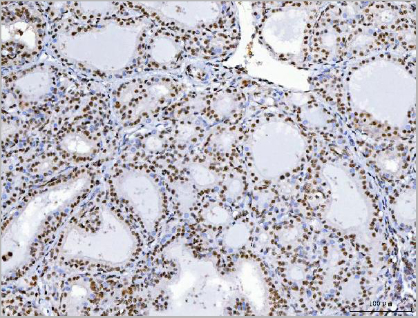
IHC (Immunohistochemistry)
(AAA19587-CHTOP-primary-antibodies-IHC-testing-7.jpg)
WB (Western Blot)
(Figure 1. Western blot analysis of C1orf77/FOP/CHTOP using anti-C1orf77/FOP/CHTOP antibody (AAA19587).Electrophoresis was performed on a 5-20% SDS-PAGE gel at 70V (Stacking gel)/90V (Resolving gel) for 2-3 hours. The sample well of each lane was loaded with 30 ug of sample under reducing conditions.Lane 1: human Hela whole cell lysates,Lane 2: monkey COS-7 whole cell lysates,Lane 3: human THP-1 whole cell lysates,Lane 4: human MOLT-4 whole cell lysates,Lane 5: human RT4 whole cell lysates,Lane 6: human HL-60 whole cell lysates,Lane 7: human MCF-7 whole cell lysates,Lane 8: rat brain tissue lysates,Lane 9: rat PC-12 whole cell lysates,Lane 10: mouse spleen tissue lysates,Lane 11: mouse brain tissue lysates,Lane 12: mouse lung tissue lysates,Lane 13: mouse L929 whole cell lysates.After electrophoresis, proteins were transferred to a nitrocellulose membrane at 150 mA for 50-90 minutes. Blocked the membrane with 5% non-fat milk/TBS for 1.5 hour at RT. The membrane was incubated with rabbit anti-C1orf77/FOP/CHTOP antigen affinity purified polyclonal antibody (#AAA19587) at 0.5 ug/mL overnight at 4 degree C, then washed with TBS-0.1%Tween 3 times with 5 minutes each and probed with a goat anti-rabbit IgG-HRP secondary antibody at a dilution of 1:5000 for 1.5 hour at RT. The signal is developed using an Enhanced Chemiluminescent detection (ECL) kit with Tanon 5200 system. A specific band was detected for C1orf77/FOP/CHTOP at approximately 28 kDa. The expected band size for C1orf77/FOP/CHTOP is at 26 kDa.)