


Mouse anti-Human HSPA8 Monoclonal Antibody | anti-HSPA8 antibody
HSPA8 Monoclonal Antibody
Preservative: 0.03% Proclin 300
Constituents: 50% Glycerol, 0.01M PBS, PH7.4
IHC: 1:100-1:500
IF: 1:100-1:300
FC/FACS: 1:100-1:300
FCM (Flow Cytometry)
(Overlay histogram showing MCF-7 cells stained with (red line). The cells were fixed with 70% Ethylalcohol (18h) and then incubated in 10% normal goat serum to block non-specific protein-protein interactions followed by the primary antibody at 1/200 for 1 h at 4 degree C. The secondary antibody used was FITC goat anti-mouse IgG(H+L) at 1/100 dilution for 30min at 4 degree C. Isotype control antibody (green line) was mouse IgG1 used under the same conditions. Acquisition of >10,000 events was performed.)
IF (Immunofluorescence)
(Immunofluorescence staining of PC-3 cells at 1:100, counter-stained with DAPI. The cells were fixed in 4% formaldehyde and blocked in 10% normal Goat Serum. The cells were then incubated with the antibody overnight at 4 degree C. Nuclear DNA was labeled in blue with DAPI. The secondary antibody was FITC-conjugated AffiniPure Goat Anti-Mouse IgG ?H+L?.)
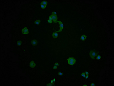
IF (Immunofluorescence)
(Immunofluorescence staining of MCF-7 cells at 1:100, counter-stained with DAPI. The cells were fixed in 4% formaldehyde and blocked in 10% normal Goat Serum. The cells were then incubated with the antibody overnight at 4 degree C. Nuclear DNA was labeled in blue with DAPI. The secondary antibody was FITC-conjugated AffiniPure Goat Anti-Mouse IgG ?H+L?.)
IF (Immunofluorescence)
(Immunofluorescence staining of Hela cells at 1:100, counter-stained with DAPI. The cells were fixed in 4% formaldehyde and blocked in 10% normal Goat Serum. The cells were then incubated with the antibody overnight at 4 degree C. Nuclear DNA was labeled in blue with DAPI. The secondary antibody was FITC-conjugated AffiniPure Goat Anti-Mouse IgG ?H+L?.)
IHC (Immunohistochemistry)
(IHC image diluted at 1:220 and staining in paraffin-embedded human prostate cancer performed on a Leica BondTM system. After dewaxing and hydration, antigen retrieval was mediated by high pressure in a citrate buffer (pH 6.0). Section was blocked with 10% normal goat serum 30min at RT. Then primary antibody (1% BSA) was incubated at 4 degree C overnight. The primary is detected by a biotinylated secondary antibody and visualized using an HRP conjugated SP system.)
IHC (Immunohistochemistry)
(IHC image diluted at 1:220 and staining in paraffin-embedded human kidney tissue performed on a Leica BondTM system. After dewaxing and hydration, antigen retrieval was mediated by high pressure in a citrate buffer (pH 6.0). Section was blocked with 10% normal goat serum 30min at RT. Then primary antibody (1% BSA) was incubated at 4 degree C overnight. The primary is detected by a biotinylated secondary antibody and visualized using an HRP conjugated SP system.)
WB (Western Blot)
(Western Blot Positive WB detected in: 20ug hela whole cell lysate HSPA8 antibody at 1:2000, 1:4000, 1:8000, 1:16000, 1:32000 Secondary Goat polyclonal to mouse IgG at 1/50000 dilution Predicted band size: 70~75 KDa Observed band size: 70~75 KDa Exposure time: 5min)
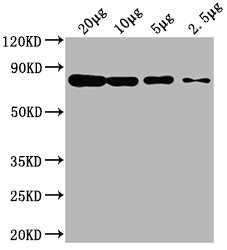
WB (Western Blot)
(Western Blot Positive WB detected in: Hela whole cell lysate at 20ug, 10ug, 5ug, 2.5ug All lanes: HSPA8 antibody at 1:2000 Secondary Goat polyclonal to mouse IgG at 1/50000 dilution Predicted band size: 70~75 KDa Observed band size: 70~75 KDa Exposure time: 5min)
WB (Western Blot)
(Western Blot Positive WB detected in: Hela whole cell lysate, K562 whole cell lysate, HepG2 whole cell lysate All lanes HSPA8 antibody at 1:2000 Secondary Goat polyclonal to mouse IgG at 1/50000 dilution Predicted band size: 70~75 KDa Observed band size: 70~75 KDa Exposure time: 5min)