


Mouse anti-Human CD45 Monoclonal Antibody | anti-CD45 antibody
CD45 Monoclonal Antibody
Preservative: 0.03% Proclin 300
Constituents: 50% Glycerol, 0.01M PBS, pH7.4
IHC: 1:200-1:600
IF: 1:100-1:300
FC: 1:200-1:600
FCM (Flow Cytometry)
(Overlay histogram showing Raji cells stained with AAA28060 (red line) at 1:500. The cells were incubated in 10% normal goat serum to block non-specific protein-protein interactions followed by the antibody (1ug/1*106cells) for 1 h at 4 degree C. The secondary antibody used was FITC-conjugated Goat Anti-Mouse IgG(H+L) at 1/100 dilution for 30min at 4 degree C. Isotype control antibody (green line) was mouse IgG2b (1ug/1*106cells) used under the same conditions. Acquisition of >10,000 events was performed.)
FCM (Flow Cytometry)
(Overlay histogram showing Jurkat cells stained with AAA28060 (red line) at 1:500. The cells were incubated in 10% normal goat serum to block non-specific protein-protein interactions followed by the antibody (1ug/1*106cells) for 1 h at 4 degree C. The secondary antibody used was FITC-conjugated Goat Anti-Mouse IgG(H+L) at 1/100 dilution for 30min at 4 degree C. Isotype control antibody (green line) was mouse IgG2b (1ug/1*106cells) used under the same conditions. Acquisition of >10,000 events was performed.)
IF (Immunofluorescence)
(Immunofluorescence staining of U937 cells with AAA28060 at 1:250, counter-stained with DAPI. The cells were incubated with the antibody overnight at 4 degree C. Nuclear DNA was labeled in blue with DAPI. The secondary antibody was FITC-conjugated AffiniPure Goat Anti-Mouse IgG (H+L).)
IF (Immunofluorescence)
(Immunofluorescence staining of Raji cells with AAA28060 at 1:250, counter-stained with DAPI. The cells were incubated with the antibody overnight at 4 degree C. Nuclear DNA was labeled in blue with DAPI. The secondary antibody was FITC-conjugated AffiniPure Goat Anti-Mouse IgG (H+L).)
IF (Immunofluorescence)
(Immunofluorescence staining of Jurkat cells with AAA28060 at 1:250, counter-stained with DAPI. The cells were incubated with the antibody overnight at 4 degree C. Nuclear DNA was labeled in blue with DAPI. The secondary antibody was FITC-conjugated AffiniPure Goat Anti-Mouse IgG (H+L).)
IHC (Immunohistchemistry)
(IHC image of AAA28060 diluted at 1:500 and staining in paraffin-embedded human lymph node tissue performed on a Leica BondTM system. After dewaxing and hydration, antigen retrieval was mediated by high pressure in a citrate buffer (pH 6.0). Section was blocked with 10% normal goat serum 30min at RT. Then primary antibody (1% BSA) was incubated at 4 degree C overnight. The primary is detected by a Goat anti-rabbit IgG labeled by HRP and visualized using 0.05% DAB.)
IHC (Immunohistochemistry)
(IHC image of AAA28060 diluted at 1:500 and staining in paraffin-embedded human tonsil tissue performed on a Leica BondTM system. After dewaxing and hydration, antigen retrieval was mediated by high pressure in a citrate buffer (pH 6.0). Section was blocked with 10% normal goat serum 30min at RT. Then primary antibody (1% BSA) was incubated at 4 degree C overnight. The primary is detected by a Goat anti-rabbit IgG labeled by HRP and visualized using 0.05% DAB.)
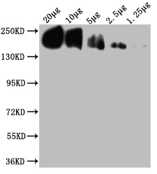
WB (Western Blot)
(Western Blot)
WB (Western Blot)
(Western Blot)
WB (Western Blot)
(Western Blot)
WB (Western Blot)
(Western Blot)


