

Rabbit p27 Kip1 Polyclonal Antibody | anti-p27Kip1 antibody
Phospho-p27 Kip1 (Thr157) Antibody
Predicted Reactivity: Bovine (89%), Sheep (89%), Rabbit (100%), Dog (100%), Chicken (89%)
Predicted Reactivity: Bovine (89%), Sheep (89%), Rabbit (100%), Dog (100%), Chicken (89%)
Tissue Specificity: Expressed in all tissues tested. Highest levels in skeletal muscle, lowest in liver and kidney.
IHC: 1:50-1:200
IF/ICC: 1:100-1:500
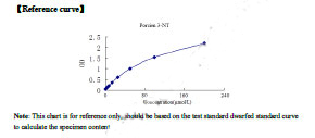
Peptide ELISA: 1:20,000-1:40,000
Note: Nuclear and cytoplasmic in quiescent cells. AKT- or RSK-mediated phosphorylation on Thr-198, binds 14-3-3, translocates to the cytoplasm and promotes cell cycle progression. Mitogen-activated UHMK1 phosphorylation on Ser-10 also results in translocation to the cytoplasm and cell cycle progression. Phosphorylation on Ser-10 facilitates nuclear export. Translocates to the nucleus on phosphorylation of Tyr-88 and Tyr-89. Colocalizes at the endosome with SNX6; this leads to lysosomal degradation (By similarity).
Application Data
(At 25 degree C. The primary antibody was diluted at 1/200 and incubated with the sample for 1 hour at 37 degree C. An Alexa Fluor 594 conjugated goat anti-rabbit IgG (H+L) Ab, diluted at 1/600, was used as the secondary antibody.)
IHC (Immunohistchemistry)
(At 1/200 staining Mouse kidney tissue sections by IHC-P. The tissue was formaldehyde fixed and a heat mediated antigen retrieval step in citrate buffer was performed. The tissue was then blocked and incubated with the antibody for 1.5 hours at 22 degree C. An HRP conjugated goat anti-rabbit antibody was used as the secondary antibody.)
IHC (Immunohistochemistry)
(At 1/200 staining Rat kidney tissue sections by IHC-P. The tissue was formaldehyde fixed and a heat mediated antigen retrieval step in citrate buffer was performed. The tissue was then blocked and incubated with the antibody for 1.5 hours at 22 degree C. An HRP conjugated goat anti-rabbit antibody was used as the secondary antibody.)
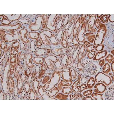
IHC (Immunohistochemistry)
(At 1/200 staining Human kidney tissue sections by IHC-P. The tissue was formaldehyde fixed and a heat mediated antigen retrieval step in citrate buffer was performed. The tissue was then blocked and incubated with the antibody for 1.5 hours at 22 degree C. An HRP conjugated goat anti-rabbit antibody was used as the secondary antibody.)
IHC (Immunohistochemistry)
(At 1/200 staining Mouse brain tissue sections by IHC-P. The tissue was formaldehyde fixed and a heat mediated antigen retrieval step in citrate buffer was performed. The tissue was then blocked and incubated with the antibody for 1.5 hours at 22 degree C. An HRP conjugated goat anti-rabbit antibody was used as the secondary antibody.)
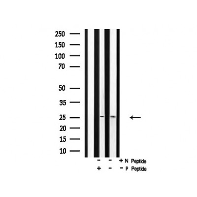
WB (Western Blot)
(Western blot analysis of p27 Kip1 (Phospho-Thr157) using HeLa whole cell lysates.-/+ means absence or presence of N peptide(non-phospho peptide) and P peptide(phospho peptide).)
WB (Western Blot)
(Western blot analysis of extracts from rat brain, using p27 Kip1 (Phospho-Thr157) Antibody. Lane 1 was treated with the blocking peptide.)