


Rabbit Mnk1 Polyclonal Antibody | anti-Mnk1 antibody
Phospho-Mnk1 (Thr250) Antibody
Predicted Reactivity: Pig (100%), Zebrafish (100%), Bovine (100%), Horse (100%), Sheep (100%), Rabbit (100%), Dog (100%), Chicken (100%), Xenopus (100%)
Predicted Reactivity: Pig (100%), Zebrafish (100%), Bovine (100%), Horse (100%), Sheep (100%), Rabbit (100%), Dog (100%), Chicken (100%), Xenopus (100%)
Tissue Specificity: Ubiquitous.
IHC: 1:50-1:200
IF/ICC: 1:100-1:500
Peptide ELISA: 1:20,000-1:40,000
Application Data
(At 25 degree C. The primary antibody was diluted at 1/200 and incubated with the sample for 1 hour at 37 degree C. An Alexa Fluor 594 conjugated goat anti-rabbit IgG (H+L) Ab, diluted at 1/600, was used as the secondary antibody.)
IHC (Immunohistochemistry)
(At 1/200 staining Human liver cancer tissue sections by IHC-P. The tissue was formaldehyde fixed and a heat mediated antigen retrieval step in citrate buffer was performed. The tissue was then blocked and incubated with the antibody for 1.5 hours at 22 degree C. An HRP conjugated goat anti-rabbit antibody was used as the secondary antibody.)
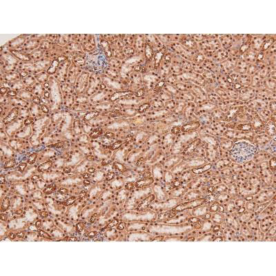
IHC (Immunohistochemistry)
(At 1/200 staining Mouse kidney tissue sections by IHC-P. The tissue was formaldehyde fixed and a heat mediated antigen retrieval step in citrate buffer was performed. The tissue was then blocked and incubated with the antibody for 1.5 hours at 22 degree C. An HRP conjugated goat anti-rabbit antibody was used as the secondary antibody.)
IHC (Immunohistochemistry)
(At 1/200 staining Rat kidney tissue sections by IHC-P. The tissue was formaldehyde fixed and a heat mediated antigen retrieval step in citrate buffer was performed. The tissue was then blocked and incubated with the antibody for 1.5 hours at 22 degree C. An HRP conjugated goat anti-rabbit antibody was used as the secondary antibody.)
WB (Western Blot)
(Western blot analysis of MNK1 (Phospho-Thr250) using TNF treated Jurkat whole cell lysates.-/+ means absence or presence of N peptide(non-phospho peptide) and P peptide(phospho peptide).)
WB (Western Blot)
(Western blot analysis of extracts from 293 cells(UV treatment), using Phospho-Mnk1 (Thr250) Antibody. The lane on the left was treated with blocking peptide.)


