 Application Data
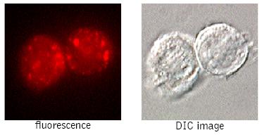
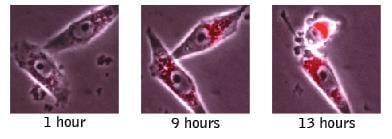
(Cathepsin K-positive THP-1 cellsfluoresce red after staining withMR-(LR)2. Red fluorophoreconcentrates inside lysosomes (Dr.Brian W. Lee, ICT).)
Application Data
(Cathepsin K-positive THP-1 cellsfluoresce red after staining withMR-(LR)2. Red fluorophoreconcentrates inside lysosomes (Dr.Brian W. Lee, ICT).)
Magic Red Cathepsin K Assay Kit
Magic Red Cathepsin K Assay Kit
Synonyms
Magic Red Cathepsin K; N/A; Magic Red Cathepsin K Assay Kit; Magic Red Cathepsin K assay kit
Reagents
MR-(LR)2
Suspension Cells
Culture cells up to 1 x 10^6 cells/mL. Establish what cell environment/treatment or cell type will yield both positive and negative controls. Cathepsin K is constitutively produced in virtually all cell types, but the level of this activity will vary by pathophysiological status. Reconstitute the Magic Red reagent with DMSO to form the stock concentrate (which can be frozen for future use). Dilute the Magic Red stock concentrate 1:10 with diH2O to form the working solution. Add ~10 - 15 uL of the working solution directly to a 300-500 uL aliquot of your cell culture for labeling. The working solution is used at approximately 1:15-1:26 to label suspension cells. Incubate 30 minutes - 4 hours at 37 degree C in the dark. If desired label DNA with Hoechst stain, and/or fix cells. Unstained cells may be labeled with Acridine Orange to reveal the lysosomes. Analyze data using a fluorescence microscope, fluorescence plate reader, or flow cytometer.
Adherent Cells
Culture cell monolayers to 80-90% confluency. Establish what cell environment/treatment or cell type will yield both positive and negative controls. Cathepsin K is constitutively produced in virtually all cell types, but the level of this activity will vary by pathophysiological status. Gently remove cell culture media from flask and rinse cell culture surface once with sterile PBS. Save any loose cells for combination with trypsinized cells in Magic Red MR-(LR)2 staining steps. Add trypsin to cell monolayer and collect disassociated cells for analysis. Dilute the newly trypsinized cells in cell culture media containing 10-20% FBS and pellet down cells in this media to concentrate cells. Reconstitute the Magic Red reagent with DMSO to form the stock concentrate (which can be frozen for future use). Dilute the Magic Red stock concentrate 1:10 with diH2O to form the working solution. Add ~ 15 uL of the working solution directly to a 500 uL aliquot of your adherent cell culture for labeling. The working solution is used at approximately 1:26 to label adherent cells. Incubate 30 minutes - 4 hours at 37 degree C in the dark. If desired label DNA with Hoechst stain, and/or fix cells. Unstained cells may be labeled with Acridine Orange to reveal the lysosomes. Analyze data using a fluorescence microscope or fluorescence plate reader.
Target
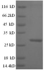
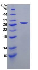
Cathepsin K
Excitation / Emission
590 nm / 620 nm
Method of Analysis
Fluorescence Microscope
Types of Samples
Cell Culture, Tissue
Preparation and Storage
Store at 2-8 degree C
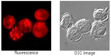
Application Data
(Cathepsin K-positive THP-1 cellsfluoresce red after staining withMR-(LR)2. Red fluorophoreconcentrates inside lysosomes (Dr.Brian W. Lee, ICT).)
Application Data
(Cathepsin K-positive THP-1 cellsfluoresce red after staining withMR-(LR)2. Red fluorophoreconcentrates inside lysosomes (Dr.Brian W. Lee, ICT).)
Related Product Information for Magic Red Cathepsin K assay kit
Background: Magic Red Cathepsin assay kits enable researchers to quantitate and monitor intracellular cathepsin B, K, or L activity over time in vitro. The Magic Red (MR) reagent is a non-cytotoxic substrate that fluoresces red upon cleavage by active cathepsin enzymes. Elevated cathepsin enzyme activity in serum or the extracellular matrix often signifies a number of gross pathological conditions. Cathepsin-mediated diseases include: Alzheimer's; numerous types of cancer; autoimmune related diseases like arthritis; and the accelerated breakdown of bone structure seen with osteoporosis1,2. Up-regulated cathepsin B and L activity has been linked to several types of cancer. These include cancer of the colon, pancreas, ovaries, breast, lung, and skin (melanoma)3-6. Upregulation of cathepsin K has been shown in lung tumors7. Increased cathepsin K activity has also been linked to degenerative bone diseases including osteopetrosis and post-menopausal osteoporosis1, 8. Cathepsins are usually characterized as members of the lysosomal cysteine protease (active site) family9 and the cathepsin family name has been synonymous with lysosomal proteolytic enzymes1. In actuality, the cathepsin family also contains members of the serine protease (cathepsin A and G) and aspartic protease (cathepsin D and E) families as well. These enzymes exist in their processed form as disulfide-linked heavy and light chain subunits with molecular weights ranging from 20-35 kDa10. Cathepsin C is the noted exception, existing as an oligomeric enzyme with a MW ~200 kDa11. Initially synthesized as inactive zymogens, cathepsins are post-translationally processed into their active configurations after passing through the endoplasmic reticulum and subsequent incorporation into the acidic environment of the lysosomes1, 11. Magic Red detection substrates utilize the photostable red fluorophore, cresyl violet. When bi-substituted via amide linkage to two cathepsin target peptide sequences, such as (leucine-arginine)2, the bi-substituted cresyl violet is nonfluorescent10. Following enzymatic cleavage at one or both arginine (R) amide linkage sites, the mono and non-substituted cresyl violet fluorophores generate red fluorescence when excited at 550-590 nm. The Magic Red cathepsin B substrate, MR-(RR)2, is comprised of cresyl violet coupled to two pairs of the amino acid sequence, arginine-arginine (RR), which is the preferential target sequence for cathepsin B. In cathepsin K substrate, MR-(LR)2, cresyl violet is coupled to two pairs of leucinearginine (LR). The MR cathepsin L substrate, MR-(FR)2, contains two pairs of phenylalanine-arginine (FR) coupled to cresyl violet. Cathepsins, like most other crucial cell survival enzymes, are somewhat permissive in the target amino acid sequence they will recognize and cleave. Although Magic Red substrates contain the amino acid target sequence preferred by a particular cathepsin enzyme, they can also recognize other active cathepsins or proteases when they are present. The encourages validation of cathepsin activity by an orthogonal technique. To use Magic Red, add the substrate directly to the cell culture media, incubate, and analyze. Because MR is cell-permeant, it easily penetrates the cell membrane and the membranes of the internal cellular organelles - no lysis or permeabilization steps are required. If cathepsin enzymes are active, they will cleave off the two dipeptide cathepsin targeting sequences and allow the cresyl violet fluorophore to become fluorescent upon excitation. The red fluorescent product will stay inside the cell and will often aggregate inside lysosomes (cathepsins are lysosomal) and other areas of low pH, such as inside the mitochondria. As protease activity progresses and more MR substrate is cleaved, the signal will intensify as the red fluorescent product accumulates within various organelles, enabling researchers to watch the color develop over time and quantify cathepsin B, K, or L activity. By varying the duration and concentration of exposure to the MR substrate, a picture can be obtained of the relative abundance of cathepsin enzymatic activity. Positive cells will fluoresce red and have pronounced red lysosomes and mitochondria. Negative cells will exhibit very low levels of background red fluorescence evenly distributed throughout the cell. This background level of substrate activity could be the result of constitutively synthesized serine proteases that target analogous amino acid sequences for hydrolysis. Please note that Magic Red substrates can undergo spontaneous hydrolysis over time, resulting in increased background fluorescence. Appropriate controls are necessary for accurate interpretation of the results. There is no interference from pro-cathepsins forms of the enzymes. If the treatment or experimental condition stimulates cathepsin activity, cells containing elevated levels of cathepsin activity will appear brighter red than cells with lower levels of cathepsin activity. The MR fluorophore, cresyl violet, fluoresces red when excited at 550-590 nm10. The red fluorescent signal can be monitored with a fluorescence microscope or plate reader. It has an optimal excitation of 592 nm and emission of 628 nm12. Hoechst 33342 is included with the kit to concurrently label nuclei after labeling with MR. It is revealed under a microscope using a UV-filter with excitation at 365 nm and emission at 480 nm. Acridine orange (AO) is also included in the kit to identify lysosomes and other intracellular organelles (Figures 3 and 4). It is revealed under a microscope using excitation at 480 nm and emission at >540 nm, or alternatively with excitation at 550 nm and emission at >610 nm.
Product Categories/Family for Magic Red Cathepsin K assay kit
References
Drake, F.H., R. Dodds, I. James, J. Connor, C. Debouck, S. Richardson, E. Lee, D. Rieman, R. Barthlow, G. Hastings, and M. Gowen. 1996. Cathepsin K, but not cathepsin B, L, or S, is abundantly expressed in human osteoclasts. J. Biol. Chem. 271: 12511-12516. Saftig, P., E. Hunziker, V. Everts, S. Jones, A. Boyde, O. Wehmeyer, A. Suter, and K. Figura. 2000. Functions of cathepsin K in bone resorption. Adv. Exp. Med. Biol. 477: 293-303. Buhling, F., N. Waldburg, A. Gerber, C. Hackel, S. Kruger, D. Reinhold, D. Bromme, E. Weber, S. Ansorge, and T. Welte. 2000. Cathepsin K expression in human lung. Adv. Exp. Med. Biol. 477: 281-286. Podgorski, I., B.E. Linebaugh, and B.F. Sloane. 2007. Cathepsin K in bone microenvironment: link between obesity and prostate cancer. Biochem. Soc. Trans. 35: 701-703. Le Gall, C. A. Bellahcene, E. Bonnelye, J.A. Gasser, V. Castronovo, J. Green, J. Zimmermann, and P. Clezardin. 2007. A cathepsin K inhibitor reduces breast cancer induced osteolysis and skeletal tumor burden. Cancer Res. 67: 9894-9902. Skoumal, M., G. Haberhauer, G. Kolarz, G. Hawa, W. Woloszczuk, and A. Klingler. 2005. Serum cathepsin K levels of patients with longstanding rheumatoid arthritis: correlation with radiological destruction. Arthritis Res. Ther. 7: R65-R70.
Customer Reviews
Loading reviews...
Share Your Experience
Similar Products


Product Notes
The Magic Red Cathepsin K (Catalog #AAA52731) is an Assay Kit and is intended for research purposes only. The product is available for immediate purchase. It is sometimes possible for the material contained within the vial of "Magic Red Cathepsin K, Assay Kit" to become dispersed throughout the inside of the vial, particularly around the seal of said vial, during shipment and storage. We always suggest centrifuging these vials to consolidate all of the liquid away from the lid and to the bottom of the vial prior to opening. Please be advised that certain products may require dry ice for shipping and that, if this is the case, an additional dry ice fee may also be required.Precautions
All products in the AAA Biotech catalog are strictly for research-use only, and are absolutely not suitable for use in any sort of medical, therapeutic, prophylactic, in-vivo, or diagnostic capacity. By purchasing a product from AAA Biotech, you are explicitly certifying that said products will be properly tested and used in line with industry standard. AAA Biotech and its authorized distribution partners reserve the right to refuse to fulfill any order if we have any indication that a purchaser may be intending to use a product outside of our accepted criteria.Disclaimer
Though we do strive to guarantee the information represented in this datasheet, AAA Biotech cannot be held responsible for any oversights or imprecisions. AAA Biotech reserves the right to adjust any aspect of this datasheet at any time and without notice. It is the responsibility of the customer to inform AAA Biotech of any product performance issues observed or experienced within 30 days of receipt of said product. To see additional details on this or any of our other policies, please see our Terms & Conditions page.Item has been added to Shopping Cart
If you are ready to order, navigate to Shopping Cart and get ready to checkout.