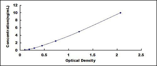
 Application Data
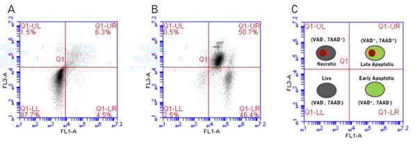
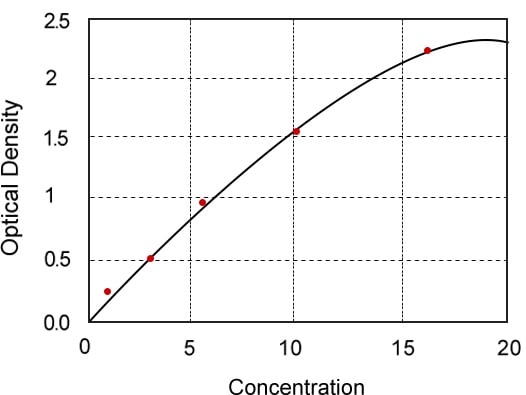
(Flow cytometry analysis of Jurkat suspension cells to quantify four populations. (A) Cells were treated with a placebo (non-induced treatment with DMSO). (B) Cells were treated with 1 uM staurosporine for 4 hours to induce apoptosis via caspase activity. Cells were then dually stained with FAM-FLICA and 7-AAD, and analyzed using an Accuri C6 flow cytometer. FAM-FLICA was analyzed on FL-1 and 7-AAD was analyzed on FL-3. The key is shown in (C). Live, unstained cells do not fluoresce (lower left quadrant). Early apoptotic cells fluoresce green with FAM-FLICA. Dually stained green and red fluorescing cells represent the population of cells in mid to late apoptosis (these cells have active caspase enzymes and compromised cell membranes). Necrotic cells fluoresce red. In the non-induced population (A), only 9.8% of cells were apoptotic (LR: 4.5% + UR: 6.3%), compared with 97.1% of the induced population (B; LR: 46.4% + UR: 50.7%).)
Application Data
(Flow cytometry analysis of Jurkat suspension cells to quantify four populations. (A) Cells were treated with a placebo (non-induced treatment with DMSO). (B) Cells were treated with 1 uM staurosporine for 4 hours to induce apoptosis via caspase activity. Cells were then dually stained with FAM-FLICA and 7-AAD, and analyzed using an Accuri C6 flow cytometer. FAM-FLICA was analyzed on FL-1 and 7-AAD was analyzed on FL-3. The key is shown in (C). Live, unstained cells do not fluoresce (lower left quadrant). Early apoptotic cells fluoresce green with FAM-FLICA. Dually stained green and red fluorescing cells represent the population of cells in mid to late apoptosis (these cells have active caspase enzymes and compromised cell membranes). Necrotic cells fluoresce red. In the non-induced population (A), only 9.8% of cells were apoptotic (LR: 4.5% + UR: 6.3%), compared with 97.1% of the induced population (B; LR: 46.4% + UR: 50.7%).)
Necrosis vs Apoptosis Assay Kit
Necrosis vs Apoptosis Assay Kit
Application Data
(Flow cytometry analysis of Jurkat suspension cells to quantify four populations. (A) Cells were treated with a placebo (non-induced treatment with DMSO). (B) Cells were treated with 1 uM staurosporine for 4 hours to induce apoptosis via caspase activity. Cells were then dually stained with FAM-FLICA and 7-AAD, and analyzed using an Accuri C6 flow cytometer. FAM-FLICA was analyzed on FL-1 and 7-AAD was analyzed on FL-3. The key is shown in (C). Live, unstained cells do not fluoresce (lower left quadrant). Early apoptotic cells fluoresce green with FAM-FLICA. Dually stained green and red fluorescing cells represent the population of cells in mid to late apoptosis (these cells have active caspase enzymes and compromised cell membranes). Necrotic cells fluoresce red. In the non-induced population (A), only 9.8% of cells were apoptotic (LR: 4.5% + UR: 6.3%), compared with 97.1% of the induced population (B; LR: 46.4% + UR: 50.7%).)
Customer Reviews
Loading reviews...
Share Your Experience
Similar Products

Product Notes
The Necrosis vs Apoptosis (Catalog #AAA52736) is an Assay Kit and is intended for research purposes only. The product is available for immediate purchase. It is sometimes possible for the material contained within the vial of "Necrosis vs Apoptosis, Assay Kit" to become dispersed throughout the inside of the vial, particularly around the seal of said vial, during shipment and storage. We always suggest centrifuging these vials to consolidate all of the liquid away from the lid and to the bottom of the vial prior to opening. Please be advised that certain products may require dry ice for shipping and that, if this is the case, an additional dry ice fee may also be required.Precautions
All products in the AAA Biotech catalog are strictly for research-use only, and are absolutely not suitable for use in any sort of medical, therapeutic, prophylactic, in-vivo, or diagnostic capacity. By purchasing a product from AAA Biotech, you are explicitly certifying that said products will be properly tested and used in line with industry standard. AAA Biotech and its authorized distribution partners reserve the right to refuse to fulfill any order if we have any indication that a purchaser may be intending to use a product outside of our accepted criteria.Disclaimer
Though we do strive to guarantee the information represented in this datasheet, AAA Biotech cannot be held responsible for any oversights or imprecisions. AAA Biotech reserves the right to adjust any aspect of this datasheet at any time and without notice. It is the responsibility of the customer to inform AAA Biotech of any product performance issues observed or experienced within 30 days of receipt of said product. To see additional details on this or any of our other policies, please see our Terms & Conditions page.Item has been added to Shopping Cart
If you are ready to order, navigate to Shopping Cart and get ready to checkout.