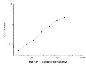
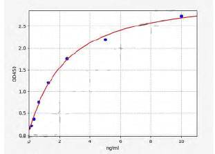
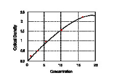
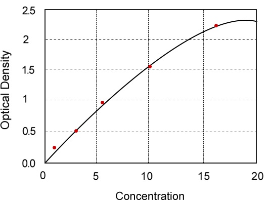
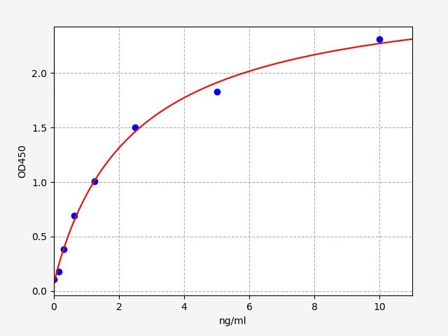
 Standard Curve (Sample)
Human Kim-1 ELISA Kit | Kim-1 elisa kit
Kim-1 Human ELISA Kit
Reactivity
Human
Applications
ELISA
Synonyms
Kim-1; N/A; Kim-1 Human ELISA Kit; Human Kim-1; Kim-1 elisa kit
Reactivity
Human
Assay Type
Sandwich
Samples
Human serum, plasma, urine, cell culture supernatant or tissue samples
Detection Range
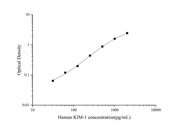
31.2 pg/ml-2000 pg/ml
Applicable Applications for Kim-1 elisa kit
ELISA
Application Notes
For quantitative detection of KIM-1 in human serum, plasma, urine, cell culture supernatant or tissue samples.
Preparation and Storage
Store at 2-8 degree C for 6 months.
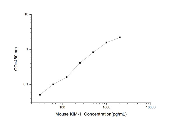
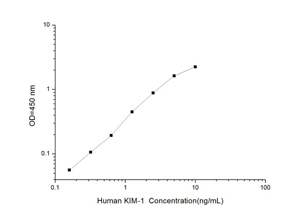
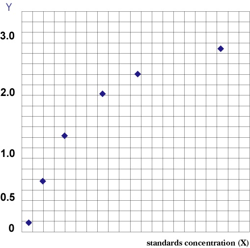
Standard Curve (Sample)
Standard Curve (Sample)
Related Product Information for Kim-1 elisa kit
Background: KIM1, also known as Hepatitis A virus cellular receptor 1, is a protein that in humans is encoded by the HAVCR1 gene, which maps to 5q33.2. It is a major cause of orally transmitted acute hepatitis, infects primate cells, but not dog or rat cells, after binding to the HAV cellular receptor (HAVCR). Infection of canine osteogenic sarcoma cells expressing HAVCR1 with HAV led Feigelstock et al. (1998) to conclude that the protein is indeed a receptor for the virus. Khademi et al. (2004) found that differential expression of TIMs by Th1 and Th2 cells may be implicated in different phases of an autoimmune disease.
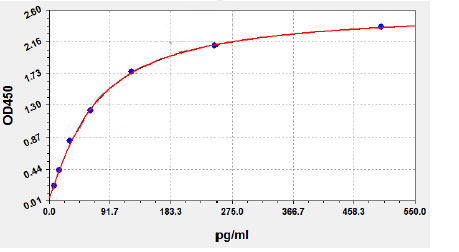
Principle of the Assay: This kit was based on standard sandwich enzyme-linked immune-sorbent assay technology. The purified anti-KIM-1 antibody was pre-coated onto 96-well plates. And the HRP conjugated anti-KIM-1 antibody was used as detection antibodies. The standards, test samples and HRP conjugated detection antibody were added to the wells subsequently, mixed and incubated, then, unbound conjugates were washed away with wash buffer. TMB substrates (A & B) were used to visualize HRP enzymatic reaction. TMB was catalyzed by HRP to produce a blue color product that changed into yellow after adding acidic stop solution. The density of yellow is proportional to the KIM-1 amount of sample captured in plate. Read the O.D. absorbance at 450nm in a microplate reader, and then the concentration of KIM-1 can be calculated.
Principle of the Assay: This kit was based on standard sandwich enzyme-linked immune-sorbent assay technology. The purified anti-KIM-1 antibody was pre-coated onto 96-well plates. And the HRP conjugated anti-KIM-1 antibody was used as detection antibodies. The standards, test samples and HRP conjugated detection antibody were added to the wells subsequently, mixed and incubated, then, unbound conjugates were washed away with wash buffer. TMB substrates (A & B) were used to visualize HRP enzymatic reaction. TMB was catalyzed by HRP to produce a blue color product that changed into yellow after adding acidic stop solution. The density of yellow is proportional to the KIM-1 amount of sample captured in plate. Read the O.D. absorbance at 450nm in a microplate reader, and then the concentration of KIM-1 can be calculated.
NCBI and Uniprot Product Information
NCBI GI #
NCBI Official Full Name
kidney injury molecule-1
Customer Reviews
Loading reviews...
Share Your Experience
Similar Products


Product Notes
The Human Kim-1 (Catalog #AAA13512) is an ELISA Kit and is intended for research purposes only. The product is available for immediate purchase. The AAA13512 ELISA Kit recognizes Human Kim-1. AAA Biotech's Kim-1 can be used in a range of immunoassay formats including, but not limited to, ELISA. For quantitative detection of KIM-1 in human serum, plasma, urine, cell culture supernatant or tissue samples. Researchers should empirically determine the suitability of the Kim-1 for an application not listed in the data sheet. Researchers commonly develop new applications and it is an integral, important part of the investigative research process. It is sometimes possible for the material contained within the vial of "Kim-1, ELISA Kit" to become dispersed throughout the inside of the vial, particularly around the seal of said vial, during shipment and storage. We always suggest centrifuging these vials to consolidate all of the liquid away from the lid and to the bottom of the vial prior to opening. Please be advised that certain products may require dry ice for shipping and that, if this is the case, an additional dry ice fee may also be required.Precautions
All products in the AAA Biotech catalog are strictly for research-use only, and are absolutely not suitable for use in any sort of medical, therapeutic, prophylactic, in-vivo, or diagnostic capacity. By purchasing a product from AAA Biotech, you are explicitly certifying that said products will be properly tested and used in line with industry standard. AAA Biotech and its authorized distribution partners reserve the right to refuse to fulfill any order if we have any indication that a purchaser may be intending to use a product outside of our accepted criteria.Disclaimer
Though we do strive to guarantee the information represented in this datasheet, AAA Biotech cannot be held responsible for any oversights or imprecisions. AAA Biotech reserves the right to adjust any aspect of this datasheet at any time and without notice. It is the responsibility of the customer to inform AAA Biotech of any product performance issues observed or experienced within 30 days of receipt of said product. To see additional details on this or any of our other policies, please see our Terms & Conditions page.Item has been added to Shopping Cart
If you are ready to order, navigate to Shopping Cart and get ready to checkout.