
Filters
▼Clonality
▼Type
▼Reactivity
▼Gene Name
▼Isotype
▼Host
▼Application
▼Clone
▼Monoclonal Antibodies
Get accurate results in your research with our Monoclonal Antibodies, which are specially made to target exactly what you require for your research, and will produce consistent, reliable performance in lab tests.
Viewing 9350-9400 of 27560 product results
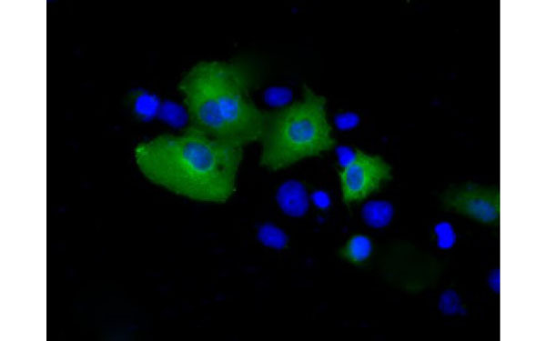 IF (Immunofluorescence)
(Immunofluorescent staining of COS7 cells transiently transfected with recombinant PSMC3 protein using PSMC3 antibody)
IF (Immunofluorescence)
(Immunofluorescent staining of COS7 cells transiently transfected with recombinant PSMC3 protein using PSMC3 antibody)
PSMC3, Monoclonal Antibody (Cat# AAA108276)
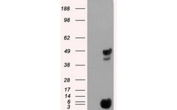
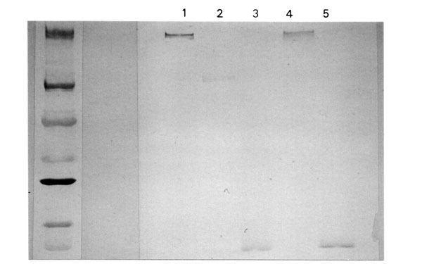 WB (Western Blot)
(Immunoblot showing C3a DesArg detection: Lane 1: purified C3, Lane 2: purifed C3 reduced/DTT, Lane 3: purified C3a, Lane 4: EDTA-plasma. Lane 5: zymosan-activated serum)
WB (Western Blot)
(Immunoblot showing C3a DesArg detection: Lane 1: purified C3, Lane 2: purifed C3 reduced/DTT, Lane 3: purified C3a, Lane 4: EDTA-plasma. Lane 5: zymosan-activated serum)
C3a desArg, Monoclonal Antibody (Cat# AAA108286)
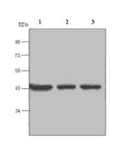
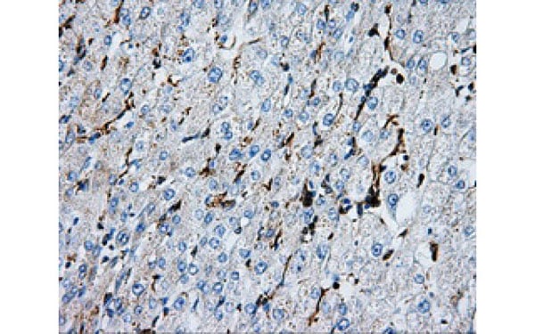
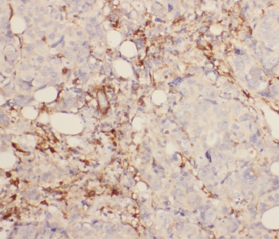
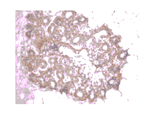 IHC (Immunohiostchemistry)
(Immunohistochemistry Staining of Human ovary tissue with beta -Actin mouse mAb(6G3) diluted at 1:200..)
IHC (Immunohiostchemistry)
(Immunohistochemistry Staining of Human ovary tissue with beta -Actin mouse mAb(6G3) diluted at 1:200..)
beta-Actin, Monoclonal Antibody (Cat# AAA108513)
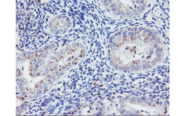
 WB (Western Blot)
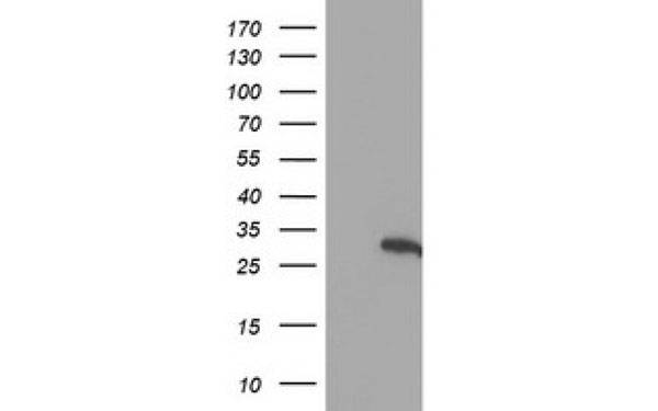
(Western Blot analysis of HEK293T cell lysates (5 ug) transfected with either recombinant HES1 protein (Right) or empty vector (Left) detected with HES1 antibody)
WB (Western Blot)
(Western Blot analysis of HEK293T cell lysates (5 ug) transfected with either recombinant HES1 protein (Right) or empty vector (Left) detected with HES1 antibody)
HES1, Monoclonal Antibody (Cat# AAA108165)
 IHC (Immunohiostchemistry)
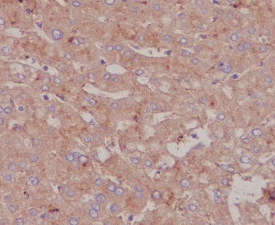
(IHC analysis of Bovine Serum Albumin using anti- BovineSerum Albumin antibody (AAA47042) on human liver. Bovine Serum Albumin was detected in paraffin-embeddedsection. Heat mediated antigen retrieval was performed incitrate buffer (pH6, epitope retrieval solution) for 20 mins.The tissue section was blocked with 10% goat serum. Thetissue section was then incubated with 1ug/ml rabbit anti-Bovine Serum Albumin Antibody (AAA47042) overnight at 4°C. Biotinylated goat anti Rabbit IgG IgG antibody was used assecondary antibody and incubated for 30 minutes at 37°C. The tissue section was developed using Strepavidin-Biotin-Complex (SABC) with DAB as the chromogen.)
IHC (Immunohiostchemistry)
(IHC analysis of Bovine Serum Albumin using anti- BovineSerum Albumin antibody (AAA47042) on human liver. Bovine Serum Albumin was detected in paraffin-embeddedsection. Heat mediated antigen retrieval was performed incitrate buffer (pH6, epitope retrieval solution) for 20 mins.The tissue section was blocked with 10% goat serum. Thetissue section was then incubated with 1ug/ml rabbit anti-Bovine Serum Albumin Antibody (AAA47042) overnight at 4°C. Biotinylated goat anti Rabbit IgG IgG antibody was used assecondary antibody and incubated for 30 minutes at 37°C. The tissue section was developed using Strepavidin-Biotin-Complex (SABC) with DAB as the chromogen.)
Serum Albumin, Monoclonal Antibody (Cat# AAA47042)
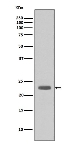 WB (Western Blot)
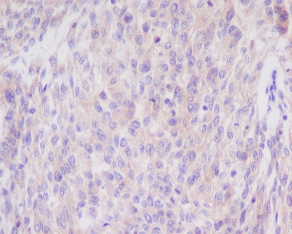
(Western blot analysis of Bim expression in A431 cell lysate (AAA47066).Electrophoresis was performed on a 5-20% SDS-PAGE gel at 70V (Stacking gel) / 90V (Resolving gel) for 2-3 hours. The sample well of each lane was loaded with 50ug of sample under reducing conditions.After Electrophoresis, proteins were transferred to a Nitrocellulose membrane at 150mA for 50-90 minutes. Blocked the membrane with 5% Non-fat Milk/ TBS for 1.5 hour at RT. The membrane was incubated with rabbit anti-BCL2L11 monoclonal antibody overnight at 4 degree C, then washed with TBS-0.1%Tween 3 times with 5 minutes each and probed with a goat anti-rabbit IgG-HRP secondary antibody at a dilution of 1:10000 for 1.5 hour at RT. The signal is developed using an Enhanced Chemiluminescent detection (ECL) kit with Tanon 5200 system. A specific band was detected for BCL2L11)
WB (Western Blot)
(Western blot analysis of Bim expression in A431 cell lysate (AAA47066).Electrophoresis was performed on a 5-20% SDS-PAGE gel at 70V (Stacking gel) / 90V (Resolving gel) for 2-3 hours. The sample well of each lane was loaded with 50ug of sample under reducing conditions.After Electrophoresis, proteins were transferred to a Nitrocellulose membrane at 150mA for 50-90 minutes. Blocked the membrane with 5% Non-fat Milk/ TBS for 1.5 hour at RT. The membrane was incubated with rabbit anti-BCL2L11 monoclonal antibody overnight at 4 degree C, then washed with TBS-0.1%Tween 3 times with 5 minutes each and probed with a goat anti-rabbit IgG-HRP secondary antibody at a dilution of 1:10000 for 1.5 hour at RT. The signal is developed using an Enhanced Chemiluminescent detection (ECL) kit with Tanon 5200 system. A specific band was detected for BCL2L11)
Bim, Monoclonal Antibody (Cat# AAA47066)
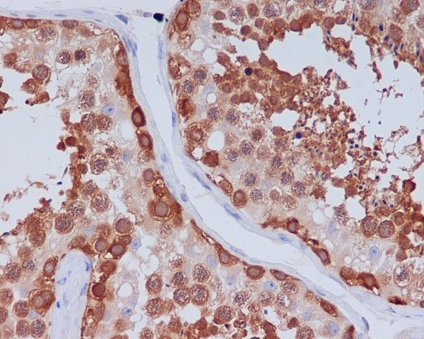
 WB (Western Blot)
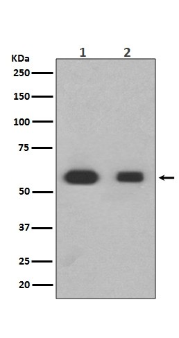
(Western blot analysis of Hsp90 beta expression in (1)HeLa cell lysate;(2)Jurkat cell lysate (AAA47072).Electrophoresis was performed on a 5-20% SDS-PAGE gel at 70V (Stacking gel) / 90V (Resolving gel) for 2-3 hours. The sample well of each lane was loaded with 50ug of sample under reducing conditions.After Electrophoresis, proteins were transferred to a Nitrocellulose membrane at 150mA for 50-90 minutes. Blocked the membrane with 5% Non-fat Milk/ TBS for 1.5 hour at RT. The membrane was incubated with rabbit anti-HSP90AB1 monoclonal antibody overnight at 4 degree C, then washed with TBS-0.1%Tween 3 times with 5 minutes each and probed with a goat anti-rabbit IgG-HRP secondary antibody at a dilution of 1:10000 for 1.5 hour at RT. The signal is developed using an Enhanced Chemiluminescent detection (ECL) kit with Tanon 5200 system. A specific band was detected for HSP90AB1)
WB (Western Blot)
(Western blot analysis of Hsp90 beta expression in (1)HeLa cell lysate;(2)Jurkat cell lysate (AAA47072).Electrophoresis was performed on a 5-20% SDS-PAGE gel at 70V (Stacking gel) / 90V (Resolving gel) for 2-3 hours. The sample well of each lane was loaded with 50ug of sample under reducing conditions.After Electrophoresis, proteins were transferred to a Nitrocellulose membrane at 150mA for 50-90 minutes. Blocked the membrane with 5% Non-fat Milk/ TBS for 1.5 hour at RT. The membrane was incubated with rabbit anti-HSP90AB1 monoclonal antibody overnight at 4 degree C, then washed with TBS-0.1%Tween 3 times with 5 minutes each and probed with a goat anti-rabbit IgG-HRP secondary antibody at a dilution of 1:10000 for 1.5 hour at RT. The signal is developed using an Enhanced Chemiluminescent detection (ECL) kit with Tanon 5200 system. A specific band was detected for HSP90AB1)
Hsp90 beta, Monoclonal Antibody (Cat# AAA47072)
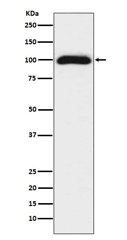 WB (Western Blot)
(Western blot analysis of CD276 expression in 293T cell lysate (AAA47100).Electrophoresis was performed on a 5-20% SDS-PAGE gel at 70V (Stacking gel) / 90V (Resolving gel) for 2-3 hours. The sample well of each lane was loaded with 50ug of sample under reducing conditions.After Electrophoresis, proteins were transferred to a Nitrocellulose membrane at 150mA for 50-90 minutes. Blocked the membrane with 5% Non-fat Milk/ TBS for 1.5 hour at RT. The membrane was incubated with rabbit anti-CD276 monoclonal antibody overnight at 4 degree C, then washed with TBS-0.1%Tween 3 times with 5 minutes each and probed with a goat anti-rabbit IgG-HRP secondary antibody at a dilution of 1:10000 for 1.5 hour at RT. The signal is developed using an Enhanced Chemiluminescent detection (ECL) kit with Tanon 5200 system. A specific band was detected for CD276)
WB (Western Blot)
(Western blot analysis of CD276 expression in 293T cell lysate (AAA47100).Electrophoresis was performed on a 5-20% SDS-PAGE gel at 70V (Stacking gel) / 90V (Resolving gel) for 2-3 hours. The sample well of each lane was loaded with 50ug of sample under reducing conditions.After Electrophoresis, proteins were transferred to a Nitrocellulose membrane at 150mA for 50-90 minutes. Blocked the membrane with 5% Non-fat Milk/ TBS for 1.5 hour at RT. The membrane was incubated with rabbit anti-CD276 monoclonal antibody overnight at 4 degree C, then washed with TBS-0.1%Tween 3 times with 5 minutes each and probed with a goat anti-rabbit IgG-HRP secondary antibody at a dilution of 1:10000 for 1.5 hour at RT. The signal is developed using an Enhanced Chemiluminescent detection (ECL) kit with Tanon 5200 system. A specific band was detected for CD276)
CD276/B7 H3, Monoclonal Antibody (Cat# AAA47100)
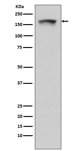
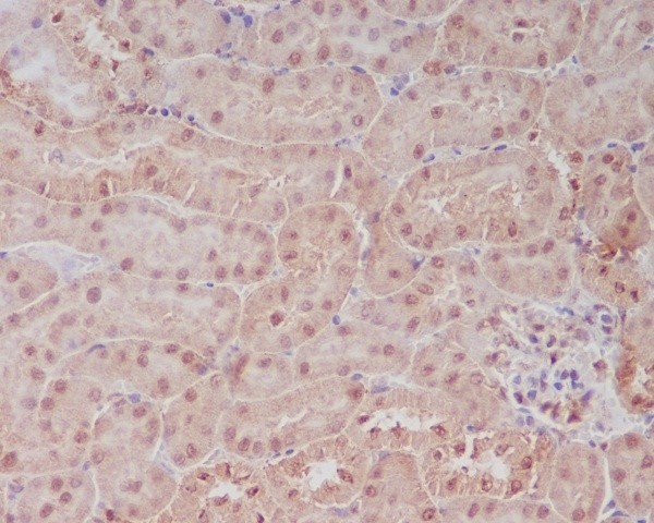 IHC (Immunohiostchemistry)
(Immunohistochemical analysis of paraffin-embedded mouse kidney, using PELP1 Antibody(AAA47114)PELP1 was detected in paraffin-embedded tissue section. Heat mediated antigen retrieval was performed in citrate buffer (pH6, epitope retrieval solution) for 20 mins. The tissue section was blocked with 10% goat serum. The tissue section was then incubated with 1ug/ml rabbit anti-PELP1 Antibody (AAA47114)overnight at 4 degree C. Biotinylated goat anti-rabbit IgG was used as secondary antibody and incubated for 30 minutes at 37 degree C. The tissue section was developed using Strepavidin-Biotin-Complex (SABC) with DAB as the chromogen.)
IHC (Immunohiostchemistry)
(Immunohistochemical analysis of paraffin-embedded mouse kidney, using PELP1 Antibody(AAA47114)PELP1 was detected in paraffin-embedded tissue section. Heat mediated antigen retrieval was performed in citrate buffer (pH6, epitope retrieval solution) for 20 mins. The tissue section was blocked with 10% goat serum. The tissue section was then incubated with 1ug/ml rabbit anti-PELP1 Antibody (AAA47114)overnight at 4 degree C. Biotinylated goat anti-rabbit IgG was used as secondary antibody and incubated for 30 minutes at 37 degree C. The tissue section was developed using Strepavidin-Biotin-Complex (SABC) with DAB as the chromogen.)
PELP1, Monoclonal Antibody (Cat# AAA47114)
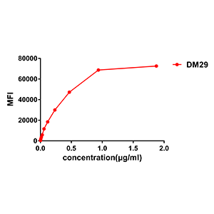
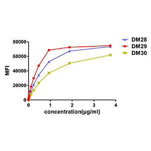
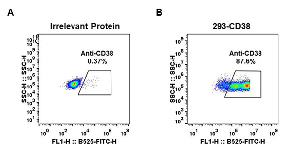 FCM/FACS (Flow Cytometry)
(Figure 3. Affinity ranking of different Rabbit anti-CD38 mAb clones by titration of different concentration onto Raji cells. The Y-axis represents the mean fluorescence intensity (MFI) while the X-axis represents the concentration of IgG used.)
FCM/FACS (Flow Cytometry)
(Figure 3. Affinity ranking of different Rabbit anti-CD38 mAb clones by titration of different concentration onto Raji cells. The Y-axis represents the mean fluorescence intensity (MFI) while the X-axis represents the concentration of IgG used.)
CD38, Monoclonal Antibody (Cat# AAA47180)
 FCM/FACS (Flow Cytometry)
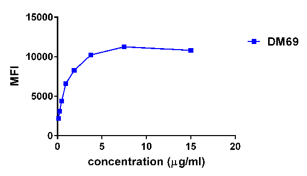
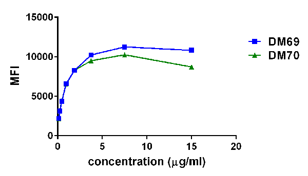
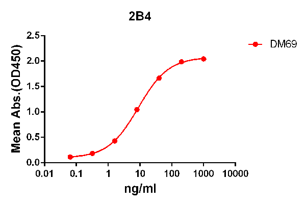
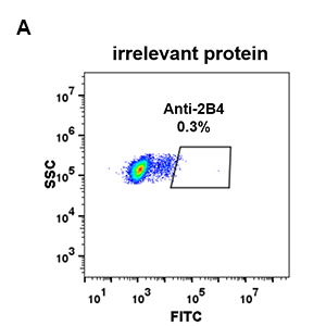
(Figure 4. Affinity ranking of different Rabbit anti-2B4 mAb clones by titration of different concentration onto THP-1 cells. The Y-axis represents the mean fluorescence intensity (MFI) while the X-axis represents the concentration of IgG used.)
FCM/FACS (Flow Cytometry)
(Figure 4. Affinity ranking of different Rabbit anti-2B4 mAb clones by titration of different concentration onto THP-1 cells. The Y-axis represents the mean fluorescence intensity (MFI) while the X-axis represents the concentration of IgG used.)
2B4, Monoclonal Antibody (Cat# AAA47301)
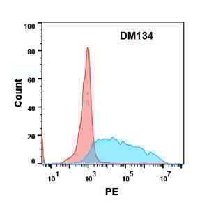 FCM/FACS (Flow Cytometry)
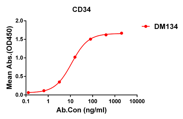
(Figure 1. ELISA plate pre-coated by 1 ug/ml (100 ul/well) Human CD34 protein, His tagged protein PME100469 can bind Rabbit anti-CD34 monoclonal antibody (clone: DM134) in a linear range of 0.2-12 ng/ml.)
FCM/FACS (Flow Cytometry)
(Figure 1. ELISA plate pre-coated by 1 ug/ml (100 ul/well) Human CD34 protein, His tagged protein PME100469 can bind Rabbit anti-CD34 monoclonal antibody (clone: DM134) in a linear range of 0.2-12 ng/ml.)
CD34, Monoclonal Antibody (Cat# AAA47456)
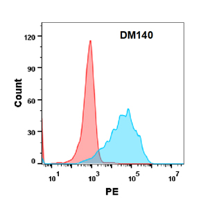 FCM/FACS (Flow Cytometry)
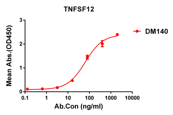
(Figure 1. ELISA plate pre-coated by 1 ug/ml (100 ul/well) Human TNFSF12 protein, hFc tagged protein PME100105 can bind Rabbit anti-TNFSF12 monoclonal antibody (clone: DM140) in a linear range of 5-200 ng/ml.)
FCM/FACS (Flow Cytometry)
(Figure 1. ELISA plate pre-coated by 1 ug/ml (100 ul/well) Human TNFSF12 protein, hFc tagged protein PME100105 can bind Rabbit anti-TNFSF12 monoclonal antibody (clone: DM140) in a linear range of 5-200 ng/ml.)
TNFSF12, Monoclonal Antibody (Cat# AAA47513)
 FCM/FACS (Flow Cytometry)
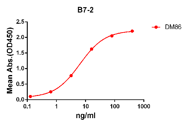
(Figure 1. ELISA plate pre-coated by 2 ug/ml (100 ul/well) Human B7-2 protein, mFc-His tagged protein PME100034 can bind Rabbit anti-B7-2 monoclonal antibody (clone: DM86) in a linear range of 1-100 ng/ml.)
FCM/FACS (Flow Cytometry)
(Figure 1. ELISA plate pre-coated by 2 ug/ml (100 ul/well) Human B7-2 protein, mFc-His tagged protein PME100034 can bind Rabbit anti-B7-2 monoclonal antibody (clone: DM86) in a linear range of 1-100 ng/ml.)
B7-2, Monoclonal Antibody (Cat# AAA47318)
 FCM/FACS (Flow Cytometry)
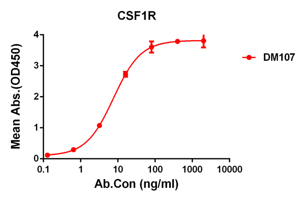
(Figure 1. ELISA plate pre-coated by 2 ug/ml (100 ul/well) Human CSF1R protein,His tagged protein PME100067 can bind Rabbit anti- CSF1R monoclonal antibody (clone: DM107) in a linear range of 0.6-40 ng/ml.)
FCM/FACS (Flow Cytometry)
(Figure 1. ELISA plate pre-coated by 2 ug/ml (100 ul/well) Human CSF1R protein,His tagged protein PME100067 can bind Rabbit anti- CSF1R monoclonal antibody (clone: DM107) in a linear range of 0.6-40 ng/ml.)
CSF1R, Monoclonal Antibody (Cat# AAA47432)
CAMPYLOBACTER FETUS, Monoclonal Antibody (Cat# AAA49173)
IgG2, Monoclonal Antibody (Cat# AAA49191)
Preparation: Purified IgG prepared by affinity chromatography on Protein G
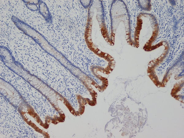
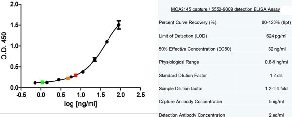
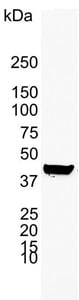 Application Data
(Western blot analysis of A549 human alveolar adenocarcinoma whole cell lysate probed with Mouse anti Human cytokeratin 19 antibody followed by HRP conjugated Goat anti Mouse IgG, visualized by chemiluminescence)
Application Data
(Western blot analysis of A549 human alveolar adenocarcinoma whole cell lysate probed with Mouse anti Human cytokeratin 19 antibody followed by HRP conjugated Goat anti Mouse IgG, visualized by chemiluminescence)
CYTOKERATIN 19, Monoclonal Antibody (Cat# AAA49195)
hCG, Monoclonal Antibody (Cat# AAA49225)
IgA, Monoclonal Antibody (Cat# AAA49235)
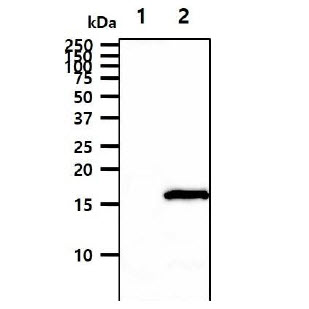
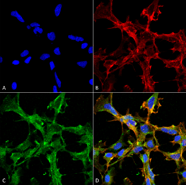
 IF (Immunofluorescence)
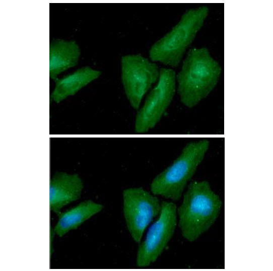
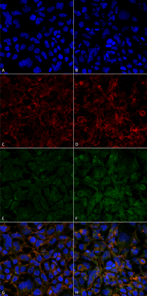
(ICC/IF analysis of MAPKSP1 in HaLa cells. The cell was stained with ATGA0503 (1:100). The secondary antibody (green) was used Alexa Fluor 488. DAPI was stained the cell nucleus (blue).)
IF (Immunofluorescence)
(ICC/IF analysis of MAPKSP1 in HaLa cells. The cell was stained with ATGA0503 (1:100). The secondary antibody (green) was used Alexa Fluor 488. DAPI was stained the cell nucleus (blue).)
MAPKSP1, Monoclonal Antibody (Cat# AAA48960)
 Application Data
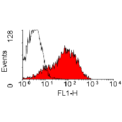
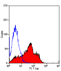
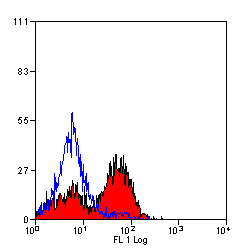
(Staining of rat spleen cells with Mouse anti Rat CD44)
Application Data
(Staining of rat spleen cells with Mouse anti Rat CD44)
CD44, Monoclonal Antibody (Cat# AAA49020)
IgG2a, Monoclonal Antibody (Cat# AAA49133)
ALDOSTERONE, Monoclonal Antibody (Cat# AAA49155)
 Application Data
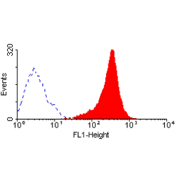
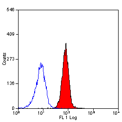
(Staining of human peripheral blood granulocytes with Mouse anti Human CD95:Alexa Fluor 647)
Application Data
(Staining of human peripheral blood granulocytes with Mouse anti Human CD95:Alexa Fluor 647)
CD95, Monoclonal Antibody (Cat# AAA49057)
 Application Data
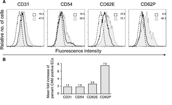
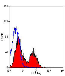
(Staining of porcine peripheral blood lymphocytes with Mouse anti Porcine CD31:FITC)
Application Data
(Staining of porcine peripheral blood lymphocytes with Mouse anti Porcine CD31:FITC)
CD31, Monoclonal Antibody (Cat# AAA49065)
 Application Data
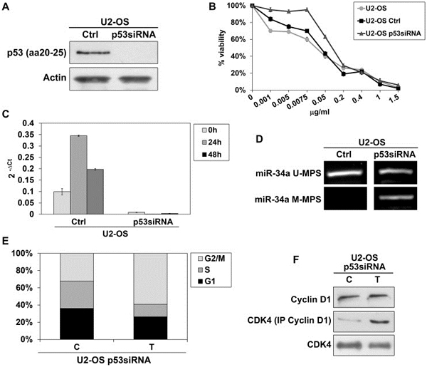
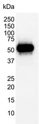
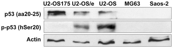
(Published customer image: p53 protein expression in OS cells. wt-p53 U2-OS, U2-OS transfected with empty vector (U2-OS/e) and p53-impaired U2-OS175 cells were positive to anti-p53 that binds the transactivation site of N-terminal domain (aa20-25), with increased expression in U2-OS175 cells. U2-OS and U2-OS/e also presented accumulation of p53 phosphorylated at Ser20 residue (p-p53). MG63 and Saos-2 were negative to both antibodies. Actina was used as loading control.From: Novello C, Pazzaglia L, Conti A, Quattrini I, Pollino S, et al. (2014) p53-Dependent Activation of microRNA-34a in Response to Etoposide-Induced DNA Damage in Osteosarcoma Cell Lines Not Impaired by Dominant Negative p53 Expression. PLoS ONE 9(12): e114757.)
Application Data
(Published customer image: p53 protein expression in OS cells. wt-p53 U2-OS, U2-OS transfected with empty vector (U2-OS/e) and p53-impaired U2-OS175 cells were positive to anti-p53 that binds the transactivation site of N-terminal domain (aa20-25), with increased expression in U2-OS175 cells. U2-OS and U2-OS/e also presented accumulation of p53 phosphorylated at Ser20 residue (p-p53). MG63 and Saos-2 were negative to both antibodies. Actina was used as loading control.From: Novello C, Pazzaglia L, Conti A, Quattrini I, Pollino S, et al. (2014) p53-Dependent Activation of microRNA-34a in Response to Etoposide-Induced DNA Damage in Osteosarcoma Cell Lines Not Impaired by Dominant Negative p53 Expression. PLoS ONE 9(12): e114757.)
p53, Monoclonal Antibody (Cat# AAA49026)
 FCM/FACS (Flow Cytometry)
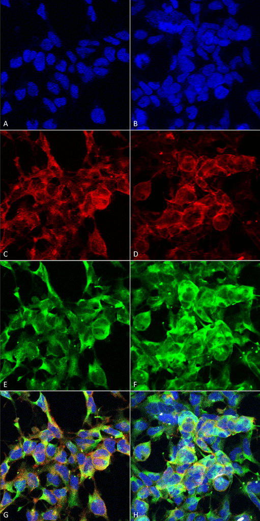
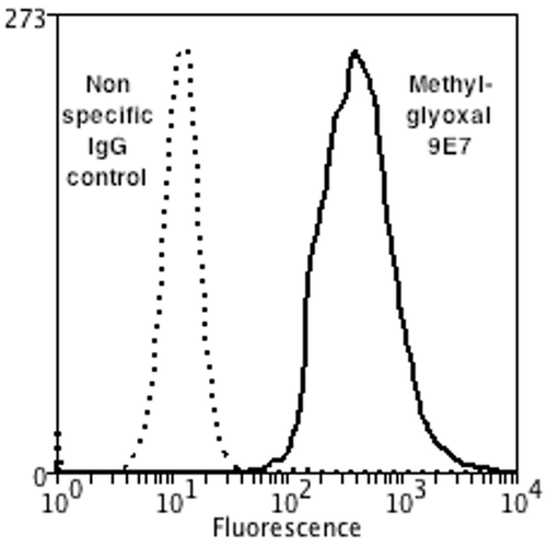
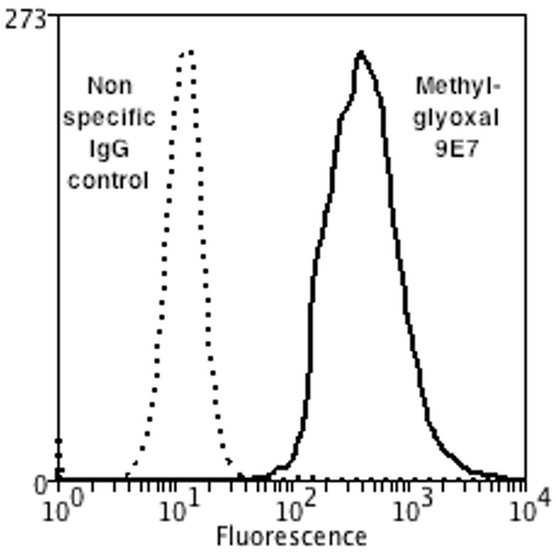
(Flow Cytometry analysis using Mouse Anti-Methylglyoxal Monoclonal Antibody, Clone 9E7. Tissue: Neuroblastoma cells (SH-SY5Y). Species: Human. Fixation: 90% Methanol. Primary Antibody: Mouse Anti-Methylglyoxal Monoclonal Antibody at 1:50 for 30 min on ice. Secondary Antibody: Goat Anti-Mouse: PE at 1:100 for 20 min at RT. Isotype Control: Non Specific IgG. Cells were subject to oxidative stress by treating with 250 uM H2O2 for 24 hours.)
FCM/FACS (Flow Cytometry)
(Flow Cytometry analysis using Mouse Anti-Methylglyoxal Monoclonal Antibody, Clone 9E7. Tissue: Neuroblastoma cells (SH-SY5Y). Species: Human. Fixation: 90% Methanol. Primary Antibody: Mouse Anti-Methylglyoxal Monoclonal Antibody at 1:50 for 30 min on ice. Secondary Antibody: Goat Anti-Mouse: PE at 1:100 for 20 min at RT. Isotype Control: Non Specific IgG. Cells were subject to oxidative stress by treating with 250 uM H2O2 for 24 hours.)
Methylglyoxal, Monoclonal Antibody (Cat# AAA104090)
 FCM/FACS (Flow Cytometry)
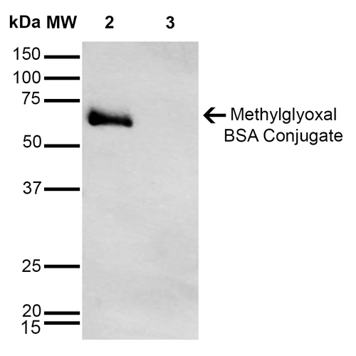
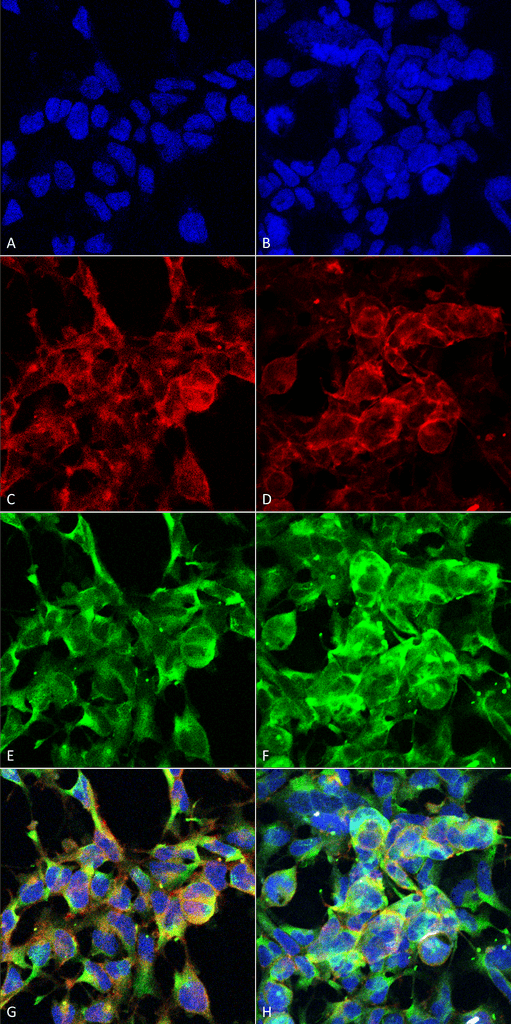
(Flow Cytometry analysis using Mouse Anti-Methylglyoxal Monoclonal Antibody, Clone 9E7. Tissue: Neuroblastoma cells (SH-SY5Y). Species: Human. Fixation: 90% Methanol. Primary Antibody: Mouse Anti-Methylglyoxal Monoclonal Antibody at 1:50 for 30 min on ice. Secondary Antibody: Goat Anti-Mouse: PE at 1:100 for 20 min at RT. Isotype Control: Non Specific IgG. Cells were subject to oxidative stress by treating with 250 uM H2O2 for 24 hours.)
FCM/FACS (Flow Cytometry)
(Flow Cytometry analysis using Mouse Anti-Methylglyoxal Monoclonal Antibody, Clone 9E7. Tissue: Neuroblastoma cells (SH-SY5Y). Species: Human. Fixation: 90% Methanol. Primary Antibody: Mouse Anti-Methylglyoxal Monoclonal Antibody at 1:50 for 30 min on ice. Secondary Antibody: Goat Anti-Mouse: PE at 1:100 for 20 min at RT. Isotype Control: Non Specific IgG. Cells were subject to oxidative stress by treating with 250 uM H2O2 for 24 hours.)
Methylglyoxal, Monoclonal Antibody (Cat# AAA104091)
 FCM/FACS (Flow Cytometry)
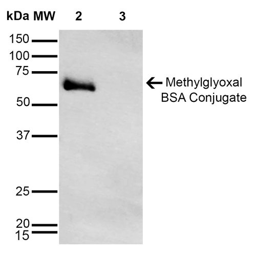
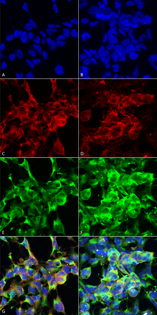
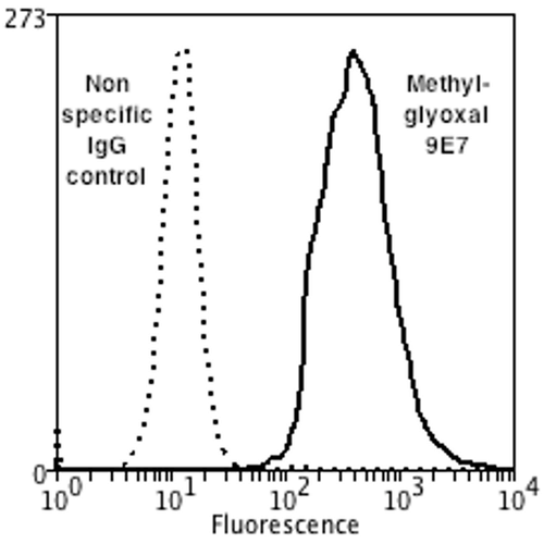
(Flow Cytometry analysis using Mouse Anti-Methylglyoxal Monoclonal Antibody, Clone 9E7. Tissue: Neuroblastoma cells (SH-SY5Y). Species: Human. Fixation: 90% Methanol. Primary Antibody: Mouse Anti-Methylglyoxal Monoclonal Antibody at 1:50 for 30 min on ice. Secondary Antibody: Goat Anti-Mouse: PE at 1:100 for 20 min at RT. Isotype Control: Non Specific IgG. Cells were subject to oxidative stress by treating with 250 uM H2O2 for 24 hours.)
FCM/FACS (Flow Cytometry)
(Flow Cytometry analysis using Mouse Anti-Methylglyoxal Monoclonal Antibody, Clone 9E7. Tissue: Neuroblastoma cells (SH-SY5Y). Species: Human. Fixation: 90% Methanol. Primary Antibody: Mouse Anti-Methylglyoxal Monoclonal Antibody at 1:50 for 30 min on ice. Secondary Antibody: Goat Anti-Mouse: PE at 1:100 for 20 min at RT. Isotype Control: Non Specific IgG. Cells were subject to oxidative stress by treating with 250 uM H2O2 for 24 hours.)
Methylglyoxal, Monoclonal Antibody (Cat# AAA104092)
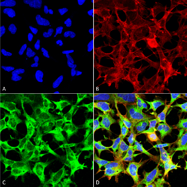
 WB (Western Blot)
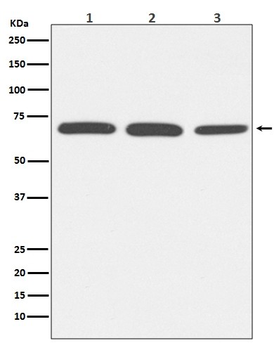
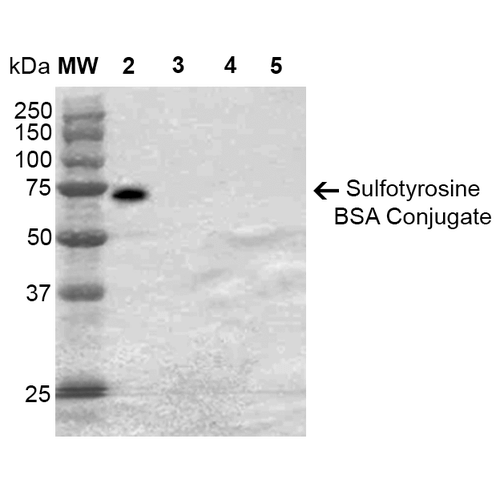
(Western Blot analysis of Sulfotyrosine-BSA Conjugate showing detection of 67 kDa Sulfotyrosine protein using Mouse Anti-Sulfotyrosine Monoclonal Antibody, Clone 7C5. Lane 1: Molecular Weight Ladder (MW). Lane 2: Sulfotyrosine-BSA. Lane 3: Tyrosine-BSA. Lane 4: Phosphotyrosine-BSA. Lane 5: BSA. Load: 2 ug. Block: 5% Skim Milk in TBST. Primary Antibody: Mouse Anti-Sulfotyrosine Monoclonal Antibody at 1:1000 for 2 hours at RT. Secondary Antibody: Goat Anti-Mouse IgG: HRP at 1:2000 for 60 min at RT. Color Development: ECL solution for 5 min in RT. Predicted/Observed Size: 67 kDa.)
WB (Western Blot)
(Western Blot analysis of Sulfotyrosine-BSA Conjugate showing detection of 67 kDa Sulfotyrosine protein using Mouse Anti-Sulfotyrosine Monoclonal Antibody, Clone 7C5. Lane 1: Molecular Weight Ladder (MW). Lane 2: Sulfotyrosine-BSA. Lane 3: Tyrosine-BSA. Lane 4: Phosphotyrosine-BSA. Lane 5: BSA. Load: 2 ug. Block: 5% Skim Milk in TBST. Primary Antibody: Mouse Anti-Sulfotyrosine Monoclonal Antibody at 1:1000 for 2 hours at RT. Secondary Antibody: Goat Anti-Mouse IgG: HRP at 1:2000 for 60 min at RT. Color Development: ECL solution for 5 min in RT. Predicted/Observed Size: 67 kDa.)
Sulfotyrosine, Monoclonal Antibody (Cat# AAA104129)
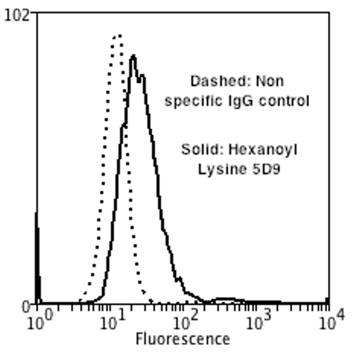
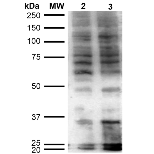 WB (Western Blot)
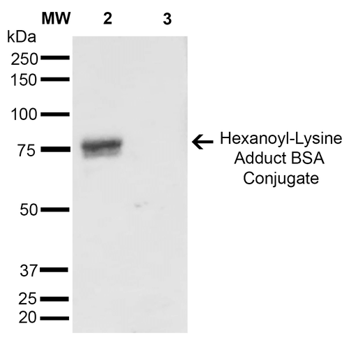
(Western Blot analysis of Human Cervical cancer cell line (HeLa) lysate showing detection of Hexanoyl-Lysine adduct protein using Mouse Anti-Hexanoyl-Lysine adduct Monoclonal Antibody, Clone 5D9. Lane 1: Molecular Weight Ladder (MW). Lane 2: HeLa cell lysate. Lane 3: H2O2 treated HeLa cell lysate. Load: 12 ug. Block: 5% Skim Milk in TBST. Primary Antibody: Mouse Anti-Hexanoyl-Lysine adduct Monoclonal Antibody at 1:1000 for 2 hours at RT. Secondary Antibody: Goat Anti-Mouse IgG: HRP at 1:2000 for 60 min at RT. Color Development: ECL solution for 5 min in RT.)
WB (Western Blot)
(Western Blot analysis of Human Cervical cancer cell line (HeLa) lysate showing detection of Hexanoyl-Lysine adduct protein using Mouse Anti-Hexanoyl-Lysine adduct Monoclonal Antibody, Clone 5D9. Lane 1: Molecular Weight Ladder (MW). Lane 2: HeLa cell lysate. Lane 3: H2O2 treated HeLa cell lysate. Load: 12 ug. Block: 5% Skim Milk in TBST. Primary Antibody: Mouse Anti-Hexanoyl-Lysine adduct Monoclonal Antibody at 1:1000 for 2 hours at RT. Secondary Antibody: Goat Anti-Mouse IgG: HRP at 1:2000 for 60 min at RT. Color Development: ECL solution for 5 min in RT.)
Hexanoyl-Lysine adduct, Monoclonal Antibody (Cat# AAA104020)
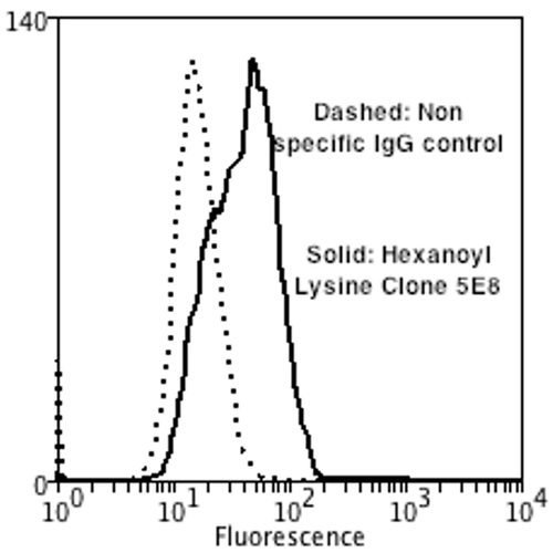
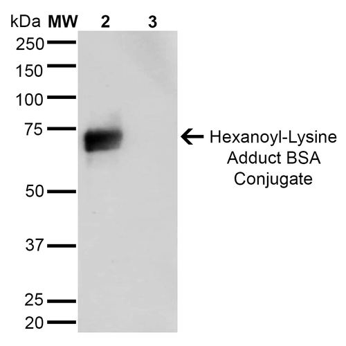
 WB (Western Blot)
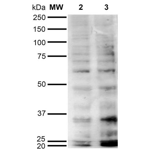
(Western Blot analysis of Human Cervical cancer cell line (HeLa) lysate showing detection of Hexanoyl-Lysine adduct protein using Mouse Anti-Hexanoyl-Lysine adduct Monoclonal Antibody, Clone 5E8. Lane 1: Molecular Weight Ladder (MW). Lane 2: HeLa cell lysate. Lane 3: H2O2 treated HeLa cell lysate. Load: 12 ug. Block: 5% Skim Milk in TBST. Primary Antibody: Mouse Anti-Hexanoyl-Lysine adduct Monoclonal Antibody at 1:1000 for 2 hours at RT. Secondary Antibody: Goat Anti-Mouse IgG: HRP at 1:2000 for 60 min at RT. Color Development: ECL solution for 5 min in RT.)
WB (Western Blot)
(Western Blot analysis of Human Cervical cancer cell line (HeLa) lysate showing detection of Hexanoyl-Lysine adduct protein using Mouse Anti-Hexanoyl-Lysine adduct Monoclonal Antibody, Clone 5E8. Lane 1: Molecular Weight Ladder (MW). Lane 2: HeLa cell lysate. Lane 3: H2O2 treated HeLa cell lysate. Load: 12 ug. Block: 5% Skim Milk in TBST. Primary Antibody: Mouse Anti-Hexanoyl-Lysine adduct Monoclonal Antibody at 1:1000 for 2 hours at RT. Secondary Antibody: Goat Anti-Mouse IgG: HRP at 1:2000 for 60 min at RT. Color Development: ECL solution for 5 min in RT.)
Hexanoyl-Lysine adduct, Monoclonal Antibody (Cat# AAA104024)
 IHC (Immunohiostchemistry)
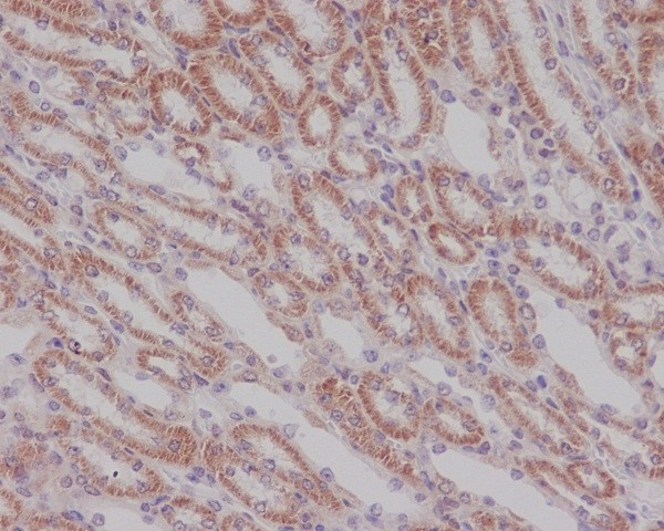
(Immunohistochemical analysis of paraffin-embedded mouse kidney, using Smad3 Antibody(AAA46858)SMAD3 was detected in paraffin-embedded tissue section. Heat mediated antigen retrieval was performed in citrate buffer (pH6, epitope retrieval solution) for 20 mins. The tissue section was blocked with 10% goat serum. The tissue section was then incubated with 1ug/ml rabbit anti-SMAD3 Antibody (AAA46858)overnight at 4 degree C. Biotinylated goat anti-rabbit IgG was used as secondary antibody and incubated for 30 minutes at 37 degree C. The tissue section was developed using Strepavidin-Biotin-Complex (SABC) with DAB as the chromogen.)
IHC (Immunohiostchemistry)
(Immunohistochemical analysis of paraffin-embedded mouse kidney, using Smad3 Antibody(AAA46858)SMAD3 was detected in paraffin-embedded tissue section. Heat mediated antigen retrieval was performed in citrate buffer (pH6, epitope retrieval solution) for 20 mins. The tissue section was blocked with 10% goat serum. The tissue section was then incubated with 1ug/ml rabbit anti-SMAD3 Antibody (AAA46858)overnight at 4 degree C. Biotinylated goat anti-rabbit IgG was used as secondary antibody and incubated for 30 minutes at 37 degree C. The tissue section was developed using Strepavidin-Biotin-Complex (SABC) with DAB as the chromogen.)
Smad3, Monoclonal Antibody (Cat# AAA46858)
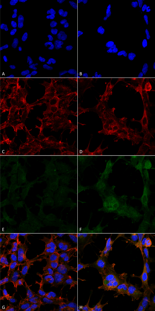
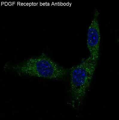 IF (Immunofluorescence)
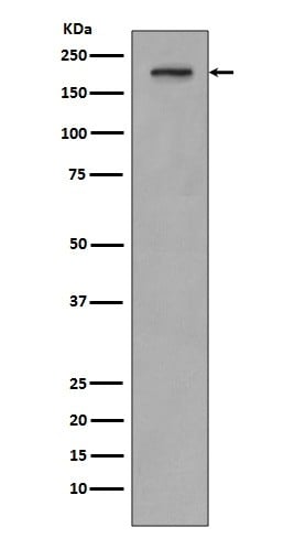
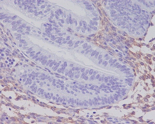
PDGF Receptor beta, Monoclonal Antibody (Cat# AAA46870)
 WB (Western Blot)
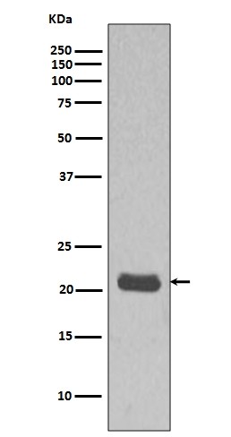
(Western blot analysis MCF7 cell lysate using HRAS Antibody (AAA46876).)
WB (Western Blot)
(Western blot analysis MCF7 cell lysate using HRAS Antibody (AAA46876).)
GTPase HRAS, Monoclonal Antibody (Cat# AAA46876)
 IHC (Immunohiostchemistry)
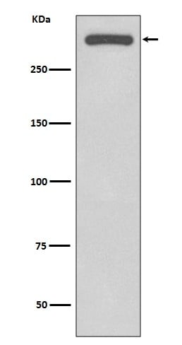
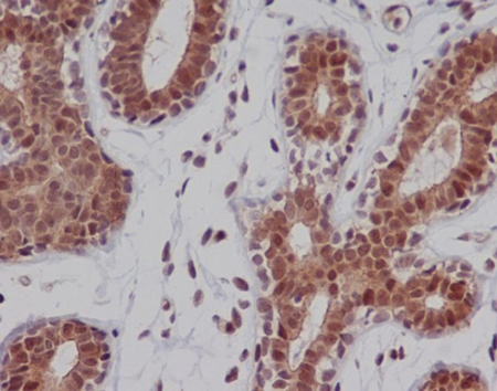
(Immunohistochemical analysis of paraffin-embedded human breast, using DNA-PKcs Antibody.)
IHC (Immunohiostchemistry)
(Immunohistochemical analysis of paraffin-embedded human breast, using DNA-PKcs Antibody.)
DNA-PKcs, Monoclonal Antibody (Cat# AAA46972)
 WB (Western Blot)
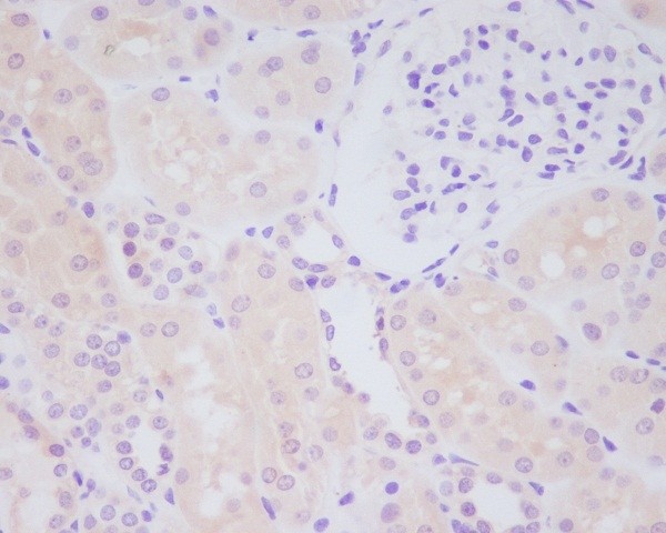
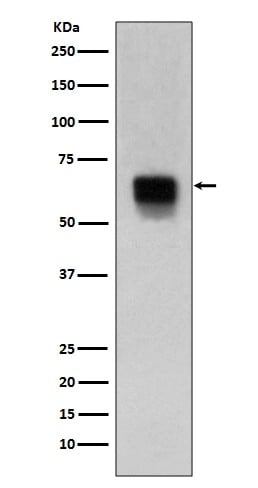
(Western blot analysis of MMP14 expression in human spleen lysate (AAA46974).Electrophoresis was performed on a 5-20% SDS-PAGE gel at 70V (Stacking gel) / 90V (Resolving gel) for 2-3 hours. The sample well of each lane was loaded with 50ug of sample under reducing conditions.After Electrophoresis, proteins were transferred to a Nitrocellulose membrane at 150mA for 50-90 minutes. Blocked the membrane with 5% Non-fat Milk/ TBS for 1.5 hour at RT. The membrane was incubated with rabbit anti-MMP14 monoclonal antibody overnight at 4 degree C, then washed with TBS-0.1%Tween 3 times with 5 minutes each and probed with a goat anti-rabbit IgG-HRP secondary antibody at a dilution of 1:10000 for 1.5 hour at RT. The signal is developed using an Enhanced Chemiluminescent detection (ECL) kit with Tanon 5200 system. A specific band was detected for MMP14)
WB (Western Blot)
(Western blot analysis of MMP14 expression in human spleen lysate (AAA46974).Electrophoresis was performed on a 5-20% SDS-PAGE gel at 70V (Stacking gel) / 90V (Resolving gel) for 2-3 hours. The sample well of each lane was loaded with 50ug of sample under reducing conditions.After Electrophoresis, proteins were transferred to a Nitrocellulose membrane at 150mA for 50-90 minutes. Blocked the membrane with 5% Non-fat Milk/ TBS for 1.5 hour at RT. The membrane was incubated with rabbit anti-MMP14 monoclonal antibody overnight at 4 degree C, then washed with TBS-0.1%Tween 3 times with 5 minutes each and probed with a goat anti-rabbit IgG-HRP secondary antibody at a dilution of 1:10000 for 1.5 hour at RT. The signal is developed using an Enhanced Chemiluminescent detection (ECL) kit with Tanon 5200 system. A specific band was detected for MMP14)
MMP14/Mt1 Mmp, Monoclonal Antibody (Cat# AAA46974)
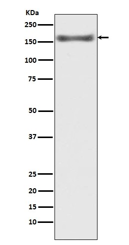
 IHC (Immunohiostchemistry)
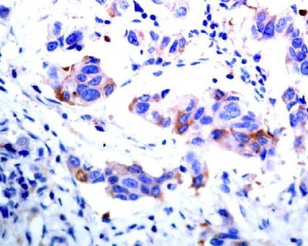
(Immunohistochemical analysis of paraffin-embedded human breast cancer, using ASK1 Antibody(AAA47001)MAP3K5 was detected in paraffin-embedded tissue section. Heat mediated antigen retrieval was performed in citrate buffer (pH6, epitope retrieval solution) for 20 mins. The tissue section was blocked with 10% goat serum. The tissue section was then incubated with 1ug/ml rabbit anti-MAP3K5 Antibody (AAA47001)overnight at 4 degree C. Biotinylated goat anti-rabbit IgG was used as secondary antibody and incubated for 30 minutes at 37 degree C. The tissue section was developed using Strepavidin-Biotin-Complex (SABC) with DAB as the chromogen.)
IHC (Immunohiostchemistry)
(Immunohistochemical analysis of paraffin-embedded human breast cancer, using ASK1 Antibody(AAA47001)MAP3K5 was detected in paraffin-embedded tissue section. Heat mediated antigen retrieval was performed in citrate buffer (pH6, epitope retrieval solution) for 20 mins. The tissue section was blocked with 10% goat serum. The tissue section was then incubated with 1ug/ml rabbit anti-MAP3K5 Antibody (AAA47001)overnight at 4 degree C. Biotinylated goat anti-rabbit IgG was used as secondary antibody and incubated for 30 minutes at 37 degree C. The tissue section was developed using Strepavidin-Biotin-Complex (SABC) with DAB as the chromogen.)
ASK1, Monoclonal Antibody (Cat# AAA47001)
 WB (Western Blot)
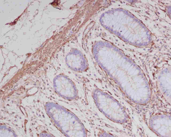
(Western blot analysis of alpha smooth muscle Actin expression in (1)A549 cell lysate; (2)C2C12 cell lysate using alpha-SMA antibody (AAA47018).Electrophoresis was performed on a 5-20% SDS-PAGE gel at 70V (Stacking gel) / 90V (Resolving gel) for 2-3 hours. The sample well of each lane was loaded with 50ug of sample under reducing conditions.After Electrophoresis, proteins were transferred to a Nitrocellulose membrane at 150mA for 50-90 minutes. Blocked the membrane with 5% Non-fat Milk/ TBS for 1.5 hour at RT. The membrane was incubated with rabbit anti-ACTA2 monoclonal antibody overnight at 4 degree C, then washed with TBS-0.1%Tween 3 times with 5 minutes each and probed with a goat anti-rabbit IgG-HRP secondary antibody at a dilution of 1:10000 for 1.5 hour at RT. The signal is developed using an Enhanced Chemiluminescent detection (ECL) kit with Tanon 5200 system. A specific band was detected for ACTA2)
WB (Western Blot)
(Western blot analysis of alpha smooth muscle Actin expression in (1)A549 cell lysate; (2)C2C12 cell lysate using alpha-SMA antibody (AAA47018).Electrophoresis was performed on a 5-20% SDS-PAGE gel at 70V (Stacking gel) / 90V (Resolving gel) for 2-3 hours. The sample well of each lane was loaded with 50ug of sample under reducing conditions.After Electrophoresis, proteins were transferred to a Nitrocellulose membrane at 150mA for 50-90 minutes. Blocked the membrane with 5% Non-fat Milk/ TBS for 1.5 hour at RT. The membrane was incubated with rabbit anti-ACTA2 monoclonal antibody overnight at 4 degree C, then washed with TBS-0.1%Tween 3 times with 5 minutes each and probed with a goat anti-rabbit IgG-HRP secondary antibody at a dilution of 1:10000 for 1.5 hour at RT. The signal is developed using an Enhanced Chemiluminescent detection (ECL) kit with Tanon 5200 system. A specific band was detected for ACTA2)
alpha smooth muscle Actin, Monoclonal Antibody (Cat# AAA47018)
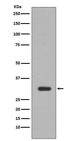 WB (Western Blot)
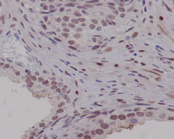
(Western blot analysis of PIM1 expression in Jurkat cell lysate (AAA47032).Electrophoresis was performed on a 5-20% SDS-PAGE gel at 70V (Stacking gel) / 90V (Resolving gel) for 2-3 hours. The sample well of each lane was loaded with 50ug of sample under reducing conditions.After Electrophoresis, proteins were transferred to a Nitrocellulose membrane at 150mA for 50-90 minutes. Blocked the membrane with 5% Non-fat Milk/ TBS for 1.5 hour at RT. The membrane was incubated with rabbit anti-PIM1 monoclonal antibody overnight at 4 degree C, then washed with TBS-0.1%Tween 3 times with 5 minutes each and probed with a goat anti-rabbit IgG-HRP secondary antibody at a dilution of 1:10000 for 1.5 hour at RT. The signal is developed using an Enhanced Chemiluminescent detection (ECL) kit with Tanon 5200 system. A specific band was detected for PIM1)
WB (Western Blot)
(Western blot analysis of PIM1 expression in Jurkat cell lysate (AAA47032).Electrophoresis was performed on a 5-20% SDS-PAGE gel at 70V (Stacking gel) / 90V (Resolving gel) for 2-3 hours. The sample well of each lane was loaded with 50ug of sample under reducing conditions.After Electrophoresis, proteins were transferred to a Nitrocellulose membrane at 150mA for 50-90 minutes. Blocked the membrane with 5% Non-fat Milk/ TBS for 1.5 hour at RT. The membrane was incubated with rabbit anti-PIM1 monoclonal antibody overnight at 4 degree C, then washed with TBS-0.1%Tween 3 times with 5 minutes each and probed with a goat anti-rabbit IgG-HRP secondary antibody at a dilution of 1:10000 for 1.5 hour at RT. The signal is developed using an Enhanced Chemiluminescent detection (ECL) kit with Tanon 5200 system. A specific band was detected for PIM1)
PIM1, Monoclonal Antibody (Cat# AAA47032)
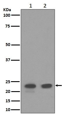 WB (Western Blot)
(Western blot analysis of CD9 expression in (1)HACAT cell lysate ;(2)HepG2 cell lysate (AAA47037).Electrophoresis was performed on a 5-20% SDS-PAGE gel at 70V (Stacking gel) / 90V (Resolving gel) for 2-3 hours. The sample well of each lane was loaded with 50ug of sample under reducing conditions.After Electrophoresis, proteins were transferred to a Nitrocellulose membrane at 150mA for 50-90 minutes. Blocked the membrane with 5% Non-fat Milk/ TBS for 1.5 hour at RT. The membrane was incubated with rabbit anti-CD9 monoclonal antibody overnight at 4 degree C, then washed with TBS-0.1%Tween 3 times with 5 minutes each and probed with a goat anti-rabbit IgG-HRP secondary antibody at a dilution of 1:10000 for 1.5 hour at RT. The signal is developed using an Enhanced Chemiluminescent detection (ECL) kit with Tanon 5200 system. A specific band was detected for CD9)
WB (Western Blot)
(Western blot analysis of CD9 expression in (1)HACAT cell lysate ;(2)HepG2 cell lysate (AAA47037).Electrophoresis was performed on a 5-20% SDS-PAGE gel at 70V (Stacking gel) / 90V (Resolving gel) for 2-3 hours. The sample well of each lane was loaded with 50ug of sample under reducing conditions.After Electrophoresis, proteins were transferred to a Nitrocellulose membrane at 150mA for 50-90 minutes. Blocked the membrane with 5% Non-fat Milk/ TBS for 1.5 hour at RT. The membrane was incubated with rabbit anti-CD9 monoclonal antibody overnight at 4 degree C, then washed with TBS-0.1%Tween 3 times with 5 minutes each and probed with a goat anti-rabbit IgG-HRP secondary antibody at a dilution of 1:10000 for 1.5 hour at RT. The signal is developed using an Enhanced Chemiluminescent detection (ECL) kit with Tanon 5200 system. A specific band was detected for CD9)
CD9, Monoclonal Antibody (Cat# AAA47037)
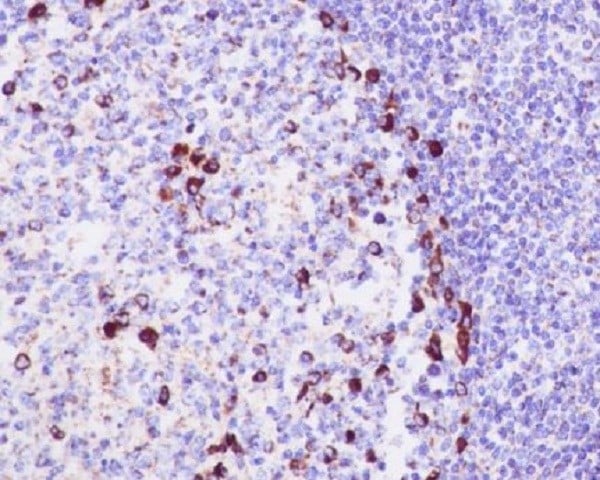
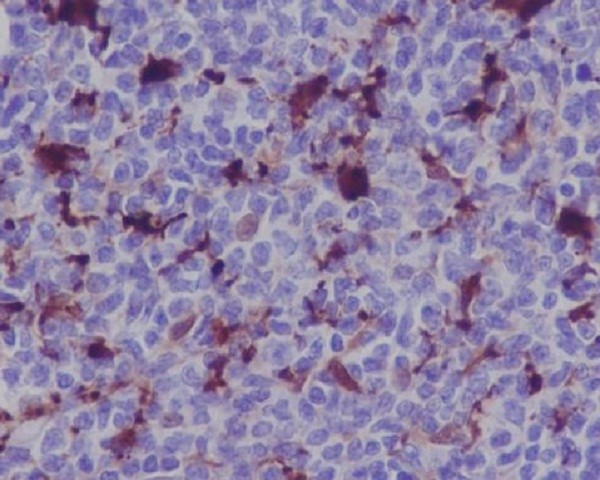 IHC (Immunohiostchemistry)
(Immunohistochemical analysis of paraffin-embedded human lymphoma, using MERTK Antibody(AAA46949)MERTK was detected in paraffin-embedded tissue section. Heat mediated antigen retrieval was performed in citrate buffer (pH6, epitope retrieval solution) for 20 mins. The tissue section was blocked with 10% goat serum. The tissue section was then incubated with 1ug/ml rabbit anti-MERTK Antibody (AAA46949)overnight at 4 degree C. Biotinylated goat anti-rabbit IgG was used as secondary antibody and incubated for 30 minutes at 37 degree C. The tissue section was developed using Strepavidin-Biotin-Complex (SABC) with DAB as the chromogen.)
IHC (Immunohiostchemistry)
(Immunohistochemical analysis of paraffin-embedded human lymphoma, using MERTK Antibody(AAA46949)MERTK was detected in paraffin-embedded tissue section. Heat mediated antigen retrieval was performed in citrate buffer (pH6, epitope retrieval solution) for 20 mins. The tissue section was blocked with 10% goat serum. The tissue section was then incubated with 1ug/ml rabbit anti-MERTK Antibody (AAA46949)overnight at 4 degree C. Biotinylated goat anti-rabbit IgG was used as secondary antibody and incubated for 30 minutes at 37 degree C. The tissue section was developed using Strepavidin-Biotin-Complex (SABC) with DAB as the chromogen.)
MERTK/Mer, Monoclonal Antibody (Cat# AAA46949)
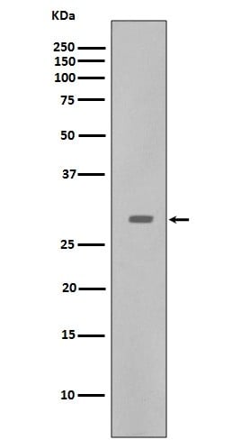
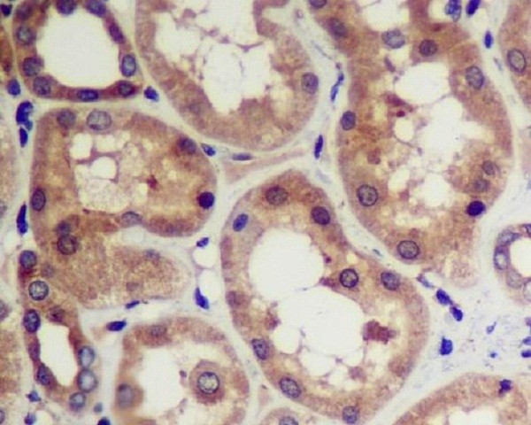 IHC (Immunohiostchemistry)
(Immunohistochemical analysis of paraffin-embedded human kidney, using CDK5 Antibody (AAA46951)CDK5 was detected in paraffin-embedded tissue section. Heat mediated antigen retrieval was performed in citrate buffer (pH6, epitope retrieval solution) for 20 mins. The tissue section was blocked with 10% goat serum. The tissue section was then incubated with 1ug/ml rabbit anti-CDK5 Antibody (AAA46951)overnight at 4 degree C. Biotinylated goat anti-rabbit IgG was used as secondary antibody and incubated for 30 minutes at 37 degree C. The tissue section was developed using Strepavidin-Biotin-Complex (SABC) with DAB as the chromogen.)
IHC (Immunohiostchemistry)
(Immunohistochemical analysis of paraffin-embedded human kidney, using CDK5 Antibody (AAA46951)CDK5 was detected in paraffin-embedded tissue section. Heat mediated antigen retrieval was performed in citrate buffer (pH6, epitope retrieval solution) for 20 mins. The tissue section was blocked with 10% goat serum. The tissue section was then incubated with 1ug/ml rabbit anti-CDK5 Antibody (AAA46951)overnight at 4 degree C. Biotinylated goat anti-rabbit IgG was used as secondary antibody and incubated for 30 minutes at 37 degree C. The tissue section was developed using Strepavidin-Biotin-Complex (SABC) with DAB as the chromogen.)
CDK5, Monoclonal Antibody (Cat# AAA46951)
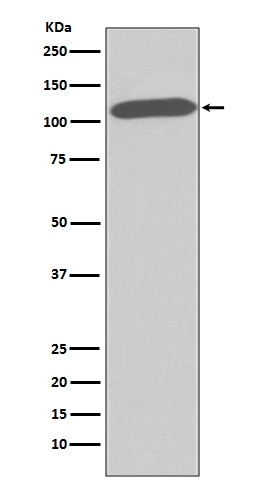 WB (Western Blot)
(Western blot analysis of CD26 expression in HUVEC cell lysate (AAA46965).Electrophoresis was performed on a 5-20% SDS-PAGE gel at 70V (Stacking gel) / 90V (Resolving gel) for 2-3 hours. The sample well of each lane was loaded with 50ug of sample under reducing conditions.After Electrophoresis, proteins were transferred to a Nitrocellulose membrane at 150mA for 50-90 minutes. Blocked the membrane with 5% Non-fat Milk/ TBS for 1.5 hour at RT. The membrane was incubated with rabbit anti-DPP4 monoclonal antibody overnight at 4 degree C, then washed with TBS-0.1%Tween 3 times with 5 minutes each and probed with a goat anti-rabbit IgG-HRP secondary antibody at a dilution of 1:10000 for 1.5 hour at RT. The signal is developed using an Enhanced Chemiluminescent detection (ECL) kit with Tanon 5200 system. A specific band was detected for DPP4)
WB (Western Blot)
(Western blot analysis of CD26 expression in HUVEC cell lysate (AAA46965).Electrophoresis was performed on a 5-20% SDS-PAGE gel at 70V (Stacking gel) / 90V (Resolving gel) for 2-3 hours. The sample well of each lane was loaded with 50ug of sample under reducing conditions.After Electrophoresis, proteins were transferred to a Nitrocellulose membrane at 150mA for 50-90 minutes. Blocked the membrane with 5% Non-fat Milk/ TBS for 1.5 hour at RT. The membrane was incubated with rabbit anti-DPP4 monoclonal antibody overnight at 4 degree C, then washed with TBS-0.1%Tween 3 times with 5 minutes each and probed with a goat anti-rabbit IgG-HRP secondary antibody at a dilution of 1:10000 for 1.5 hour at RT. The signal is developed using an Enhanced Chemiluminescent detection (ECL) kit with Tanon 5200 system. A specific band was detected for DPP4)
CD26, Monoclonal Antibody (Cat# AAA46965)
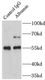
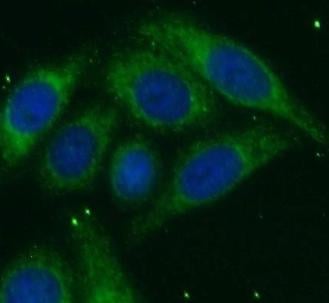
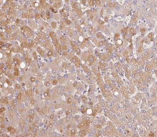
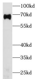 WB (Western Blot)
(human plasma (diluted 5000 fold) was subjected to SDS PAGE followed by western blot with AAA102676 (Albumin Antibody) at dilution of 1:10000)
WB (Western Blot)
(human plasma (diluted 5000 fold) was subjected to SDS PAGE followed by western blot with AAA102676 (Albumin Antibody) at dilution of 1:10000)
Albumin, Monoclonal Antibody (Cat# AAA102676)
Purification: Protein A+G purification
 WB (Western Blot)
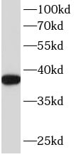
(human brain tissue were subjected to SDS PAGE followed by western blot with AAA102689 (ANXA2 antibody) at dilution of 1:1000)
WB (Western Blot)
(human brain tissue were subjected to SDS PAGE followed by western blot with AAA102689 (ANXA2 antibody) at dilution of 1:1000)
Annexin A2, Monoclonal Antibody (Cat# AAA102689)
Protein A+G purification
 WB (Western Blot)
(Western Blot analysis of Citrulline-BSA Conjugate showing detection of 67 kDa Citrulline protein using Mouse Anti-Citrulline Monoclonal Antibody, Clone 2D3-1B9. Lane 1: Molecular Weight Ladder (MW). Lane 2: BSA (0.5 ug). Lane 3: BSA (2.0 ug). Lane 4: Citrulline-BSA (0.5 ug). Lane 5: Citrulline-BSA (2.0 ug). Block: 5% Skim Milk in TBST. Primary Antibody: Mouse Anti-Citrulline Monoclonal Antibody at 1:1000 for 2 hours at RT. Secondary Antibody: Goat Anti-Mouse IgG: HRP at 1:2000 for 60 min at RT. Color Development: ECL solution for 5 min in RT. Predicted/Observed Size: 67 kDa.)
WB (Western Blot)
(Western Blot analysis of Citrulline-BSA Conjugate showing detection of 67 kDa Citrulline protein using Mouse Anti-Citrulline Monoclonal Antibody, Clone 2D3-1B9. Lane 1: Molecular Weight Ladder (MW). Lane 2: BSA (0.5 ug). Lane 3: BSA (2.0 ug). Lane 4: Citrulline-BSA (0.5 ug). Lane 5: Citrulline-BSA (2.0 ug). Block: 5% Skim Milk in TBST. Primary Antibody: Mouse Anti-Citrulline Monoclonal Antibody at 1:1000 for 2 hours at RT. Secondary Antibody: Goat Anti-Mouse IgG: HRP at 1:2000 for 60 min at RT. Color Development: ECL solution for 5 min in RT. Predicted/Observed Size: 67 kDa.)
Citrulline, Monoclonal Antibody (Cat# AAA103963)
 WB (Western Blot)
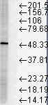
(Western Blot analysis of Human Cervical cancer cell line (HeLa) lysate showing detection of FKBP51 protein using Mouse Anti-FKBP51 Monoclonal Antibody, Clone Hi51B. Load: 15 ug. Block: 1.5% BSA for 30 minutes at RT. Primary Antibody: Mouse Anti-FKBP51 Monoclonal Antibody at 1:1000 for 2 hours at RT. Secondary Antibody: Sheep Anti-Mouse IgG: HRP for 1 hour at RT.)
WB (Western Blot)
(Western Blot analysis of Human Cervical cancer cell line (HeLa) lysate showing detection of FKBP51 protein using Mouse Anti-FKBP51 Monoclonal Antibody, Clone Hi51B. Load: 15 ug. Block: 1.5% BSA for 30 minutes at RT. Primary Antibody: Mouse Anti-FKBP51 Monoclonal Antibody at 1:1000 for 2 hours at RT. Secondary Antibody: Sheep Anti-Mouse IgG: HRP for 1 hour at RT.)
FKBP51, Monoclonal Antibody (Cat# AAA103420)
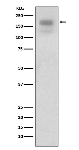
 WB (Western Blot)
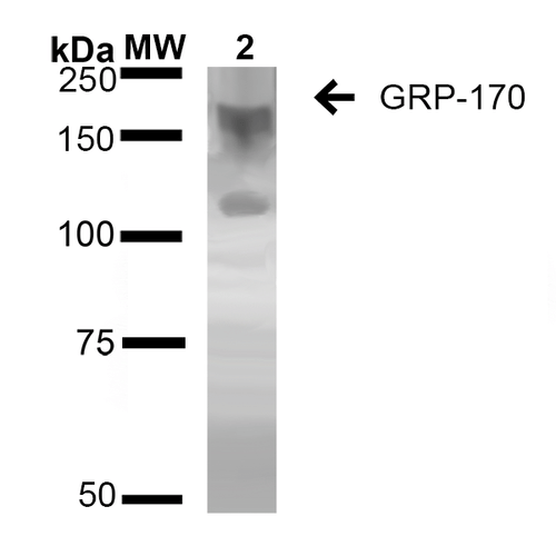
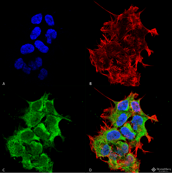
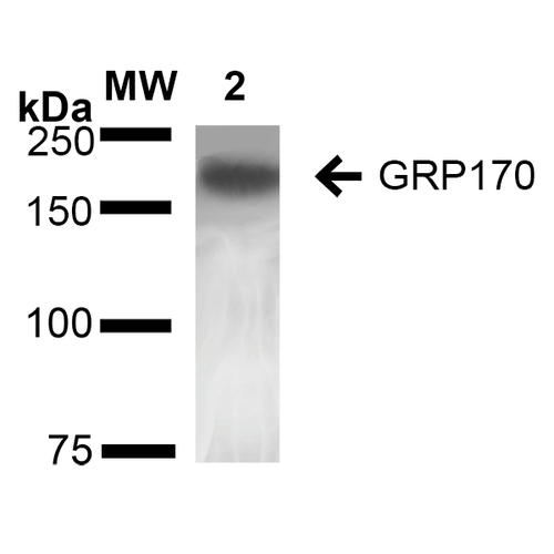
(Western Blot analysis of Rat Liver showing detection of ~170 kDa GRP170 protein using Mouse Anti-GRP170 Monoclonal Antibody, Clone 6E3-2C3 . Lane 1: Molecular Weight Ladder (MW). Lane 2: Rat Liver cell lysate. Load: 20 ug. Block: 2% BSA and 2% Skim Milk in 1X TBST. Primary Antibody: Mouse Anti-GRP170 Monoclonal Antibody at 1:1000 for 16 hours at 4 degree C. Secondary Antibody: Goat Anti-Mouse IgG: HRP at 1:100 for 60 min at RT. Color Development: ECL solution for 6 min in RT. Predicted/Observed Size: ~170 kDa.)
WB (Western Blot)
(Western Blot analysis of Rat Liver showing detection of ~170 kDa GRP170 protein using Mouse Anti-GRP170 Monoclonal Antibody, Clone 6E3-2C3 . Lane 1: Molecular Weight Ladder (MW). Lane 2: Rat Liver cell lysate. Load: 20 ug. Block: 2% BSA and 2% Skim Milk in 1X TBST. Primary Antibody: Mouse Anti-GRP170 Monoclonal Antibody at 1:1000 for 16 hours at 4 degree C. Secondary Antibody: Goat Anti-Mouse IgG: HRP at 1:100 for 60 min at RT. Color Development: ECL solution for 6 min in RT. Predicted/Observed Size: ~170 kDa.)
GRP170, Monoclonal Antibody (Cat# AAA103793)
What are Monoclonal Antibodies?
Monoclonal antibodies are specialized laboratory-produced proteins developed for binding to specific biological antigens or other molecular targets. Since they come from a single cell (or clone), they are especially consistent and accurate in the data they are involved in producing.
This type of antibody material has been shown to be a powerful tool in finding and subsequently destroying harmful cells in an organism, such as those found in cancers or various autoimmune diseases. This makes them excellent aids in medical testing and research, which is why they are so widely used.
AAA Biotech offers a comprehensive range of high-quality monoclonal antibodies that perform effectively in various laboratory tests, including (amongst others) ELISA, western blotting, immunohistochemistry, and flow cytometry. All of the products in our catalog are thoroughly quality tested to make sure that they are reliable and will consistently perform well in your research.
What Are The Uses of Monoclonal Antibodies
Monoclonal antibodies are used in many lab tests, including (amongst others) ELISA, western blotting, immunohistochemistry, and flow cytometry.
ELISA is a test that helps detect a specific substance/analyte in a sample. It uses antibodies (often monoclonal) bound to a solid surface (such as the well of a microplate) to “capture” the substance/analyte in the sample and immobilize it so that the detection antibody component can then bind to it and produce a signal, which can then be measured.
Western blotting identifies specific proteins in a sample. The sample is first separated on a gel, and then antibodies are applied that will typically bind to the target, which will all be localized to a single band in a lane.
Immunohistochemistry helps locate specific proteins in cells or tissue samples using antibodies.
Flow cytometry looks at and sorts cells. It uses antibodies that are conjugated to reporter molecules called “fluorophores”, which, under special lights, emit light themselves, which can then be measured by a detector instrument. For a deeper understanding of these techniques, explore our complete guide to monoclonal antibodies and their benefits.
How Monoclonal Antibodies Are Used as Medicine?
Please note that all of the products listed in AAA Biotech’s also known as AAA Bio or AAABio catalog are strictly for research-use only (RUO).
Monoclonal antibodies can also be used as therapeutic/medical treatments, particularly in the context of cancers. They are designed to find and bind to specific cells or proteins, helping the immune system recognize and attack the cancer. These treatments work in different ways, such as:
- Radioimmunotherapy attaches a small amount of radioactive molecule to the antibody, so it delivers the radiation directly to the cancer cells that the antibody is specifically binding to.
- Antibody-directed enzyme prodrug therapy uses antibodies that are specifically bound to special enzymes. These enzymes activate a harmless drug in the body and turn it into a cancer-killing drug only near the cancer cells—this helps avoid harming healthy cells.
- Immunoliposomes are tiny “bubbles” filled with medicine/drug and coated with antibodies. They carry the drug straight to the cancer cells.
Why Buy Monoclonal Antibodies From Us?
At AAA Biotech, we provide high-performance monoclonal antibodies designed to support a wide range of research needs.
1. Validated for Versatile Applications
The antibodies in our catalog are extensively validated and compatible with multiple techniques, including (but not limited to) ELISA, flow cytometry (FC), immunocytochemistry (ICC), immunofluorescence (IF), immunohistochemistry (IHC), immunoprecipitation (IP), and western blotting (WB).
2. Wide Selection & Specialized Options
We offer antibodies for common and rare species, that are available in various conjugated forms, and also in recombinant formats. Essentially, there is almost anything one might need to meet their experimental model’s requirements.
3. High-Quality Proteins
Our proteins meet high purity standards—90% or more as confirmed by SDS-PAGE. Many are available with tags like His, Flag, GST, or MBP, and we also supply native and biologically active proteins for functional studies.
Frequently Asked Questions
1. Are your monoclonal antibodies validated for specific applications?
Yes, our antibodies are tested and validated for use in methods such as ELISA, western blot, IHC, flow cytometry, and more. Refer to specific product pages or datasheets for individual product information.
2. How do I choose the right monoclonal antibody for my application?
Review the product details directly for application validation, species reactivity, and target information. You may also contact our support team at any time for help.
3. How quickly can I receive my order?
Most orders are processed and shipped within 1–3 business days, depending on product availability and your shipping location.