

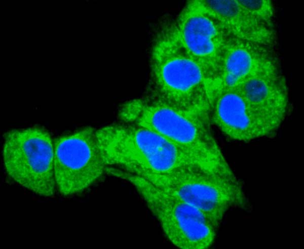


 ICC (Immunocytochemistry)
(ICC staining Calnexin in PANC-1 cells (green). The nuclear counter stain is DAPI (blue). Cells were fixed in paraformaldehyde, permeabilised with 0.25% Triton X100/PBS.)
ICC (Immunocytochemistry)
(ICC staining Calnexin in PANC-1 cells (green). The nuclear counter stain is DAPI (blue). Cells were fixed in paraformaldehyde, permeabilised with 0.25% Triton X100/PBS.)
Rabbit anti-Human, Rat Calnexin Monoclonal Antibody | anti-CANX antibody
Calnexin Antibody
IHC: 1:50-1:200
ICC: 1:100-1:500
FC/FACS: 1:50-1:100
ICC (Immunocytochemistry)
(ICC staining Calnexin in PANC-1 cells (green). The nuclear counter stain is DAPI (blue). Cells were fixed in paraformaldehyde, permeabilised with 0.25% Triton X100/PBS.)
ICC (Immunocytochemistry)
(ICC staining Calnexin in PANC-1 cells (green). The nuclear counter stain is DAPI (blue). Cells were fixed in paraformaldehyde, permeabilised with 0.25% Triton X100/PBS.)
ICC (Immunocytochemistry)
(ICC staining Calnexin in HepG2 cells (green). The nuclear counter stain is DAPI (blue). Cells were fixed in paraformaldehyde, permeabilised with 0.25% Triton X100/PBS.)
ICC (Immunocytochemistry)
(ICC staining Calnexin in HepG2 cells (green). The nuclear counter stain is DAPI (blue). Cells were fixed in paraformaldehyde, permeabilised with 0.25% Triton X100/PBS.)
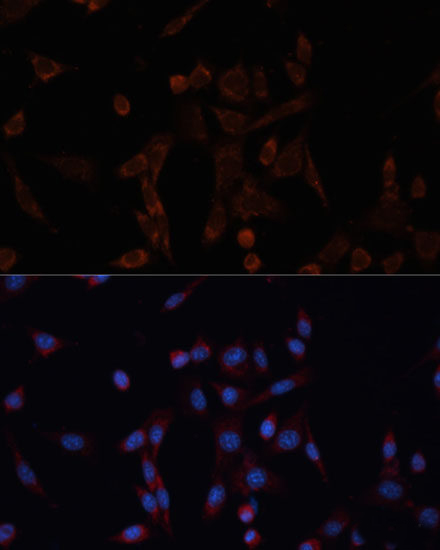
ICC (Immunocytochemistry)
(ICC staining Calnexin in Hela cells (green). The nuclear counter stain is DAPI (blue). Cells were fixed in paraformaldehyde, permeabilised with 0.25% Triton X100/PBS.)
ICC (Immunocytochemistry)
(ICC staining Calnexin in Hela cells (green). The nuclear counter stain is DAPI (blue). Cells were fixed in paraformaldehyde, permeabilised with 0.25% Triton X100/PBS.)
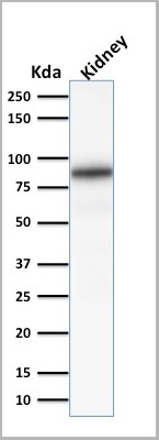
IHC (Immunohistochemistry)
(Immunohistochemical analysis of paraffin-embedded human kidney tissue using anti-Calnexin antibody. Counter stained with hematoxylin.)
IHC (Immunohistochemistry)
(Immunohistochemical analysis of paraffin-embedded human kidney tissue using anti-Calnexin antibody. Counter stained with hematoxylin.)
IHC (Immunohistchemistry)
(Immunohistochemical analysis of paraffin-embedded rat kidney tissue using anti-Calnexin antibody. Counter stained with hematoxylin.)
IHC (Immunohistchemistry)
(Immunohistochemical analysis of paraffin-embedded rat kidney tissue using anti-Calnexin antibody. Counter stained with hematoxylin.)
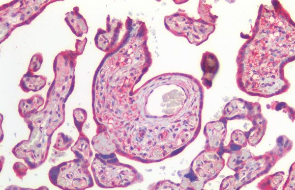
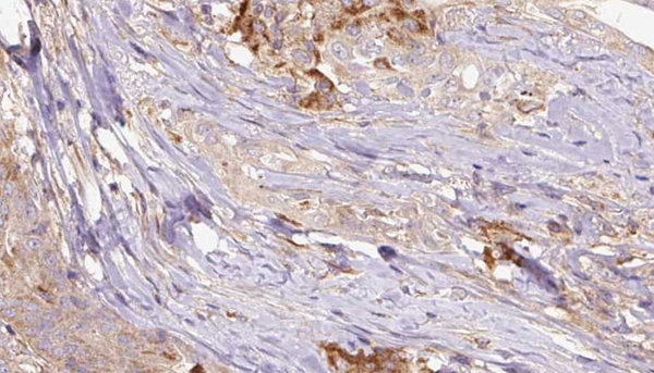
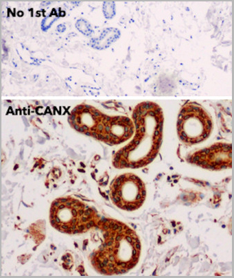
IHC (Immunohistochemistry)
(Immunohistochemical analysis of paraffin-embedded human liver cancer tissue using anti-Calnexin antibody. Counter stained with hematoxylin.)
IHC (Immunohistochemistry)
(Immunohistochemical analysis of paraffin-embedded human liver cancer tissue using anti-Calnexin antibody. Counter stained with hematoxylin.)
IHC (Immunohistochemistry)
(Immunohistochemical analysis of paraffin-embedded rat pancreas tissue using anti-Calnexin antibody. Counter stained with hematoxylin.)
IHC (Immunohistochemistry)
(Immunohistochemical analysis of paraffin-embedded rat pancreas tissue using anti-Calnexin antibody. Counter stained with hematoxylin.)
IHC (Immunohistochemistry)
(Immunohistochemical analysis of paraffin-embedded rat heart tissue using anti-Calnexin antibody. Counter stained with hematoxylin.)
IHC (Immunohistochemistry)
(Immunohistochemical analysis of paraffin-embedded rat heart tissue using anti-Calnexin antibody. Counter stained with hematoxylin.)
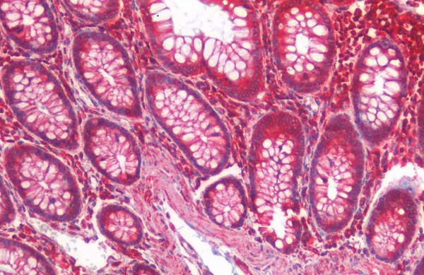
IHC (Immunohistochemistry)
(Immunohistochemical analysis of paraffin-embedded human pancreas tissue using anti-Calnexin antibody. Counter stained with hematoxylin.)
IHC (Immunohistochemistry)
(Immunohistochemical analysis of paraffin-embedded human pancreas tissue using anti-Calnexin antibody. Counter stained with hematoxylin.)
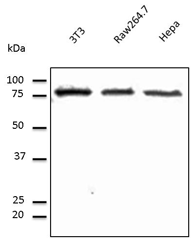
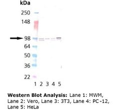
WB (Western Blot)
(Western blot analysis of Calnexin on Hela cells lysates using anti-Calnexin antibody at 1/1, 000 dilution.)
WB (Western Blot)
(Western blot analysis of Calnexin on Hela cells lysates using anti-Calnexin antibody at 1/1, 000 dilution.)
NCBI and Uniprot Product Information
Customer Reviews
Loading reviews...
Share Your Experience
Similar Products


Product Notes
The CANX canx (Catalog #AAA30180) is an Antibody produced from Rabbit and is intended for research purposes only. The product is available for immediate purchase. The Calnexin Antibody reacts with Human, Rat and may cross-react with other species as described in the data sheet. AAA Biotech's Calnexin can be used in a range of immunoassay formats including, but not limited to, WB (Western Blot), ICC (Immunocytochemistry), IF (Immunofluorescence), IHC (Immunohistochemistry), FCM/FACS (Flow Cytometry). WB: 1:1000-5000 IHC: 1:50-1:200 ICC: 1:100-1:500 FC/FACS: 1:50-1:100. Researchers should empirically determine the suitability of the CANX canx for an application not listed in the data sheet. Researchers commonly develop new applications and it is an integral, important part of the investigative research process. It is sometimes possible for the material contained within the vial of "Calnexin, Monoclonal Antibody" to become dispersed throughout the inside of the vial, particularly around the seal of said vial, during shipment and storage. We always suggest centrifuging these vials to consolidate all of the liquid away from the lid and to the bottom of the vial prior to opening. Please be advised that certain products may require dry ice for shipping and that, if this is the case, an additional dry ice fee may also be required.Precautions
All products in the AAA Biotech catalog are strictly for research-use only, and are absolutely not suitable for use in any sort of medical, therapeutic, prophylactic, in-vivo, or diagnostic capacity. By purchasing a product from AAA Biotech, you are explicitly certifying that said products will be properly tested and used in line with industry standard. AAA Biotech and its authorized distribution partners reserve the right to refuse to fulfill any order if we have any indication that a purchaser may be intending to use a product outside of our accepted criteria.Disclaimer
Though we do strive to guarantee the information represented in this datasheet, AAA Biotech cannot be held responsible for any oversights or imprecisions. AAA Biotech reserves the right to adjust any aspect of this datasheet at any time and without notice. It is the responsibility of the customer to inform AAA Biotech of any product performance issues observed or experienced within 30 days of receipt of said product. To see additional details on this or any of our other policies, please see our Terms & Conditions page.Frequently Asked Questions
What is calnexin used as a marker for in cell biology studies?
Calnexin is an endoplasmic reticulum (ER) chaperone protein and an accepted marker for ER localization. It facilitates quality control of newly synthesized glycoproteins through transient interaction with N-linked glycans. In cell biology, calnexin marks the ER compartment, monitors ER stress responses and unfolded protein response (UPR) activation, and evaluates protein trafficking and ER-associated degradation pathways in cellular stress models.
Can this calnexin antibody detect both human and rat proteins?
Verify cross-reactivity specifications on the AAA Biotech datasheet, as species reactivity varies by antibody clone. Many calnexin antibodies recognize human, mouse, and rat epitopes due to sequence conservation (~90% identity). Confirm reactivity through Western blot or immunofluorescence pre-validation on your target species. Some antibodies may have species-specific limitations requiring optimization or alternative clones.
Is calnexin antibody suitable as an ER (endoplasmic reticulum) marker in immunofluorescence?
Yes, calnexin antibodies are widely validated for immunofluorescence (IF) ER marking. Use formaldehyde or methanol fixation; calnexin tolerates both. The antibody produces characteristic reticular ER staining patterns when proper permeabilization is performed. Optimal results require permeabilization buffers (0.1-0.5% Triton X-100) to access intracellular epitopes. Co-staining with other ER markers validates specificity.
Can this antibody be used for Western blot to detect calnexin expression?
Yes, calnexin antibodies are suitable for Western blotting to quantify calnexin protein expression (~90 kDa). Calnexin is highly stable across tissues and conditions, serving as a reliable loading control. Use standard wet transfer protocols; expected molecular weight aids band identification. Recommend 1:1000-1:5000 primary antibody dilutions. Calnexin levels remain relatively constant, making it an excellent housekeeping protein reference.
Does fixation method affect calnexin staining quality in IHC/IF?
Yes, fixation critically impacts staining. Formaldehyde fixation (formalin-fixed paraffin-embedded) requires heat-induced epitope retrieval (HIER) to unmask cross-linked epitopes. Methanol or ethanol fixation preserves antigenicity better without retrieval. Fixation duration influences epitope accessibility; prolonged fixation increases cross-linking, requiring longer retrieval times. Optimize fixation parameters for your specific application.
Is calnexin expression affected by ER stress or unfolded protein response?
Interestingly, calnexin expression decreases during ER stress despite initial mRNA upregulation. Paradoxically, calnexin protein levels decline during unfolded protein response (UPR) activation via PERK, ATF6, and IRE1α pathways. This counterintuitive reduction may impair protein quality control during prolonged stress. Calnexin depletion heightens susceptibility to ER stress, making it a sensitive UPR biomarker.
Item has been added to Shopping Cart
If you are ready to order, navigate to Shopping Cart and get ready to checkout.