


 Application Data
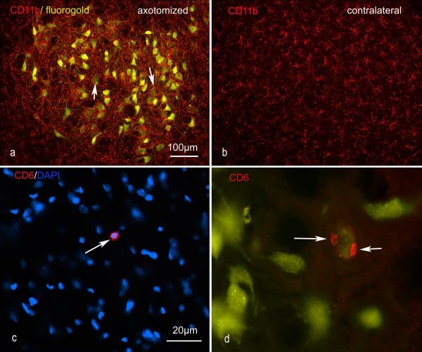
(Published customer image: Representative images of the inflammatory changes in the facial nucleus during axonal regeneration, one week following facial nerve transaction. a, b: CD11b immunoreactivity for microglia is increased in the axotomized facial nucleus, and microglia enwrap the facial motor neurons, e.g. at arrows. The regenerating neurons were retrogradely labelled with fluorogold. c, d: CD6- positive T-cells accumulated in the injured motor nucleus (arrows). They had little cytoplasm but dense nuclei (c) and were sometimes clustered around neurons retrogradely labelled with fluorogold (d). The scale bar in (a) also applies to (b) and that in (c) also applies to (d).From: Shokouhi et al. BMC Neuroscience 2010 11:13.)
Application Data
(Published customer image: Representative images of the inflammatory changes in the facial nucleus during axonal regeneration, one week following facial nerve transaction. a, b: CD11b immunoreactivity for microglia is increased in the axotomized facial nucleus, and microglia enwrap the facial motor neurons, e.g. at arrows. The regenerating neurons were retrogradely labelled with fluorogold. c, d: CD6- positive T-cells accumulated in the injured motor nucleus (arrows). They had little cytoplasm but dense nuclei (c) and were sometimes clustered around neurons retrogradely labelled with fluorogold (d). The scale bar in (a) also applies to (b) and that in (c) also applies to (d).From: Shokouhi et al. BMC Neuroscience 2010 11:13.)
Mouse CD11b Monoclonal Antibody | anti-CD11b antibody
MOUSE ANTI RAT CD11b
Purified IgG - liquid
Immunohistology - Frozen: Minimum Dilution: 1/50; Maximum Dilution: 1/100
Flow Cytometry: Minimum Dilution: 1/50; Maximum Dilution: 1/100
Preparation
Shelf Life: 18 months from date of despatch.
Application Data
(Published customer image: Representative images of the inflammatory changes in the facial nucleus during axonal regeneration, one week following facial nerve transaction. a, b: CD11b immunoreactivity for microglia is increased in the axotomized facial nucleus, and microglia enwrap the facial motor neurons, e.g. at arrows. The regenerating neurons were retrogradely labelled with fluorogold. c, d: CD6- positive T-cells accumulated in the injured motor nucleus (arrows). They had little cytoplasm but dense nuclei (c) and were sometimes clustered around neurons retrogradely labelled with fluorogold (d). The scale bar in (a) also applies to (b) and that in (c) also applies to (d).From: Shokouhi et al. BMC Neuroscience 2010 11:13.)
Application Data
(Published customer image: CD11b immunoreactive (red) microglia in the motor cortex following cervical corticospinal tract injury. Representative images of motor cortex - a, b: one week (deconvolved images); c, d: two weeks following corticospinal tract injury. The corticospinal neurons were retrogradely labelled with fluorogold applied to the spinal cord at the time of injury. There is little sign of microglial activation. The inset in (c) shows a CD6-positive T-cell in the motor cortex, but no accumulation of such cells was detected.From: Shokouhi BN, Wong BZ, Siddiqui S, Lieberman AR, Campbell G, Tohyama K, Anderson PN. Microglial responses around intrinsic CNS neurons are correlated with axonal regeneration. BMC Neurosci. 2010 Feb 5;11:13.)
Application Data
(Staining of rat peritoneal macrophages with Mouse anti Rat CD11b)
Application Data
(Staining of rat peritoneal macrophages with Mouse anti Rat CD11b)
Application Data
(Published customer image: Microglia in the red nucleus (a-d and inset) and motor cortex (e, f) 4 weeks after nerve graft implantation into the rubrospinal tract and cervical dorsal columns respectively. Neurons with regenerating axons were retrogradely labelled with fluorogold. Microglia established close contacts with regenerating rubrospinal neurons (arrows), which also expressed ATF3 (inset). No retrogradely labelled corticospinal neurons were detected (e and f) and there was no microglial activation in the motor cortex. (a-c) are non-deconvolved and (d-f plus inset) are deconvolved images. The scale bar in (a) also applies to (b).From: Shokouhi BN, Wong BZ, Siddiqui S, Lieberman AR, Campbell G, Tohyama K, Anderson PN. Microglial responses around intrinsic CNS neurons are correlated with axonal regeneration. BMC Neurosci. 2010 Feb 5;11:13.)
Application Data
(Immunoperoxidase staining of rat lymph node cryosection with Mouse anti Rat CD11b antibody followed by horseradish peroxidase conjugated Goat anti Mouse IgG2a as a detection reagent. High power)
Application Data
(Immunoperoxidase staining of rat lymph node cryosection with Mouse anti Rat CD11b antibody followed by horseradish peroxidase conjugated Goat anti Mouse IgG2a as a detection reagent. High power)
Application Data
(Staining of rat peritoneal macrophages cells with Mouse anti Rat CD11b:Alexa Fluor 488)
Application Data
(Staining of rat peritoneal macrophages cells with Mouse anti Rat CD11b:Alexa Fluor 488)
Application Data
(Published customer image: Rubrospinal injury produces little microglial activation and no accumulation of T-cells in the red nucleus. a, b: Representative images show there is little difference in CD11b immunoreactivity for microglia in the red nucleus (*) one week following axotomy. c, d: confocal images of beta thymosin immunoreactive microglia in the red nucleus 3 weeks after injury. e: a rare CD6-positive T lymphocyte in the red nucleus of an unoperated rat. f: one week after injury no T lymphocytes can be identified in the red nucleus around the axotomized rubrospinal neurons, which can be recognised by their expression of ATF3 (green). Neuronal cytoplasm has been visualised by high gain in the red signal; this does not represent CD6 signal.From: Shokouhi BN, Wong BZ, Siddiqui S, Lieberman AR, Campbell G, Tohyama K, Anderson PN. Microglial responses around intrinsic CNS neurons are correlated with axonal regeneration. BMC Neurosci. 2010 Feb 5;11:13.)
Application Data
(Staining of rat peritoneal macrophages with Mouse anti Rat CD11b:RPE-Alexa Fluor 647)
Application Data
(Staining of rat peritoneal macrophages with Mouse anti Rat CD11b:RPE-Alexa Fluor 647)
Application Data
(Immunofluoescence staining of rat lymph node cryosection with Mouse anti Rat CD11b antibody , red in A and Mouse anti Rat CD8 , green in B. C is the merged image with nuclei counter-stained in blue using DAPI. High power)
Application Data
(Immunofluoescence staining of rat lymph node cryosection with Mouse anti Rat CD11b antibody , red in A and Mouse anti Rat CD8 , green in B. C is the merged image with nuclei counter-stained in blue using DAPI. High power)
Application Data
(Immunofluoescence staining of rat lymph node cryosection with Mouse anti Rat CD11b antibody , red in A and Mouse anti Rat CD8 , green in B. C is the merged image with nuclei counter-stained in blue using DAPI. Low power)
Application Data
(Immunofluoescence staining of rat lymph node cryosection with Mouse anti Rat CD11b antibody , red in A and Mouse anti Rat CD8 , green in B. C is the merged image with nuclei counter-stained in blue using DAPI. Low power)
NCBI and Uniprot Product Information
Similar Products


Product Notes
The CD11b cd11b (Catalog #AAA11969) is an Antibody produced from Mouse and is intended for research purposes only. The product is available for immediate purchase. AAA Biotech's CD11b can be used in a range of immunoassay formats including, but not limited to, Immunohistology Frozen, Flow cytometry (FC/FACS), Immunofluorescence (IF), Immunoprecipitation (IP). Flow Cytometry: Use 10ul of the suggested working dilution to label 106 cells in 100ul. Immunohistology - Frozen: Minimum Dilution: 1/50; Maximum Dilution: 1/100 Flow Cytometry: Minimum Dilution: 1/50; Maximum Dilution: 1/100. Researchers should empirically determine the suitability of the CD11b cd11b for an application not listed in the data sheet. Researchers commonly develop new applications and it is an integral, important part of the investigative research process. It is sometimes possible for the material contained within the vial of "CD11b, Monoclonal Antibody" to become dispersed throughout the inside of the vial, particularly around the seal of said vial, during shipment and storage. We always suggest centrifuging these vials to consolidate all of the liquid away from the lid and to the bottom of the vial prior to opening. Please be advised that certain products may require dry ice for shipping and that, if this is the case, an additional dry ice fee may also be required.Precautions
All products in the AAA Biotech catalog are strictly for research-use only, and are absolutely not suitable for use in any sort of medical, therapeutic, prophylactic, in-vivo, or diagnostic capacity. By purchasing a product from AAA Biotech, you are explicitly certifying that said products will be properly tested and used in line with industry standard. AAA Biotech and its authorized distribution partners reserve the right to refuse to fulfill any order if we have any indication that a purchaser may be intending to use a product outside of our accepted criteria.Disclaimer
Though we do strive to guarantee the information represented in this datasheet, AAA Biotech cannot be held responsible for any oversights or imprecisions. AAA Biotech reserves the right to adjust any aspect of this datasheet at any time and without notice. It is the responsibility of the customer to inform AAA Biotech of any product performance issues observed or experienced within 30 days of receipt of said product. To see additional details on this or any of our other policies, please see our Terms & Conditions page.Item has been added to Shopping Cart
If you are ready to order, navigate to Shopping Cart and get ready to checkout.