
 Application Data
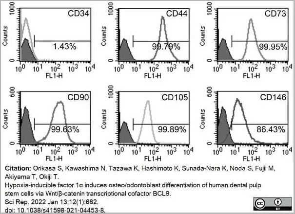
(Published customer image:Mouse anti Human CD146 antibody, clone OJ79c used to assess CD146 expression on cultured mesenchymal stem cells by flow cytometry.Image caption:hDPSCs express typical mesenchymal stem cell (MSC) markers and exhibit multi-differentiation potential. (a) Typical MSC markers (CD44, CD73, CD90, CD105, CD146) are highly expressed in hDPSCs, and hDPSCs are mostly negative for a hematopoietic marker (CD34). hDPSCs possess neurogenic, adipogenic, chondrogenic, and osteogenic differentiation potential, as determined by the expression of neurogenic markers (GFAP and NF-M)From: Orikasa S, Kawashima N, Tazawa K, Hashimoto K, Sunada-Nara K, Noda S, Fujii M, Akiyama T, Okiji T.Hypoxia-inducible factor 1? induces osteo/odontoblast differentiation of human dental pulp stem cells via Wnt/?-catenin transcriptional cofactor BCL9.)
Application Data
(Published customer image:Mouse anti Human CD146 antibody, clone OJ79c used to assess CD146 expression on cultured mesenchymal stem cells by flow cytometry.Image caption:hDPSCs express typical mesenchymal stem cell (MSC) markers and exhibit multi-differentiation potential. (a) Typical MSC markers (CD44, CD73, CD90, CD105, CD146) are highly expressed in hDPSCs, and hDPSCs are mostly negative for a hematopoietic marker (CD34). hDPSCs possess neurogenic, adipogenic, chondrogenic, and osteogenic differentiation potential, as determined by the expression of neurogenic markers (GFAP and NF-M)From: Orikasa S, Kawashima N, Tazawa K, Hashimoto K, Sunada-Nara K, Noda S, Fujii M, Akiyama T, Okiji T.Hypoxia-inducible factor 1? induces osteo/odontoblast differentiation of human dental pulp stem cells via Wnt/?-catenin transcriptional cofactor BCL9.)
Mouse CD146 Monoclonal Antibody | anti-CD146 antibody
MOUSE ANTI HUMAN CD146:RPE
ALEXA FLUOR 647, FITC, Preservative Free, Purified, RPE
Phosphate buffered saline
0.09% Sodium Azide (NaN3)
1% Bovine Serum Albumin
5% Sucrose
Care should be taken during reconstitution as the protein may appear as a film at the bottom of the vial. We recommend that the vial is gently mixed after reconstitution.
Application Data
(Published customer image:Mouse anti Human CD146 antibody, clone OJ79c used to assess CD146 expression on cultured mesenchymal stem cells by flow cytometry.Image caption:hDPSCs express typical mesenchymal stem cell (MSC) markers and exhibit multi-differentiation potential. (a) Typical MSC markers (CD44, CD73, CD90, CD105, CD146) are highly expressed in hDPSCs, and hDPSCs are mostly negative for a hematopoietic marker (CD34). hDPSCs possess neurogenic, adipogenic, chondrogenic, and osteogenic differentiation potential, as determined by the expression of neurogenic markers (GFAP and NF-M)From: Orikasa S, Kawashima N, Tazawa K, Hashimoto K, Sunada-Nara K, Noda S, Fujii M, Akiyama T, Okiji T.Hypoxia-inducible factor 1? induces osteo/odontoblast differentiation of human dental pulp stem cells via Wnt/?-catenin transcriptional cofactor BCL9.)
Application Data
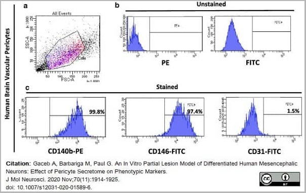
(Published customer image:FITC conjugated Mouse anti Human CD146 antibody, clone OJ79c used to evaluate CD146 expression on human brain vascular pericytes by flow cytometry.Image caption:Characterisation of human brain vascular pericytes by flow cytometry. (A) Cells were identified according to forward scatter (FSC) and side scatter (SSC) intensity. (B) Histograms of unstained human brain pericytes. (C) Histograms represent the fluorescence intensity of human brain pericytes after incubation with CD140b, CD146, and CD31 antibodies, respectively. (3 independent experiments, 3 replicates))
Application Data
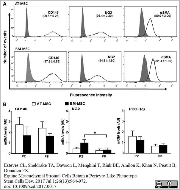
(Published customer image:Alexa-Fluor®647 conjugated Mouse anti Human CD146 antibody, clone OJ79c used to assess CD146 nexpression on adipose tissue-derived and mone marrow-derived mesenchymal stem cells by flow cytometry.Image caption:(A) Flow cytometry histograms showing the proportions of AT-MSCs and BM-MSCs (upper and lower panels, respectively) that are positive for staining with the antibodies CD146-AF647, NG2-APC, and ?SMA (with secondary AF405 conjugated). Isotype controls are shown by the gray peak and unstained curves are omitted for simplicity. (B) Results of qPCR analysis of CD146, NG2, and PDGFR? in AT-MSCs and BM-MSCs at passages 2 (P2) and 8 (P8). All results are shown as mean?±?SEM; n?=?3 animals. *P?)
Application Data
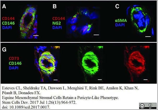
(Published customer image:FITC conjugated Mouse anti CD146 antibody, clone OJ79c used to demonstrate CD146 expressing equine pericytes by immunofluorescence.Image caption:IHC of equine AT sections showing staining for the pericyte markers, CD146 (A), NG2 (B), ?SMA (C),(red; A, B ). Colocalization of MSC markers CD146 and CD73; F, G, in yellow (left panel) from the overlap of green (middle panel) and red (right panel) individual antibody fluorescence. DAPI was used to stain nuclei. Scale bar, 10?&mu,m, is indicated by white bars. IHC, immunohistochemistry; MSC, mesenchymal stem/stromal cell. l.)
Application Data
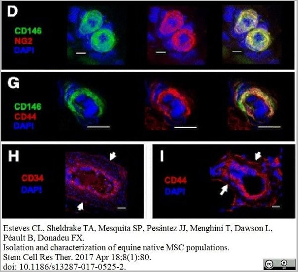
(Published customer image:Mouse anti Human CD146 antibody, clone OJ79c used for the demonstration of equine CD146 expressing cells by immunofluorescence.Image caption:Immunohistochemistry of pericyte, adventitial cell, and MSC markers in equine tissues. d, Dual staining (right panel) with the pericyte markers NG2 and CD146 (d) in adipose tissue. g, Colocalization (right panel) of the MSC and pericyte markers CD44 and CD146 (g) in adipose tissue. d–g Individual staining is shown (left and middle panels). h, i Immunodetection of the adventitial cell marker CD34 (h) and the MSC marker CD44 (i) in the outer layer of blood vessels (arrows) in adipose tissue. 4?,6-Diamidino-2-phenylindole (DAPI) was used to stain cell nuclei. White scale bars?=?10 um.)
Application Data
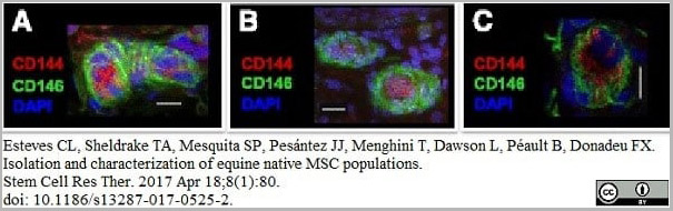
(Published customer image:Mouse anti Human CD146 antibody, clone OJ79c used for the demonstration of equine CD146 expressing cells by immunofluorescence.Image caption:Immunohistochemistry of pericyte, adventitial cell, and MSC markers in equine tissues. a–c Pericytes stained for CD146 surrounding CD144+ endothelial cells in adipose tissue (a), testis (b), and skeletal muscle (c). 4?,6-Diamidino-2-phenylindole (DAPI) was used to stain cell nuclei. White scale bars?=?10 um.)
Application Data
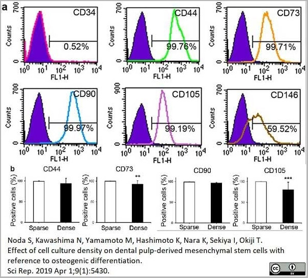
(Published customer image:Mouse anti Human CD146 antibody, clone OJ79c used for the evaluation of CD146 expression on dental pulp derived cells by flow cytometry.Image caption:Cell surface markers. (a) Cell surface markers before separation into sparse (sDPSCs) and dense (dDPSCs) groups. A representative case among seven donors is shown. (b) MSC marker expression in sparse (sDPSCs) and dense (dDPSCs) groups. **p?=?0.0079 and ***p?=?0.0006 (Mann-Whitney U test). The error bar is SD (n?=?7). Solid colored histograms represent IgG? treated cells for control, and open colored histograms represent fluorophore labeled antibody treated cells.)
Application Data
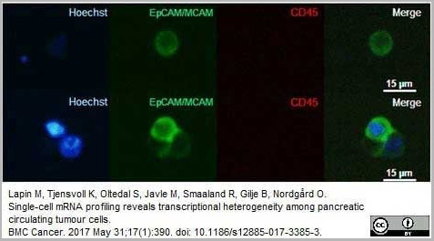
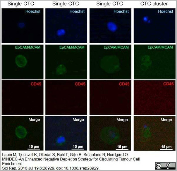
(Published customer image:Mouse anti Human CD146 antibody, clone OJ79c used to identify MCAM expressing circulating tumor cells by immunofluorescence.Image caption:Thumbnail gallery of analysed CTCs. Representative fluorescence images of a CTC (top row) and a CTC cluster (bottom row) isolated from the peripheral blood of a patient with pancreatic cancer. (Left column) Hoechst stain (blue) identifies nuclei; (second column) EpCAM-MCAM (green) identifies membranes of CTCs; (third column) CD45 (red) identifies leucocytes; (right column) superimposed images confirms intact cells. Images were acquired with 20× magnification. To enhance visibility, we adjusted the brightness and contrast equally for all microscopic images, and these adjustments were applied to the entire image)
Application Data
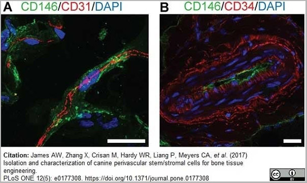
(Published customer image:Mouse anti Human CD146 antibody, clone OJ79c used to identify canine pericytes in situ by immunofluorescence staining of adipose cryosections.Image caption:Canine PSC in situ identification.Marker expression (A) Canine adipose tissue stained with pericytes marker CD146 (green), endothelial marker CD31 (red) (63×), and (B) adventitial cell marker, CD34 (red), CD146 (green) (40×). DAPI nuclear counterstain appears blue. Scale bar: 25uM.)
Application Data
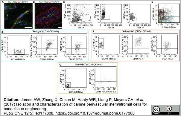
(Published customer image:Mouse anti Human CD146 antibody, clone OJ79c used to identify canine pericytes in situ by immunofluorescence and isolated from adipose tissue by flow cytometry.Image caption:Canine PSC in situ identification.Marker expression and isolation by fluorescence activated cell sorting (FACS). (A) Canine adipose tissue stained with pericytes marker CD146 (green), endothelial marker CD31 (red) (63×), and (B) adventitial cell marker, CD34 (red), CD146 (green) (40×). DAPI nuclear counterstain appears blue. (C) Cell size. (D) Purified PSC consist of distinct CD34?CD146+ pericytes and CD34+CD146? adventitial cells. (E) CD146+CD34- pericytes and (F) CD146-CD34+ adventitial cells express characteristic MSC markers, including CD44 and CD90. In comparison, (G) CD146-CD34- “non-PSC” are predominantly negative for CD44 and CD90. Scale bar: 25uM.)
Application Data
(Published customer image:FITC conjugated Mouse anti Human CD146 antibody, clone oJ79c used to identify MCAM expression on circulating tumor cells in patients by immunofluorescence.Image caption:Representative fluorescence images of CTCs and a CTC cluster isolated from peripheral blood of patients with metastatic pancreatic cancer.Enriched blood samples were stained with Hoechst 33343 (nuclei, blue), EpCAM (green), MCAM (green), and CD45 (red).)
Application Data
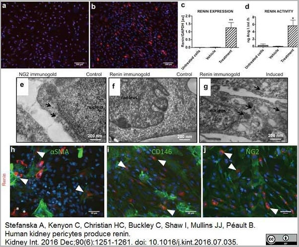
(Published customer image:Mouse anti Human CD146 antibody, clone oJ79c used to identify CD146 expressing pericytes in human fetal kidneys by immunofluorescence.Image caption:CD146+NG2+ ?SMA+/? renal pericytes express and secrete enzymatically active renin in vitro. Renal pericyte primary cultures at passage 2 were stained for renin (red). (a) Control cells (vehicle: medium + vehicle; untreated cells: medium) show no renin immunoreactivity. Forskolin and isobutyl-1-methylxanthine (cyclic adenosine monophosphate induction) treatment in pericytes (b) caused robust renin staining, (c) increased renin mRNA expression, and (d) renin activity. One-way analysis of variance followed by Bonferroni's post hoc comparisons were used to test statistical significance. Data are shown as mean ± SEM (n = 3, *P)
Application Data
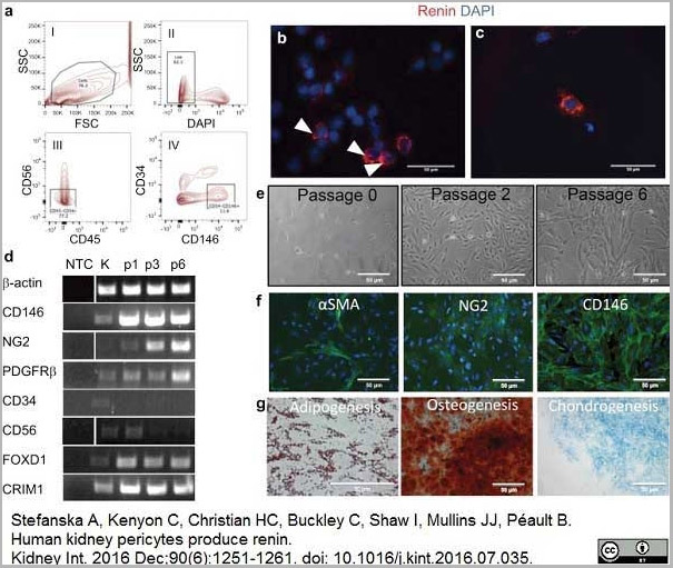
(Published customer image:Mouse anti Human CD146 antibody, clone oJ79c used to identify CD146 expressing pericytes in human fetal kidneys by flow cytometry and to demonstrate CD146 positivity in pericytes cultured in vitro by immunofluorescence.Image caption:Isolation and in vitro characterization of pericytes from a human fetal kidney. (a) FACS of renal pericytes purified from a 16-week human fetal kidney. Dot plots show backgating strategy to obtain CD146+CD34?CD56?CD45? pericytes. Red-color dots show gated populations and the percentages of the parent gates. The sorted pericyte population contained no CD45+, CD56+, or CD34+ cells. (b) Cytospins of freshly sorted pericytes were immunostained for renin (red, arrowheads). (c) Granular, intracellular expression of renin (red) can be observed for up to 48 hours in occasional cells of renal pericyte primary cultures. Positively stained cells are native JG cells, which retain renin expression for a short term in vitro. (d) Representative results of gene expression profiles of renal pericyte primary cultures (n ? 3). Pericytes showing expression of the pericyte markers CD146, nerve/glial antigen 2 (NG2), and platelet-derived growth factor receptor-? (PDGFR?), were negative for hematopoietic CD45 and metanephric mesenchyme CD56 expressions. In addition, pericyte cultures were enriched in Foxd1 (stromal marker) and CRIM1 (pericyte/parietal cell/podocyte marker). (e) Morphology of cultured CD146+CD34?CD56?CD45? pericytes. Bright field images of renal pericyte primary cultures are shown after plating at passages (p) 0, 2, and 6. Cells displayed a spindle-shaped morphology at p0 and p2 but eventually acquired fibroblastic-like appearance at p6. (f) Cultured kidney pericytes at p2 show some ?-smooth muscle actin (?SMA) (green) positive staining, whereas most of the cells are positive for NG2 (green) and all cultured pericytes are CD146+ (green). (g) Kidney pericyte primary cultures underwent mesodermal lineage induction. After 14 days of differentiation, primary cultures were confirmed to differentiate along adipogenic (Oil red O staining), chondrogenic (Alcian blue staining), and osteogenic lineages (Alizarin red staining). Control cells were incubated with growth medium (not shown). Original magnifications are shown on the images. DAPI, 4',6-diamidino-2-phenylindole; FACS, fluorescence-activated cell sorter; FSC, forward scatter; JG, juxtaglomerular cell; K, kidney (positive control); NTC, no template control; SSC, side scatter.)
Application Data
(Published customer image:Mouse anti Human CD146 antibody, clone oJ79c used to identify CD146 expressing pericytes in human fetal kidneys by immunofluorescence.Image caption:Renin is colocalized with pericyte markers in the human fetal kidney. Triple staining of fetal kidney demonstrates that (a) renin immunoreactivity (red) coincides with pericyte marker CD146 (green) expression in afferent arterioles. CD146 staining is also present in the mesangium (open arrowhead). Characteristically, renin staining had a striped pattern. The inset (a') shows an afferent arteriole at high magnification. Renin and CD146 stainings overlap on the abluminal side of the vessel (arrowheads), whereas immunoreactivity for CD144 (grey), an endothelial cell marker, is visible inside blood vessels (arrow). (b) Nerve/glial antigen 2 (NG2) (green) staining is found in the mesangium and afferent arteriole. Renin (red) is present in the JG area. Inset (b') shows renin+ cells coexpressing NG2 (arrowheads) at higher magnification. JG, juxtaglomerular cell.From: Stefanska A, Kenyon C, Christian HC, Buckley C, Shaw I, Mullins JJ, Péault B.Human kidney pericytes produce renin.)
Application Data
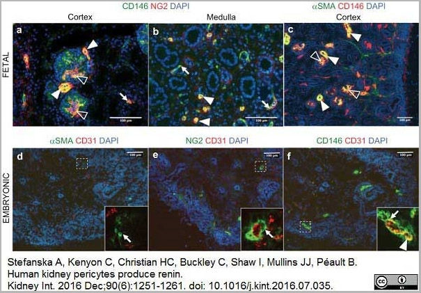
(Published customer image:Mouse anti Human CD146 antibody, clone oJ79c used to identify CD146 expressing pericytes in human fetal and embryonic kidneys by immunofluorescenceImage caption:Identification of pericytes in human embryonic and fetal kidneys. (a) Fetal pericytes were labeled with antibodies against CD146 (green) and NG2 (red) in a 15-week-old kidney cortex. CD146+NG2+ pericytes were found in peritubular capillaries (arrow), mesangial cells (open arrowheads), and afferent arterioles (arrowheads). (b) In the kidney medulla, pericytes encircled peritubular capillaries (arrows) and the vasa recta (arrowheads). (c) Double staining for CD146 (red) and smooth muscle ?-actin (?SMA) (green) shows that in the cortex, pericytes coexpress ?SMA in the mesangium (open arrowheads) and afferent arterioles (arrowheads). In embryonic pericytes, sections of 7-week-old kidneys were stained for pericyte markers: CD146 (green), nerve/glial antigen 2 (NG2) (green), and ?SMA (green), with the endothelial cell marker CD31 (red) (on consecutive sections). (d) ?SMA immunoreactivity was sparse; only single vascular smooth muscle cells were found (inset, arrow). (e) NG2+ cells surrounded endothelial cells (inset, arrow). (f) CD146 staining was perivascular (inset, arrow) and in endothelial cells (inset, arrowhead). DAPI, 4',6-diamidino-2-phenylindole.)
Application Data
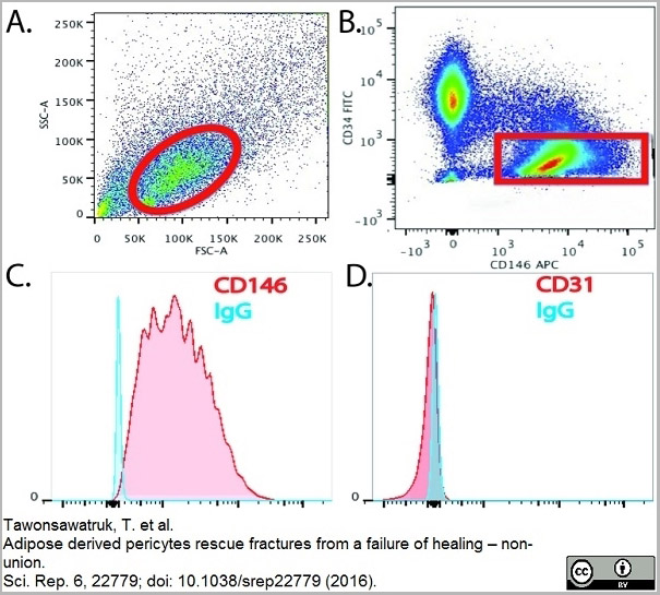
(Published customer image:AlexaFluor 647 conjugated Mouse anti Human CD146 antibody, clone OF79c used for the detection of CD146 expressing cells by flow cytometry.Image caption:(A) Pericytes can be purified from the SVF of adipose tissue by FACS. Cells (circled in red) are selected on an initial FSC v SSC dot plot. (B) Following removal of dead cells, haematopoietic (CD45+) and endothelial cells (CD31+), pericytes (red box) can be selected based on their unique phenotype (CD146+, CD34?, CD31?, CD45?). (C,D) Pericytes maintain a stable phenotype over extended periods of culture (CD146+, CD31?).)
Application Data
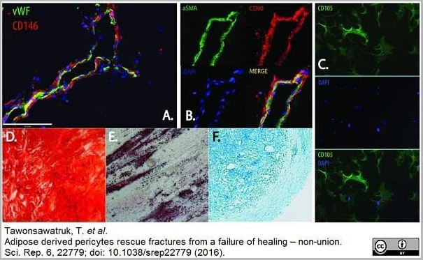
(Published customer image:AlexaFluor 647 conjugated Mouse anti Human CD146 antibody, clone OF79c used for the detection of CD146 expressing cells by immunofluorescence.Image caption:(A) Pericytes expressing CD146 reside on the abluminal surface of endothelial cells expressing vWF. (B) ?SMA expressing pericytes co-express MSC markers including CD90. (C) In-vitro pericytes express MSC markers including CD105. Cultured pericytes are multipotent as seen by their ability to differentiate into bone (D) (Alizarin red staining ×10), fat (E) (Oil Red O staining × 10), cartilage (F) (Alcian blue staining × 5).)
Application Data
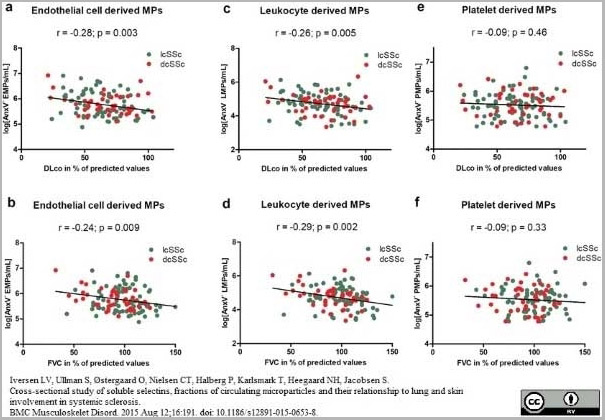
(Published customer image:Mouse anti HumanCD146 antibody, clone OJ79c used for the identification of endothelial cell derived non-annexin V binding, circulating microparticles by flow cytometry.Image caption:Plasma levels of annexin V non-binding, cell-derived microparticles (AnxV? MPs) correlated to lung function parameters. Endothelial cell derived MPs and leukocyte derived MPs (log counts/mL) correlated with both carbon monoxide diffusing capacity in percent of predicted values (DLco, %) (r = -0.28; p = 0.003, and r = -0.26; p = 0.005) respectively (a and b) and forced vital capacity in percent of predicted values (FVC, %) (r = -0.24; p = 0.009, and r = -0.29; p = 0.002) respectively (c and d). No correlations between AnxV? platelet derived MPs and lung function parameters were found (e and f). Correlation analysis was performed by use of Pearson's r test using log transformed microparticle data.From: Iversen LV, Ullman S, Østergaard O, Nielsen CT, Halberg P, Karlsmark T, Heegaard NH, Jacobsen S.Cross-sectional study of soluble selectins, fractions of circulating microparticles and their relationship to lung and skin involvement in systemic sclerosis.BMC Musculoskelet Disord. 2015 Aug 12;16:191.This is from an open access article distributed under the terms of the Creative Commons Attribution License.)
Application Data
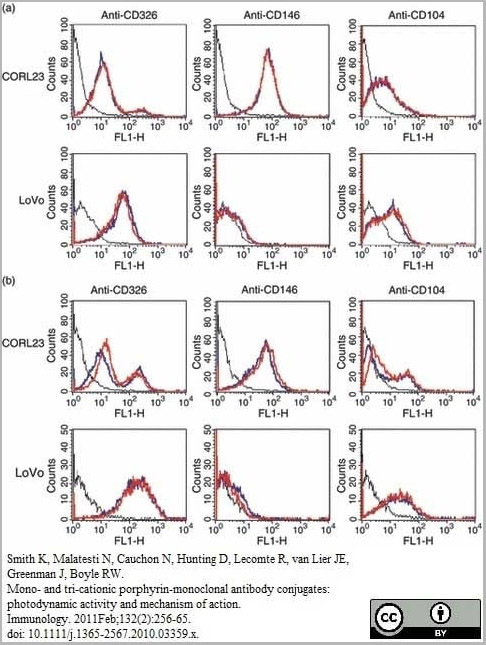
(Published customer image;Mouse anti Human CD146 antibody, clone OJ79c used for the evaluation of CD146 expression on cell lines and compared to binding of porphyrin conjugated antibodies by flow cytometry.Image captionConjugated and unconjugated anti-CD326, anti-CD146 and anti-CD104 were analysed by flow cytometry for binding to CORL23 and LoVo cells [black line, negative control; blue line, unconjugated antibody; red line, immunoconjugates with porphyrin 1 (a) or porphyrin 2 (b)].From: Smith K, Malatesti N, Cauchon N, Hunting D, Lecomte R, van Lier JE, Greenman J, Boyle RW.Mono- and tri-cationic porphyrin-monoclonal antibody conjugates: photodynamic activity and mechanism of action.)
Application Data
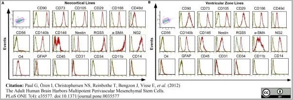
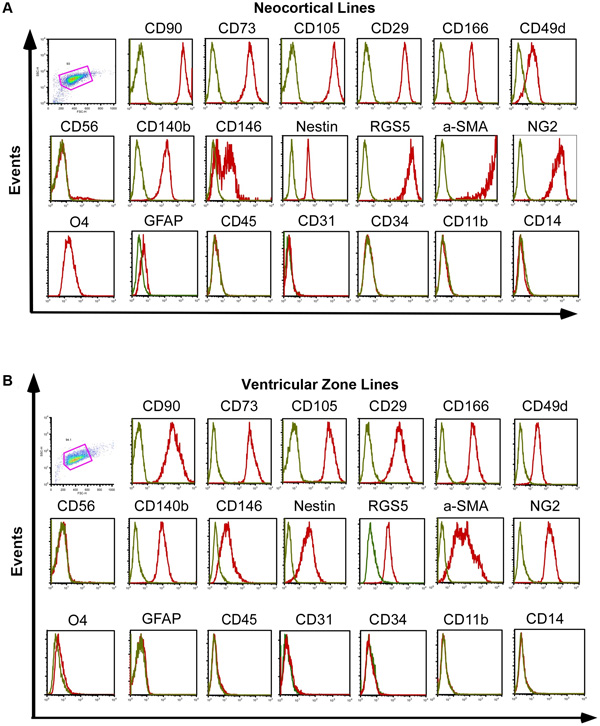
(Published customer image:Mouse anti Human CD146 antibody, clone OJ79c used for the evaluation of CD146 expression on brain derived cell lines by flow cytometry.Image caption:Brain-derived cell lines express markers for mesenchymal stem cells and pericytes but not for glial or neuronal precursors. (A) Representative histogram of flow cytometry analysis of cortical line and (B) ventricular zone line. Progenitors from all donors and both regions highly express MSC (CD90, CD73, CD105, CD29, CD166 and CD49d) and pericyte markers (CD140b/PDGFR-?, RGS5, CD146, Nestin, ?-SMA and NG2). They do not express hematopoietic (CD45), endothelial (CD31, CD34), microglial (CD14, CD11), glial or neuronal precursor cell markers (GFAP, O4) or myofibroblast markers (CD56), (green = isotype, red = respective marker).From: Paul G, Özen I, Christophersen NS, Reinbothe T, Bengzon J, et al. (2012)The Adult Human Brain Harbors Multipotent Perivascular Mesenchymal Stem Cells.PLoS ONE 7(4): e35577.)
Application Data
(Published customer image:Mouse anti Human CD146 antibody, clone OJ79c used for the evaluation of CD146 expression on brain derived cell lines by flow cytometry.Image caption:Brain-derived cell lines express markers for mesenchymal stem cells and pericytes but not for glial or neuronal precursors. (A) Representative histogram of flow cytometry analysis of cortical line and (B) ventricular zone line. Progenitors from all donors and both regions highly express MSC (CD90, CD73, CD105, CD29, CD166 and CD49d) and pericyte markers (CD140b/PDGFR-?, RGS5, CD146, Nestin, ?-SMA and NG2). They do not express hematopoietic (CD45), endothelial (CD31, CD34), microglial (CD14, CD11), glial or neuronal precursor cell markers (GFAP, O4) or myofibroblast markers (CD56), (green = isotype, red = respective marker).From: Paul G, Özen I, Christophersen NS, Reinbothe T, Bengzon J, et al. (2012)The Adult Human Brain Harbors Multipotent Perivascular Mesenchymal Stem Cells.PLoS ONE 7(4): e35577.)
Customer Reviews
Loading reviews...
Share Your Experience
Similar Products


Product Notes
The CD146 (Catalog #AAA12293) is an Antibody produced from Mouse and is intended for research purposes only. The product is available for immediate purchase. The MOUSE ANTI HUMAN CD146:RPE reacts with Human, Dog, Pig and may cross-react with other species as described in the data sheet. AAA Biotech's CD146 can be used in a range of immunoassay formats including, but not limited to, FCM/FACS (Flow Cytometry). FC/FACS: Max Neat. Researchers should empirically determine the suitability of the CD146 for an application not listed in the data sheet. Researchers commonly develop new applications and it is an integral, important part of the investigative research process. It is sometimes possible for the material contained within the vial of "CD146, Monoclonal Antibody" to become dispersed throughout the inside of the vial, particularly around the seal of said vial, during shipment and storage. We always suggest centrifuging these vials to consolidate all of the liquid away from the lid and to the bottom of the vial prior to opening. Please be advised that certain products may require dry ice for shipping and that, if this is the case, an additional dry ice fee may also be required.Precautions
All products in the AAA Biotech catalog are strictly for research-use only, and are absolutely not suitable for use in any sort of medical, therapeutic, prophylactic, in-vivo, or diagnostic capacity. By purchasing a product from AAA Biotech, you are explicitly certifying that said products will be properly tested and used in line with industry standard. AAA Biotech and its authorized distribution partners reserve the right to refuse to fulfill any order if we have any indication that a purchaser may be intending to use a product outside of our accepted criteria.Disclaimer
Though we do strive to guarantee the information represented in this datasheet, AAA Biotech cannot be held responsible for any oversights or imprecisions. AAA Biotech reserves the right to adjust any aspect of this datasheet at any time and without notice. It is the responsibility of the customer to inform AAA Biotech of any product performance issues observed or experienced within 30 days of receipt of said product. To see additional details on this or any of our other policies, please see our Terms & Conditions page.Item has been added to Shopping Cart
If you are ready to order, navigate to Shopping Cart and get ready to checkout.