
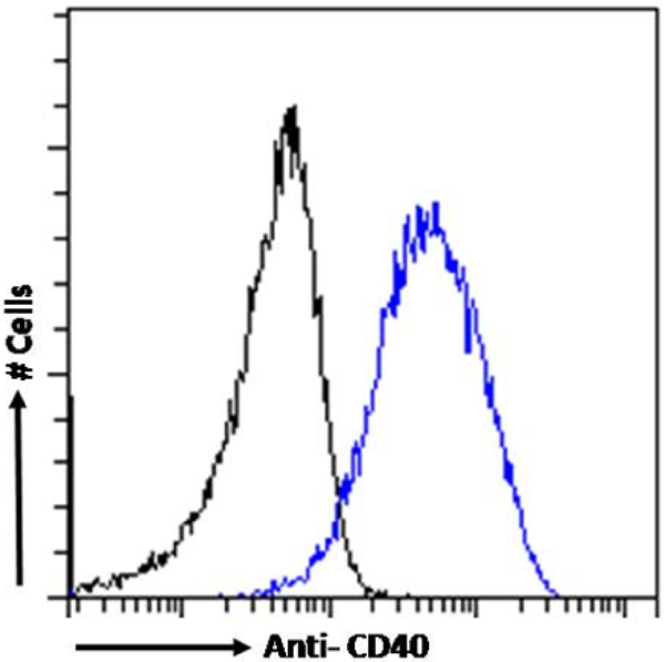
 Application Data
(Staining of human peripheral blood lymphocytes with Mouse anti Human CD40:RPE)
Application Data
(Staining of human peripheral blood lymphocytes with Mouse anti Human CD40:RPE)
Mouse CD40 Monoclonal Antibody | anti-CD40 antibody
MOUSE ANTI HUMAN CD40:RPE
Flow Cytometry: Maximum Dilution: Neat
Perservative Stabilisers
Fusion Partners
Shelf Life: 12 months from date of reconstitution.
Application Data
(Staining of human peripheral blood lymphocytes with Mouse anti Human CD40:RPE)
Application Data
(Staining of human peripheral blood lymphocytes with Mouse anti Human CD40:RPE)
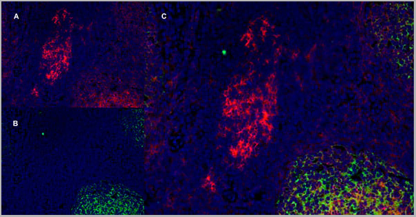
Application Data
(Immunofluorescence staining of a human tonsil cryostetion with Mouse anti Human CD40 antibody, clone LOB7/6 , red in A and Mouse anti Human CD21 antibody, clone LB21 , green in B. The merged image is C with nuclei counterstained blue using DAPI. Low power)
Application Data
(Immunofluorescence staining of a human tonsil cryostetion with Mouse anti Human CD40 antibody, clone LOB7/6 , red in A and Mouse anti Human CD21 antibody, clone LB21 , green in B. The merged image is C with nuclei counterstained blue using DAPI. Low power)
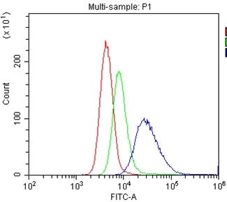
Application Data
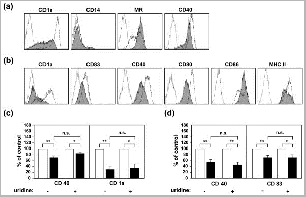
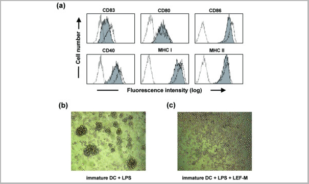
(Published customer image: LEF-M interferes with DC differentiation. (a) Monocytes were cultured for 5 days with granulocyte -macrophage colony-stimulating factor (GM-CSF; 50 ng/ml) plus IL-4 (10 ng/ml) in the absence or presence of 150 umol/l of the active metabolite of leflunomide (LEF-M). Subsequently, surface marker expression was determined using fluorescence-activated cell sorting (FACS) analysis. Open profiles with dotted line represent staining pattern with an isotype control antibody, open profiles with fine line indicate the staining pattern of differentiated control dendritic cells (DCs) stained with the indicated mAbs, whereas solid grey profiles show staining of DCs differentiated in the presence of LEF-M. (b) Myeloid precursor cells differentiated in the presence of LEF-M are resistant to maturation. Cells were treated as described above and then stimulated with lipopolysaccharide (LPS; 100 ng/ml) for 48 hours. Open profiles with dotted line represent staining pattern with an isotype control antibody, open profiles with fine line indicate staining of activated control DCs, and solid grey profiles show staining of DCs differentiated in the presence of LEF-M and subsequently exposed to LPS. Data are representative of at least four independent experiments. (c,d) The effects of LEF-M on DC differentiation are independent of pyrimidine depletion. The respective change in mean flourescence intensity (MFI) are shown (c) after the differentiation phase for CD40 and CD1a and (d) after subsequent maturation with 100 ng/ml LPS for CD40 and CD83 with and without 50 umol/l uridine. White bars represent control DCs, and black bars indicate LEF-M-treated cells. Shown are mean percentage control responses +/- standard error of the mean, calculated from five to eight independent experiments. Student's t-tests were calculated for control versus LEF-M-treated DCs and for LEF-M-treated DCs versus without uridine addition, as indicated. *P < 0.05, **P < 0.01.From: Kirsch BM, Zeyda M, Stuhlmeier K, Grisar J, Smolen JS, Watschinger B, Stulnig TM, H¶rl WH, Zlabinger GJ, S¤emann MD. The active metabolite of leflunomide, A77 1726, interferes with dendritic cell function. Arthritis Res Ther. 2005;7(3):R694-703.)
Application Data
(Published customer image: LEF-M interferes with DC differentiation. (a) Monocytes were cultured for 5 days with granulocyte -macrophage colony-stimulating factor (GM-CSF; 50 ng/ml) plus IL-4 (10 ng/ml) in the absence or presence of 150 umol/l of the active metabolite of leflunomide (LEF-M). Subsequently, surface marker expression was determined using fluorescence-activated cell sorting (FACS) analysis. Open profiles with dotted line represent staining pattern with an isotype control antibody, open profiles with fine line indicate the staining pattern of differentiated control dendritic cells (DCs) stained with the indicated mAbs, whereas solid grey profiles show staining of DCs differentiated in the presence of LEF-M. (b) Myeloid precursor cells differentiated in the presence of LEF-M are resistant to maturation. Cells were treated as described above and then stimulated with lipopolysaccharide (LPS; 100 ng/ml) for 48 hours. Open profiles with dotted line represent staining pattern with an isotype control antibody, open profiles with fine line indicate staining of activated control DCs, and solid grey profiles show staining of DCs differentiated in the presence of LEF-M and subsequently exposed to LPS. Data are representative of at least four independent experiments. (c,d) The effects of LEF-M on DC differentiation are independent of pyrimidine depletion. The respective change in mean flourescence intensity (MFI) are shown (c) after the differentiation phase for CD40 and CD1a and (d) after subsequent maturation with 100 ng/ml LPS for CD40 and CD83 with and without 50 umol/l uridine. White bars represent control DCs, and black bars indicate LEF-M-treated cells. Shown are mean percentage control responses +/- standard error of the mean, calculated from five to eight independent experiments. Student's t-tests were calculated for control versus LEF-M-treated DCs and for LEF-M-treated DCs versus without uridine addition, as indicated. *P < 0.05, **P < 0.01.From: Kirsch BM, Zeyda M, Stuhlmeier K, Grisar J, Smolen JS, Watschinger B, Stulnig TM, H¶rl WH, Zlabinger GJ, S¤emann MD. The active metabolite of leflunomide, A77 1726, interferes with dendritic cell function. Arthritis Res Ther. 2005;7(3):R694-703.)
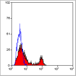
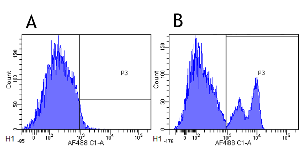
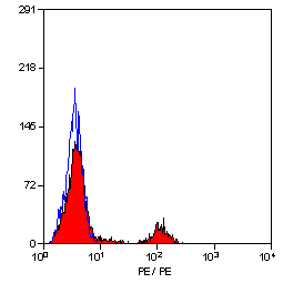
Application Data
(Staining of human peripheral blood lymphocytes with Mouse anti Human CD40: Alexa Fluor 488)
Application Data
(Staining of human peripheral blood lymphocytes with Mouse anti Human CD40: Alexa Fluor 488)
Application Data
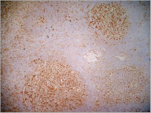
(Immunoperoxidase staining of a human tonsil cryosection with Mouse anti Human CD40 antibody, clone LOB7/6 followed by the Histar Detection system . High power)
Application Data
(Immunoperoxidase staining of a human tonsil cryosection with Mouse anti Human CD40 antibody, clone LOB7/6 followed by the Histar Detection system . High power)
Application Data
(Published customer image: Treatment with LEF-M during maturation of immature DCs leads to a differentially affected phenotype. Monocytes were cultured for 5 days with granulocyte -macrophage colony-stimulating factor (GM-CSF; 50 ng/ml) plus IL-4 (10 ng/ml). (a) On day 5 these immature dendritic cells (DCs; 5 x 10⁵/ml) were activated with lipopolysaccharide (LPS; 100 ng/ml) in the absence or presence of 150 umol/l of the active metabolite of leflunomide (LEF-M) for 48 hours. Surface marker expression was determined by fluorescence-activated cell sorting analysis. Open profiles with dotted line represent the staining pattern with an isotype control, open profiles with fine line indicate the staining pattern of DC exposed to LPS with the indicated monoclonal antibodies, and solid grey profiles show staining of DCs matured in the presence of LEF-M. The results shown are representative of five independent experiments. (b,c) Effect of LEF-M on maturation-associated clustering of DCs; immature DCs were stimulated with LPS in the (panel b) absence or (panel c) presence of 150 umol/l LEF-M. After 8 hours of cultivation, cells were analyzed by inspecting photomicrographs obtained by light microscopy. Similar results were obtained in four additional experiments. MHC, major histocompatibility complex.From: Kirsch BM, Zeyda M, Stuhlmeier K, Grisar J, Smolen JS, Watschinger B, Stulnig TM, H¶rl WH, Zlabinger GJ, S¤emann MD. The active metabolite of leflunomide, A77 1726, interferes with dendritic cell function. Arthritis Res Ther. 2005;7(3):R694-703.)
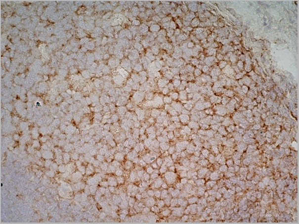
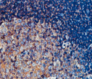
Application Data
(Immunoperoxidase staining of a human tonsil cryosection with Mouse anti Human CD40 antibody, clone LOB7/6 followed by the Histar Detection system . Low power)
Application Data
(Immunoperoxidase staining of a human tonsil cryosection with Mouse anti Human CD40 antibody, clone LOB7/6 followed by the Histar Detection system . Low power)
Application Data
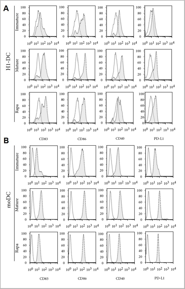
(Published customer image: Effect of rapamycin (Rapa) on the phenotype and function of H1-DC. DCs were either untreated, matured in response to the maturation cocktail or treated with Rapa for 3 days prior to maturation. (a) H1-DC stained for the expression of the maturation marker CD83, the costimulatory molecules CD86 and CD40, as well as the inhibitory receptor PD-L1. Dead cells were excluded from analysis using 7-AAD. Open histograms represent the level of background staining using appropriate isotype-matched controls. Data from one of 3 independent experiments are shown. (b) Phenotypic analysis of control populations of moDC treated and stained in parallel with rapamycin. (c) Effect of rapamycin on the allostimulatory capacity of DC in the allogeneic MLR. DCs were mitotically-inactivated using mitomycin C and plated in triplicate at a top dose of 104 cells per well of a 96-well round-bottomed plate; na¯ve CD4+ T cells were plated at 5 x 10⁴?cells/well. Cells were incubated for 5 days before pulsing with 3H-thymidine overnight. Graphs show the mean of triplicate cultures +/- S.D. Data are shown from one experiment, representative of 3 independent experiments.From: Silk KM, Leishman AJ, Nishimoto KP, Reddy A, Fairchild PJ. Rapamycin conditioning of dendritic cells differentiated from human ES cells promotes a tolerogenic phenotype. J Biomed Biotechnol. 2012;2012:172420.)
Application Data
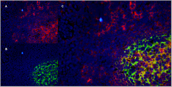
(Immunofluorescence staining of a human tonsil cryostetion with Mouse anti Human CD40 antibody, clone LOB7/6 , red in A and Mouse anti Human CD21 antibody, clone LB21 , green in B. The merged image is C with nuclei counterstained blue using DAPI. High power)
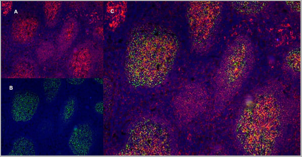
Application Data
(Immunofluorescence staining of a human tonsil cryostetion with Mouse anti Human CD40 antibody, clone LOB7/6 , red in A and Mouse anti Human CD21 antibody, clone LB21 , green in B. The merged image is C with nuclei counterstained blue using DAPI. High power)
Application Data
(Immunofluorescence staining of a human tonsil cryostetion with Mouse anti Human CD40 antibody, clone LOB7/6 , red in A and Mouse anti Human CD21 antibody, clone LB21 , green in B. The merged image is C with nuclei counterstained blue using DAPI. Medium power)
Application Data
(Immunofluorescence staining of a human tonsil cryostetion with Mouse anti Human CD40 antibody, clone LOB7/6 , red in A and Mouse anti Human CD21 antibody, clone LB21 , green in B. The merged image is C with nuclei counterstained blue using DAPI. Medium power)
NCBI and Uniprot Product Information
Customer Reviews
Loading reviews...
Share Your Experience
Similar Products


Product Notes
The CD40 cd40 (Catalog #AAA12089) is an Antibody produced from Mouse and is intended for research purposes only. The product is available for immediate purchase. AAA Biotech's CD40 can be used in a range of immunoassay formats including, but not limited to, FCM/FACS (Flow Cytometry). Flow Cytometry: Use 10ul of the suggested working dilution to label 106 cells in 100ul. Flow Cytometry: Maximum Dilution: Neat. Researchers should empirically determine the suitability of the CD40 cd40 for an application not listed in the data sheet. Researchers commonly develop new applications and it is an integral, important part of the investigative research process. It is sometimes possible for the material contained within the vial of "CD40, Monoclonal Antibody" to become dispersed throughout the inside of the vial, particularly around the seal of said vial, during shipment and storage. We always suggest centrifuging these vials to consolidate all of the liquid away from the lid and to the bottom of the vial prior to opening. Please be advised that certain products may require dry ice for shipping and that, if this is the case, an additional dry ice fee may also be required.Precautions
All products in the AAA Biotech catalog are strictly for research-use only, and are absolutely not suitable for use in any sort of medical, therapeutic, prophylactic, in-vivo, or diagnostic capacity. By purchasing a product from AAA Biotech, you are explicitly certifying that said products will be properly tested and used in line with industry standard. AAA Biotech and its authorized distribution partners reserve the right to refuse to fulfill any order if we have any indication that a purchaser may be intending to use a product outside of our accepted criteria.Disclaimer
Though we do strive to guarantee the information represented in this datasheet, AAA Biotech cannot be held responsible for any oversights or imprecisions. AAA Biotech reserves the right to adjust any aspect of this datasheet at any time and without notice. It is the responsibility of the customer to inform AAA Biotech of any product performance issues observed or experienced within 30 days of receipt of said product. To see additional details on this or any of our other policies, please see our Terms & Conditions page.Item has been added to Shopping Cart
If you are ready to order, navigate to Shopping Cart and get ready to checkout.