 IF (Immunofluorescence)
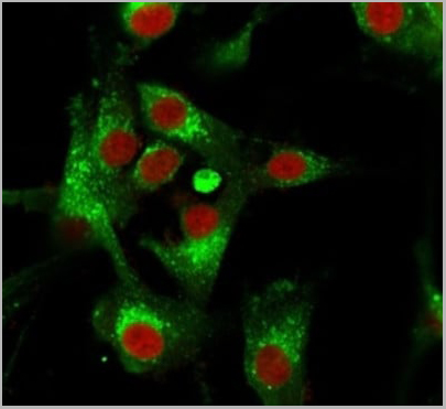
(Immunofluorescence Analysis of PFA-fixed U87MG cells stained using CD63 Mouse Monoclonal Antibody (MX-49.129.5) followed by goat anti-mouse IgG-CF488 (green). CF640R phalloidin (red).)
IF (Immunofluorescence)
(Immunofluorescence Analysis of PFA-fixed U87MG cells stained using CD63 Mouse Monoclonal Antibody (MX-49.129.5) followed by goat anti-mouse IgG-CF488 (green). CF640R phalloidin (red).)
Mouse anti-Human, Mouse CD63 Monoclonal Antibody | anti-CD63 antibody
CD63 (Late Endosomes Marker) Mouse Monoclonal Antibody
Immunofluorescence (0.5-1ug/ml)
Western Blot (0.5-1ug/ml)
Immunohistochemistry (Formalin-fixed) (1-2ug/ml for 30 minutes at RT)
(Staining of formalin-fixed tissues requires heating tissue sections in 10mM Tris with 1mM EDTA, pH 9.0, for 45 min at 95 °C followed by cooling at RT for 20 minutes)
Optimal dilution for a specific application should be determined.
IF (Immunofluorescence)
(Immunofluorescence Analysis of PFA-fixed U87MG cells stained using CD63 Mouse Monoclonal Antibody (MX-49.129.5) followed by goat anti-mouse IgG-CF488 (green). CF640R phalloidin (red).)
IF (Immunofluorescence)
(Immunofluorescence Analysis of PFA-fixed U87MG cells stained using CD63 Mouse Monoclonal Antibody (MX-49.129.5) followed by goat anti-mouse IgG-CF488 (green). CF640R phalloidin (red).)
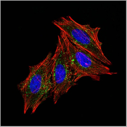
IF (Immunofluorescence)
(IF staining of HeLa cells using AF488 labeled CD63 Monoclonal Antibody (MX-49.129.5) (green). F-actin filaments are labeled with Dylight 554 phalloidin (red). Nuclei stained with DAPI (blue).)
IF (Immunofluorescence)
(IF staining of HeLa cells using AF488 labeled CD63 Monoclonal Antibody (MX-49.129.5) (green). F-actin filaments are labeled with Dylight 554 phalloidin (red). Nuclei stained with DAPI (blue).)
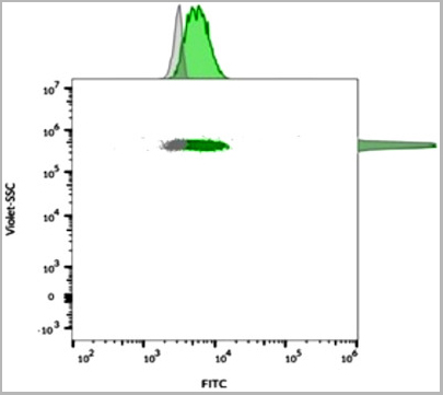
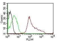
FCM (Flow Cytometry)
(Flow cytometric analysis of bead-bound exosomes derived from MCF-7 cells. CD63 Mouse Monoclonal Antibody (MX-49.129.5) followed by goat anti-mouse IgG-CF568 (orange); unstained exosomes (gray).)
FCM (Flow Cytometry)
(Flow cytometric analysis of bead-bound exosomes derived from MCF-7 cells. CD63 Mouse Monoclonal Antibody (MX-49.129.5) followed by goat anti-mouse IgG-CF568 (orange); unstained exosomes (gray).)
FCM (Flow Cytometry)
(Flow cytometric analysis of bead-bound exosomes derived from MCF-7 cells. CD63 Mouse Monoclonal Antibody (MX-49.129.5) followed by goat anti-mouse IgG-CF488 (green); unstained exosomes (gray).)
FCM (Flow Cytometry)
(Flow cytometric analysis of bead-bound exosomes derived from MCF-7 cells. CD63 Mouse Monoclonal Antibody (MX-49.129.5) followed by goat anti-mouse IgG-CF488 (green); unstained exosomes (gray).)
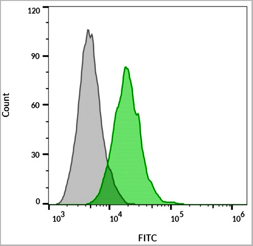
FCM (Flow Cytometry)
(Flow cytometric analysis of MCF-7 cells. CD63 Mouse Monoclonal Antibody (MX-49.129.5) followed by goat anti-mouse IgG-CF488 (green); unstained cells (gray).)
FCM (Flow Cytometry)
(Flow cytometric analysis of MCF-7 cells. CD63 Mouse Monoclonal Antibody (MX-49.129.5) followed by goat anti-mouse IgG-CF488 (green); unstained cells (gray).)
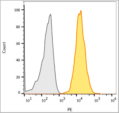
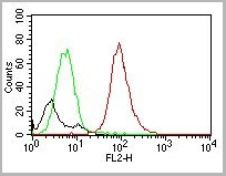
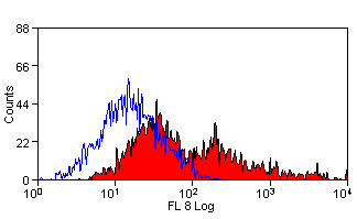
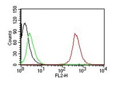
FCM (Flow Cytometry)
(Flow Cytometry of NIH/3T3 Cells.Black: Cells alone; Green: Isotype Control; Red: PE-labeled CD63 Monoclonal Antibody (MX-49.129.5).)
FCM (Flow Cytometry)
(Flow Cytometry of NIH/3T3 Cells.Black: Cells alone; Green: Isotype Control; Red: PE-labeled CD63 Monoclonal Antibody (MX-49.129.5).)
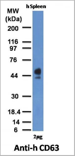
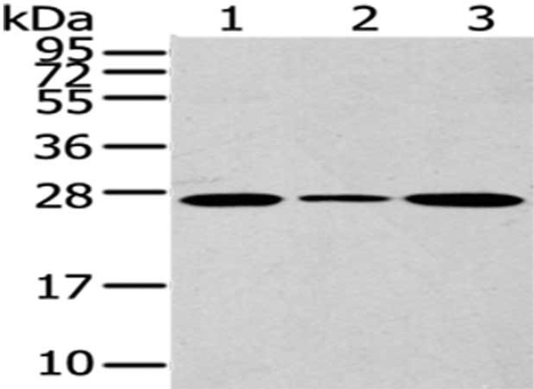
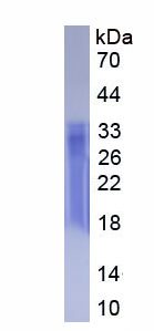
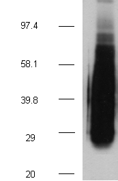
WB (Western Blot)
(Western Blot of human Spleen Lysate with CD63 Monoclonal Antibody (MX-49.129.5))
WB (Western Blot)
(Western Blot of human Spleen Lysate with CD63 Monoclonal Antibody (MX-49.129.5))
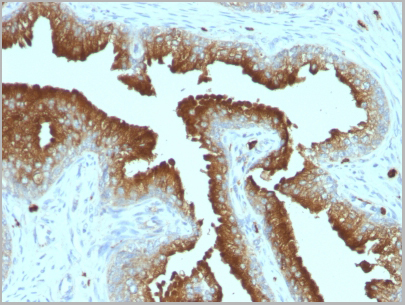
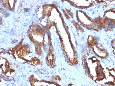
IHC (Immunohistochemistry)
(Formalin-fixed, paraffin-embedded human Prostate Carcinoma stained with CD63 Monoclonal Antibody (MX-49.129.5))
IHC (Immunohistochemistry)
(Formalin-fixed, paraffin-embedded human Prostate Carcinoma stained with CD63 Monoclonal Antibody (MX-49.129.5))
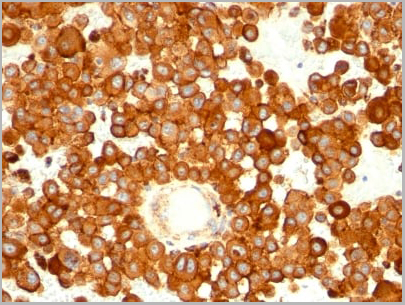
IHC (Immunohistochemistry)
(Formalin-fixed, paraffin-embedded mouse spleen stained with CD63 Monoclonal Antibody (MX-49.129.5))
IHC (Immunohistochemistry)
(Formalin-fixed, paraffin-embedded mouse spleen stained with CD63 Monoclonal Antibody (MX-49.129.5))
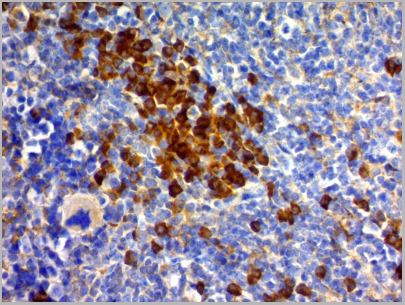
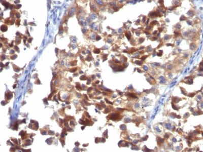
IHC (Immunohistochemistry)
(Formalin-fixed, paraffin-embedded human Melanoma stained with CD63 Monoclonal Antibody (MX-49.129.5))
IHC (Immunohistochemistry)
(Formalin-fixed, paraffin-embedded human Melanoma stained with CD63 Monoclonal Antibody (MX-49.129.5))
NCBI and Uniprot Product Information
Customer Reviews
Loading reviews...
Share Your Experience
Similar Products


Product Notes
The CD63 cd63 (Catalog #AAA13803) is an Antibody produced from Mouse and is intended for research purposes only. The product is available for immediate purchase. The CD63 (Late Endosomes Marker) Mouse Monoclonal Antibody reacts with Human, Mouse and may cross-react with other species as described in the data sheet. AAA Biotech's CD63 can be used in a range of immunoassay formats including, but not limited to, FCM/FACS (Flow Cytometry), IF (Immunofluorescence), WB (Western Blot), IHC (Immunohistochemistry). Flow Cytometry (0.5-1ug/million cells) Immunofluorescence (0.5-1ug/ml) Western Blot (0.5-1ug/ml) Immunohistochemistry (Formalin-fixed) (1-2ug/ml for 30 minutes at RT) (Staining of formalin-fixed tissues requires heating tissue sections in 10mM Tris with 1mM EDTA, pH 9.0, for 45 min at 95 °C followed by cooling at RT for 20 minutes) Optimal dilution for a specific application should be determined. Researchers should empirically determine the suitability of the CD63 cd63 for an application not listed in the data sheet. Researchers commonly develop new applications and it is an integral, important part of the investigative research process. It is sometimes possible for the material contained within the vial of "CD63, Monoclonal Antibody" to become dispersed throughout the inside of the vial, particularly around the seal of said vial, during shipment and storage. We always suggest centrifuging these vials to consolidate all of the liquid away from the lid and to the bottom of the vial prior to opening. Please be advised that certain products may require dry ice for shipping and that, if this is the case, an additional dry ice fee may also be required.Precautions
All products in the AAA Biotech catalog are strictly for research-use only, and are absolutely not suitable for use in any sort of medical, therapeutic, prophylactic, in-vivo, or diagnostic capacity. By purchasing a product from AAA Biotech, you are explicitly certifying that said products will be properly tested and used in line with industry standard. AAA Biotech and its authorized distribution partners reserve the right to refuse to fulfill any order if we have any indication that a purchaser may be intending to use a product outside of our accepted criteria.Disclaimer
Though we do strive to guarantee the information represented in this datasheet, AAA Biotech cannot be held responsible for any oversights or imprecisions. AAA Biotech reserves the right to adjust any aspect of this datasheet at any time and without notice. It is the responsibility of the customer to inform AAA Biotech of any product performance issues observed or experienced within 30 days of receipt of said product. To see additional details on this or any of our other policies, please see our Terms & Conditions page.Item has been added to Shopping Cart
If you are ready to order, navigate to Shopping Cart and get ready to checkout.