


 IF (Immunofluorescence)
(MILIN1 is expressed in association with lymphatic vessels. Cryostat sections of normal mouse tissues stained with anti-EMILIN1 (green) antibodies. In all mouse tissues and organs examined, EMILIN1 was uniformly distributed in the stroma. In the skin, EMILIN1 staining colocalizes with LYVE-1-positive lymphatic vessels surrounding hair follicles. In the small intestine, EMILIN1 colocalizes with LYVE-1-positive lacteals and submucosal lymphatic vessels. At higher magnification, in the lung and lymph nodes, it is more evident that EMILIN1 is distributed at the abluminal surfaces of LECs. In the lymph node, EMILIN1-positive fibers connecting LECs to the surrounding ECM are evident.)
IF (Immunofluorescence)
(MILIN1 is expressed in association with lymphatic vessels. Cryostat sections of normal mouse tissues stained with anti-EMILIN1 (green) antibodies. In all mouse tissues and organs examined, EMILIN1 was uniformly distributed in the stroma. In the skin, EMILIN1 staining colocalizes with LYVE-1-positive lymphatic vessels surrounding hair follicles. In the small intestine, EMILIN1 colocalizes with LYVE-1-positive lacteals and submucosal lymphatic vessels. At higher magnification, in the lung and lymph nodes, it is more evident that EMILIN1 is distributed at the abluminal surfaces of LECs. In the lymph node, EMILIN1-positive fibers connecting LECs to the surrounding ECM are evident.)
EMILIN-1 (Emilin1) Antibody – Rat Monoclonal
Rat Anti-Mouse Emilin-1
Product Overview
Optimal dilutions should determined by each laboratory for each application
IF (Immunofluorescence)
(MILIN1 is expressed in association with lymphatic vessels. Cryostat sections of normal mouse tissues stained with anti-EMILIN1 (green) antibodies. In all mouse tissues and organs examined, EMILIN1 was uniformly distributed in the stroma. In the skin, EMILIN1 staining colocalizes with LYVE-1-positive lymphatic vessels surrounding hair follicles. In the small intestine, EMILIN1 colocalizes with LYVE-1-positive lacteals and submucosal lymphatic vessels. At higher magnification, in the lung and lymph nodes, it is more evident that EMILIN1 is distributed at the abluminal surfaces of LECs. In the lymph node, EMILIN1-positive fibers connecting LECs to the surrounding ECM are evident.)
IF (Immunofluorescence)
(Positive staining of EMILIN1 (green) in mouse lymphatic endothelial cells (LAEC). Nuclei are stained in blue. Scale bar 38 um)
IF (Immunofluorescence)
(Positive staining of EMILIN1 (green) in mouse lymphatic endothelial cells (LAEC). Nuclei are stained in blue. Scale bar 38 um)
IF (Immunofluorescence)
(Positive staining of EMILIN1 (green) in mouse fibroblasts (NIH 3T3). Actin cytoskeleton is stained in red, nuclei in blue. Scale bar 28 um)
IF (Immunofluorescence)
(Positive staining of EMILIN1 (green) in mouse fibroblasts (NIH 3T3). Actin cytoskeleton is stained in red, nuclei in blue. Scale bar 28 um)
IF (Immunofluorescence)
(Positive staining for EMILIN1 (red) in mouse skin. Blue, nuclei;Scale bar: 37.00 um)
IF (Immunofluorescence)
(Positive staining for EMILIN1 (red) in mouse skin. Blue, nuclei;Scale bar: 37.00 um)
IF (Immunofluorescence)
(Positive staining for EMILIN1 green) in mouse lymph node. In red, lymphatic vessels positive for Lyve-1. Yellow represents the closeassociation of EMILIN1 with lymphatic vessel structures. Scale bar: 37.00 ?m)
IF (Immunofluorescence)
(Positive staining for EMILIN1 green) in mouse lymph node. In red, lymphatic vessels positive for Lyve-1. Yellow represents the closeassociation of EMILIN1 with lymphatic vessel structures. Scale bar: 37.00 ?m)
IF (Immunofluorescence)
(Positive staining for EMILIN1 (green) in mouse lung tissue is both detected in lung parenchymal fibers and associated with lymphatic vessel structures. Lyve-1 (red) is used as lymphatic vessel marker. Blue, nuclei.Scale bar: 37.00 um)
IF (Immunofluorescence)
(Positive staining for EMILIN1 (green) in mouse lung tissue is both detected in lung parenchymal fibers and associated with lymphatic vessel structures. Lyve-1 (red) is used as lymphatic vessel marker. Blue, nuclei.Scale bar: 37.00 um)
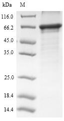
WB (Western Blot)
(Western blot on recombinant protein obtained from supernatant of cells expressing human EMILIN1 (hE1), mouse EMILIN1 (mE1), mouse EMILIN2 (mE2).)
WB (Western Blot)
(Western blot on recombinant protein obtained from supernatant of cells expressing human EMILIN1 (hE1), mouse EMILIN1 (mE1), mouse EMILIN2 (mE2).)
NCBI and Uniprot Product Information
Customer Reviews
Loading reviews...
Share Your Experience
Similar Products


Additional Details
Product Notes
The Emilin1 emilin1 (Catalog #AAA14943) is an Antibody produced from Rat and is intended for research purposes only. The product is available for immediate purchase. The Rat Anti-Mouse Emilin-1 reacts with Mouse and may cross-react with other species as described in the data sheet. AAA Biotech's Emilin-1 can be used in a range of immunoassay formats including, but not limited to, WB (Western Blot), IHC (Immunohistochemistry). Western Blot: Use at 2-10 ug/ml IF/IHC: Frozen sections Optimal dilutions should determined by each laboratory for each application. Researchers should empirically determine the suitability of the Emilin1 emilin1 for an application not listed in the data sheet. Researchers commonly develop new applications and it is an integral, important part of the investigative research process. It is sometimes possible for the material contained within the vial of "Emilin-1, Monoclonal Antibody" to become dispersed throughout the inside of the vial, particularly around the seal of said vial, during shipment and storage. We always suggest centrifuging these vials to consolidate all of the liquid away from the lid and to the bottom of the vial prior to opening. Please be advised that certain products may require dry ice for shipping and that, if this is the case, an additional dry ice fee may also be required.Precautions
All products in the AAA Biotech catalog are strictly for research-use only, and are absolutely not suitable for use in any sort of medical, therapeutic, prophylactic, in-vivo, or diagnostic capacity. By purchasing a product from AAA Biotech, you are explicitly certifying that said products will be properly tested and used in line with industry standard. AAA Biotech and its authorized distribution partners reserve the right to refuse to fulfill any order if we have any indication that a purchaser may be intending to use a product outside of our accepted criteria.Disclaimer
Though we do strive to guarantee the information represented in this datasheet, AAA Biotech cannot be held responsible for any oversights or imprecisions. AAA Biotech reserves the right to adjust any aspect of this datasheet at any time and without notice. It is the responsibility of the customer to inform AAA Biotech of any product performance issues observed or experienced within 30 days of receipt of said product. To see additional details on this or any of our other policies, please see our Terms & Conditions page.Item has been added to Shopping Cart
If you are ready to order, navigate to Shopping Cart and get ready to checkout.