
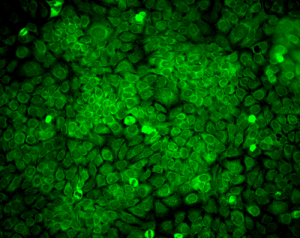
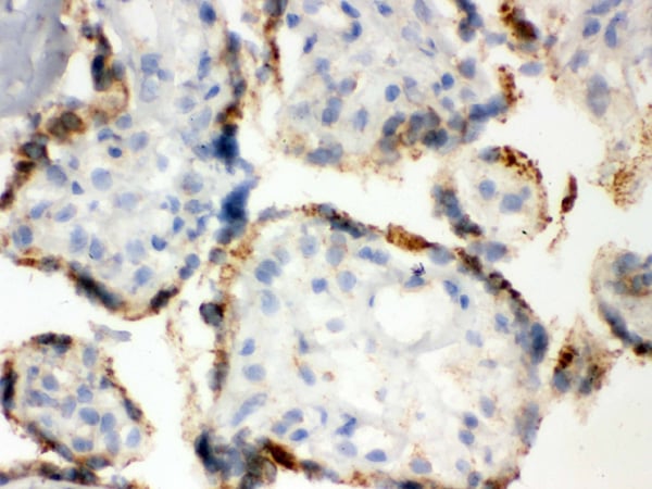
 ICC (Immunocytochemistry)
(Immunocytochemistry/Immunofluorescence analysis using Mouse Anti-Erp57 Monoclonal Antibody, Clone Map.ERP57. Tissue: HaCaT cells. Species: Human. Fixation: Cold 100% methanol for 10 minutes at -20 degree C. Primary Antibody: Mouse Anti-Erp57 Monoclonal Antibody at 1:100 for 1 hour at RT. Secondary Antibody: FITC Goat Anti-Mouse (green) at 1:50 for 1 hour at RT. Localization: Cytoplasmic and perinuclear staining.)
ICC (Immunocytochemistry)
(Immunocytochemistry/Immunofluorescence analysis using Mouse Anti-Erp57 Monoclonal Antibody, Clone Map.ERP57. Tissue: HaCaT cells. Species: Human. Fixation: Cold 100% methanol for 10 minutes at -20 degree C. Primary Antibody: Mouse Anti-Erp57 Monoclonal Antibody at 1:100 for 1 hour at RT. Secondary Antibody: FITC Goat Anti-Mouse (green) at 1:50 for 1 hour at RT. Localization: Cytoplasmic and perinuclear staining.)
Mouse Erp57 (Grp58) Monoclonal Antibody | anti-PDIA3 antibody
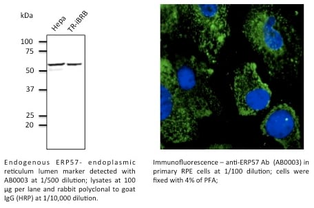
Erp57 (Grp58) Antibody: RPE
ICC (Immunocytochemistry)
(Immunocytochemistry/Immunofluorescence analysis using Mouse Anti-Erp57 Monoclonal Antibody, Clone Map.ERP57. Tissue: HaCaT cells. Species: Human. Fixation: Cold 100% methanol for 10 minutes at -20 degree C. Primary Antibody: Mouse Anti-Erp57 Monoclonal Antibody at 1:100 for 1 hour at RT. Secondary Antibody: FITC Goat Anti-Mouse (green) at 1:50 for 1 hour at RT. Localization: Cytoplasmic and perinuclear staining.)
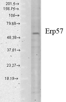
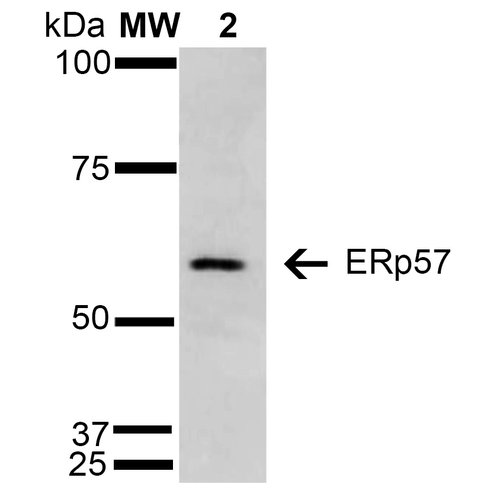
WB (Western Blot)
(Western Blot analysis of Human cell lysates showing detection of Erp57 protein using Mouse Anti-Erp57 Monoclonal Antibody, Clone Map.ERP57. Load: 15 ug. Block: 1.5% BSA for 30 minutes at RT. Primary Antibody: Mouse Anti-Erp57 Monoclonal Antibody at 1:1000 for 2 hours at RT. Secondary Antibody: Sheep Anti-Mouse IgG: HRP for 1 hour at RT.)
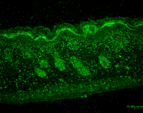
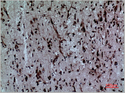
IHC (Immunohistochemistry)
(Immunohistochemistry analysis using Mouse Anti-Erp57 Monoclonal Antibody, Clone Map.ERP57. Tissue: backskin. Species: Mouse. Fixation: Bouin's Fixative and paraffin-embedded. Primary Antibody: Mouse Anti-Erp57 Monoclonal Antibody at 1:100 for 1 hour at RT. Secondary Antibody: FITC Goat Anti-Mouse (green) at 1:50 for 1 hour at RT. Localization: Epidermis and Hair Follicles.)
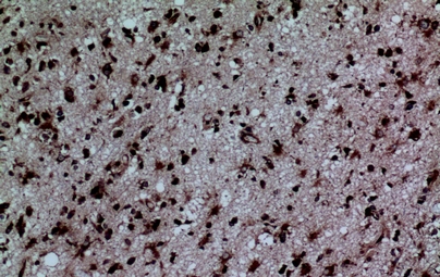
Scientific Background: ERp57, also known as Glucose Regulated Protein 58 (Grp58), Hormone-Induced Protein-70 (HIP-70) and microsomal Carnitine Palmitoyltransferase, is a member of the protein disulfide isomerase family, containing two canonical CXHC tetrapeptide active site motifs (1-5). It has quite a few diverse roles. It functions as an accessory oxidoreductase involved in disulfide bond formation. In the ER, ERp57 interacts with membrane bound calnexin and soluble calreticulin (lectin chaperones) via their praline rich P-domain arms. Lectin chaperones bind nascent non-native glycoproteins, and position ERp57 to act upon the immature or misfolded glycoproteins that possess mono-glycosylated side chains. ERp57 deletion impairs posttranslational phases of influenza hema-glutinin folding, and causes accelerated release of MHC-I molecules, resulting in the coupling of sub-optimal peptides and reduced expression and stability on the cell surface (6). ERp57 also contains two thioredoxin active-site sequences, CGHC and an estrogen-binding domain. ERp57 is induced by both estrogen and leuteinizing-hormone-releasing hormone in the hippocampus (7).
1. Lynes, E.M. et al. (2013). Palmitoylation is the Switch that Assigns Calnexin to Quality Control or ER Calcium Signaling. J Cell Sci. Advance Online Article July 10, 2013. doi: 10.1242/jcs.125856
NCBI and Uniprot Product Information
Customer Reviews
Loading reviews...
Share Your Experience
Similar Products


Product Notes
The PDIA3 pdia3 (Catalog #AAA103101) is an Antibody produced from Mouse and is intended for research purposes only. The product is available for immediate purchase. The Erp57 (Grp58) Antibody: RPE reacts with Human, Mouse, Bovine, Canine, Guinea pig, Hamster, Monkey, Pig, Rabbit, Rat and may cross-react with other species as described in the data sheet. AAA Biotech's Erp57 (Grp58) can be used in a range of immunoassay formats including, but not limited to, IHC (Immunohistochemistry), IP (Immunoprecipitation), WB (Western Blot). Researchers should empirically determine the suitability of the PDIA3 pdia3 for an application not listed in the data sheet. Researchers commonly develop new applications and it is an integral, important part of the investigative research process. It is sometimes possible for the material contained within the vial of "Erp57 (Grp58), Monoclonal Antibody" to become dispersed throughout the inside of the vial, particularly around the seal of said vial, during shipment and storage. We always suggest centrifuging these vials to consolidate all of the liquid away from the lid and to the bottom of the vial prior to opening. Please be advised that certain products may require dry ice for shipping and that, if this is the case, an additional dry ice fee may also be required.Precautions
All products in the AAA Biotech catalog are strictly for research-use only, and are absolutely not suitable for use in any sort of medical, therapeutic, prophylactic, in-vivo, or diagnostic capacity. By purchasing a product from AAA Biotech, you are explicitly certifying that said products will be properly tested and used in line with industry standard. AAA Biotech and its authorized distribution partners reserve the right to refuse to fulfill any order if we have any indication that a purchaser may be intending to use a product outside of our accepted criteria.Disclaimer
Though we do strive to guarantee the information represented in this datasheet, AAA Biotech cannot be held responsible for any oversights or imprecisions. AAA Biotech reserves the right to adjust any aspect of this datasheet at any time and without notice. It is the responsibility of the customer to inform AAA Biotech of any product performance issues observed or experienced within 30 days of receipt of said product. To see additional details on this or any of our other policies, please see our Terms & Conditions page.Item has been added to Shopping Cart
If you are ready to order, navigate to Shopping Cart and get ready to checkout.