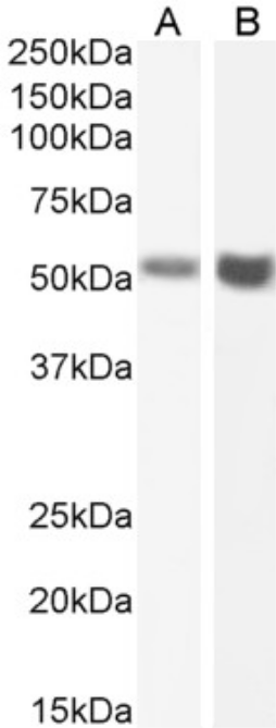
 WB (Western Blot)
( Western Blot using anti-Fas antibody R-125224 (Ab00802) Human testis (A) and human ovary (B) lysate samples (35ug protein in RIPA buffer) were resolved on a 10% SDS PAGE gel and blots probed with the chimeric rabbit IgG version of R-125224 (Ab00802-23.0) at 2 ug/ml before detection using an anti-rabbit secondary antibody. A primary incubation of 1h was used and protein was detected by chemiluminescence. The expected running size for unmodified Fas is 37.7kDa, but this protein is glycosylated at several positions leading to the observed running size.)
WB (Western Blot)
( Western Blot using anti-Fas antibody R-125224 (Ab00802) Human testis (A) and human ovary (B) lysate samples (35ug protein in RIPA buffer) were resolved on a 10% SDS PAGE gel and blots probed with the chimeric rabbit IgG version of R-125224 (Ab00802-23.0) at 2 ug/ml before detection using an anti-rabbit secondary antibody. A primary incubation of 1h was used and protein was detected by chemiluminescence. The expected running size for unmodified Fas is 37.7kDa, but this protein is glycosylated at several positions leading to the observed running size.)
Fas recombinant antibody
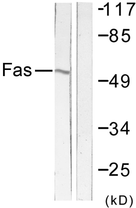
Anti-Fas [R-125224]
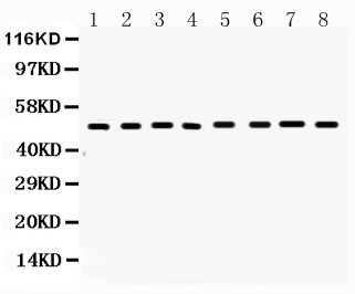
WB (Western Blot)
( Western Blot using anti-Fas antibody R-125224 (Ab00802) Human testis (A) and human ovary (B) lysate samples (35ug protein in RIPA buffer) were resolved on a 10% SDS PAGE gel and blots probed with the chimeric rabbit IgG version of R-125224 (Ab00802-23.0) at 2 ug/ml before detection using an anti-rabbit secondary antibody. A primary incubation of 1h was used and protein was detected by chemiluminescence. The expected running size for unmodified Fas is 37.7kDa, but this protein is glycosylated at several positions leading to the observed running size.)
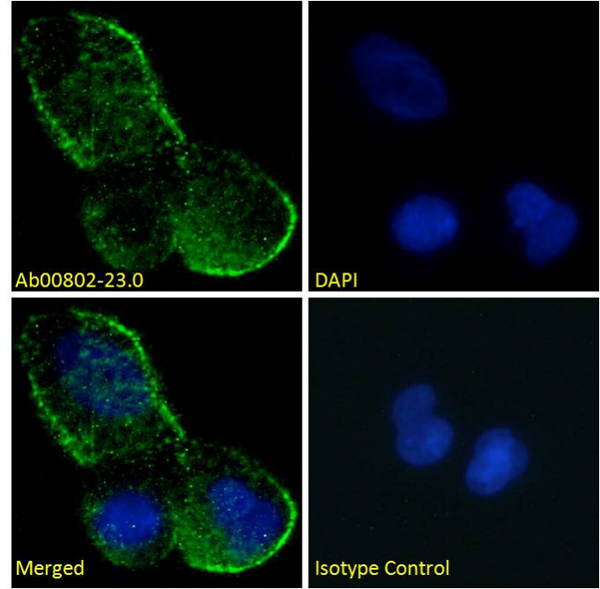
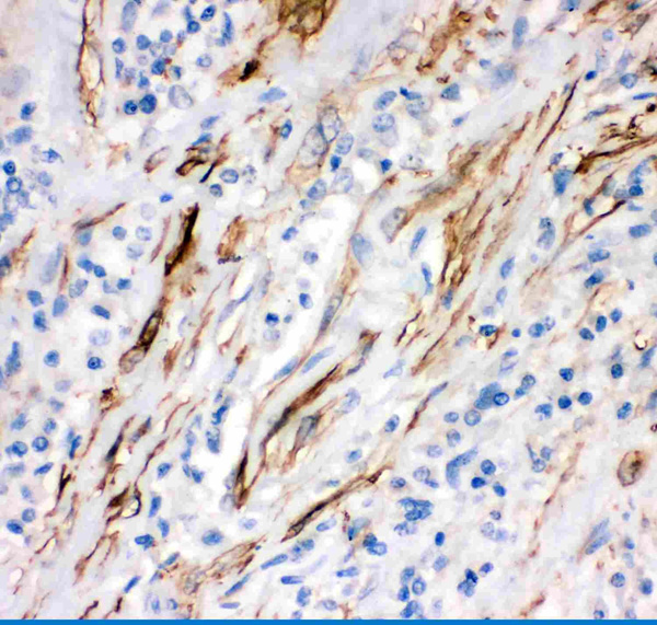
IF (Immunofluorescence)
( Immunofluorescence staining of fixed MCF7 cells with anti-Fas antibody R-125224 (Ab00802) Immunofluorescence analysis of paraformaldehyde fixed MCF7 cells permeabilized with 0.15% Triton and stained with the chimeric mouse IgG1 version of R-125224 (Ab00802-23.0) at 10 ug/ml for 1h followed by Alexa Fluor® 488 secondary antibody (2 ug/ml), showing membrane staining. The nuclear stain is DAPI (blue). Panels show from left-right, top-bottom Ab00802-23.0, DAPI, merged channels and an isotype control. The isotype control was stained with an anti-unknown specificity antibody (Ab178-23.0) followed by Alexa Fluor® 488 secondary antibody.)
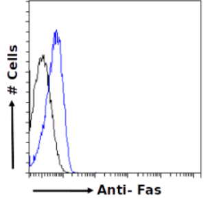
FCM/FACS (Flow Cytometry)
( Flow-cytometry using the anti-Fas antibody R-125224 (Ab00802) Jurkat cells were fixed using 2% PFA, permeabilised using 0.5% Triton and stained with unimmunized rabbit IgG antibody (MOPC-21; isotype control, black line) or the rabbit IgG-chimeric version of R-125224 (Ab00802-23.0, blue line) at a dilution of 1:100 for 1h at RT. After washing, bound antibody was detected using a goat anti-rabbit IgG AlexaFluor® 488 antibody at a dilution of 1:1000 and cells analyzed using a FACSCanto flow-cytometer.)
R-125224 is generated by the humanization of the murine HFE7A anti-Fas antibody by grafting the CDR regions to the framework regions of the human 8E10 antibody and substituting key framework residues from the murine antibody into the 8E10 sequence. The original HFE7A was derived from a hybridoma cell line generated by the fusion of NS1 myeloma cells with splenocytes from Fas-deficient mice which had been immunized with partially purified recombinant human Fas-AIC2A chimera protein consisting of the extracellular region of human Fas antigen (aa-16 to 150) and the extracellular region of the murine IL-3 receptor AIC2 (aa 3-423). The HFE7A hybridoma was selected after screening by flow cytometry for the production of antibodies with the ability to bind to the WR19L12a transformed murine T cell lymphoma cell line expressing human Fas or the L5178YA1 cell line expressing murine Fas, but not to the parental WR19L or L5178Y cells.
NCBI and Uniprot Product Information
Customer Reviews
Loading reviews...
Share Your Experience
Similar Products

Product Notes
The Fas fas (Catalog #AAA72007) is a Recombinant Antibody and is intended for research purposes only. The product is available for immediate purchase. The Anti-Fas [R-125224] reacts with Human and may cross-react with other species as described in the data sheet. AAA Biotech's Fas can be used in a range of immunoassay formats including, but not limited to, FCM/FACS (Flow Cytometry), ELISA. Researchers should empirically determine the suitability of the Fas fas for an application not listed in the data sheet. Researchers commonly develop new applications and it is an integral, important part of the investigative research process. It is sometimes possible for the material contained within the vial of "Fas, Monoclonal Recombinant Antibody" to become dispersed throughout the inside of the vial, particularly around the seal of said vial, during shipment and storage. We always suggest centrifuging these vials to consolidate all of the liquid away from the lid and to the bottom of the vial prior to opening. Please be advised that certain products may require dry ice for shipping and that, if this is the case, an additional dry ice fee may also be required.Precautions
All products in the AAA Biotech catalog are strictly for research-use only, and are absolutely not suitable for use in any sort of medical, therapeutic, prophylactic, in-vivo, or diagnostic capacity. By purchasing a product from AAA Biotech, you are explicitly certifying that said products will be properly tested and used in line with industry standard. AAA Biotech and its authorized distribution partners reserve the right to refuse to fulfill any order if we have any indication that a purchaser may be intending to use a product outside of our accepted criteria.Disclaimer
Though we do strive to guarantee the information represented in this datasheet, AAA Biotech cannot be held responsible for any oversights or imprecisions. AAA Biotech reserves the right to adjust any aspect of this datasheet at any time and without notice. It is the responsibility of the customer to inform AAA Biotech of any product performance issues observed or experienced within 30 days of receipt of said product. To see additional details on this or any of our other policies, please see our Terms & Conditions page.Item has been added to Shopping Cart
If you are ready to order, navigate to Shopping Cart and get ready to checkout.