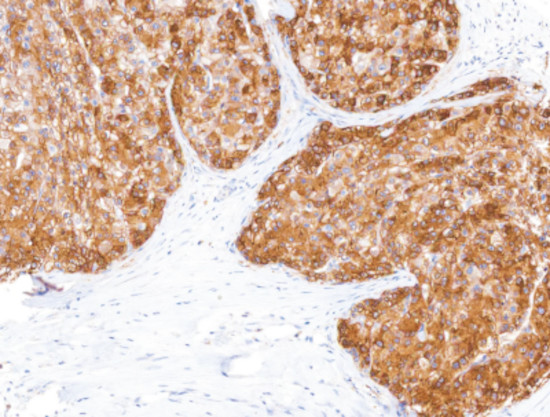
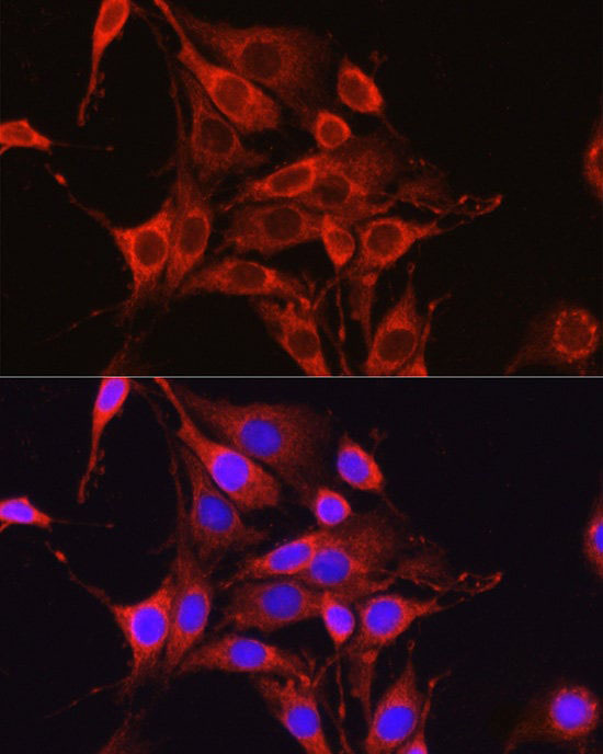
 IHC (Immunohistochemistry)
(Glutamine Synthetase on Liver Cancer)
IHC (Immunohistochemistry)
(Glutamine Synthetase on Liver Cancer)
Mouse anti-Human Glutamine Synthetase Monoclonal Antibody
Glutamine Synthetase
Reactivity
Human
Applications
Immunohistochemistry
Synonyms
Glutamine Synthetase, Antibody; Glutamine Synthetase; anti-Glutamine Synthetase antibody
Host
Mouse
Reactivity
Human
Clonality
Monoclonal
Isotype
IgG2a
Clone Number
IHC586
Form/Format
Tris Buffer, pH 7.3 - 7.7, with 1% BSA and <0.1% Sodium Azide
Applicable Applications for anti-Glutamine Synthetase antibody
IHC (Immunohistochemistry)
Intended Use
The Glutamine Synthetase [IHC586] antibody is intended for qualified laboratories to qualitatively identify by light microscopy, the presence of Glutamine Synthetase expression in formalin-fixed, paraffin-embedded (FFPE) tissue sections using immunohistochemistry assay. Use of this antibody is indicated when there is a need to know the existence of the protein specific cell type and expression levels requested by a qualified pathologist.
Reconstitution, Mixing, Dilution, and Titration
The prediluted antibody does not require any mixing, dilution, reconstitution, or titration; the antibody is ready-to-use and optimized for staining. Any further dilution may affect the quality of the staining signal or antibody-antigen interaction. The concentrated antibody requires dilution using an Antibody Diluent Buffer, to the recommended working dilution range listed in the table above, prior to use.
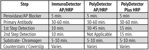
Instructions For Use
Automated Staining with Leica Biosystems Bond-MAX Platform:
This primary antibody has been optimized and validated using the Leica Bond-MAX Fully Automated IHC & ISH Stainer, applying IHC Protocol F. Heat-induced epitope retrieval (HIER) is recommended using ER2 for 20-30 minutes. Antibody concentrate dilution range is 1:50-1:200.
Automated Staining with Ventana BenchMark ULTRA Platform:
This primary antibody has been optimized and validated using the Ventana BenchMark ULTRA IHC/ISH System.
Antibody concentrate dilution range is 1:25-1:100.
Recommended protocol parameters are as follows:
a) Detection Kit: OptiView DAB IHC
b) Pretreatment Protocol: CC1 32-48 minutes, 100°C
c) Primary Antibody: 32 minutes, 36°C
Quality Control Procedures and Interpretation of Results
The immunohistochemical staining process results in a colorimetric reaction at the site of the antigen, localized by the primary antibody. A qualified pathologist must interpret the tissue specimen results only after the positive and negative control tissues have been analyzed. It is recommended to include a set of tissue controls with each staining run to monitor for antibody, tissue, and reagent performance. Tissue sections may contain both positive and negative staining elements. In these cases and where applicable, these sections may serve as both the positive and negative tissue control.
Positive Control Tissue
A positive control tissue should be processed in the same manner as the specimen and run with each test condition to provide control for variables such as tissue processing, fixation, and staining. It should function to provide validity to the specimen results obtained and can consist of fresh autopsy, biopsy, or surgical tissue. Once stained, the positive control tissue should analyzed first to ensure that the antibody and all reagents are performing as intended. Counterstaining will result in a blue coloration, which may range from pale to dark depending on the length of the incubation time and potency of the hematoxylin. If positive staining is not observed, the positive control tissue must be deemed invalid and the results obtained with the tissue specimen must also be treated as such. Positive tissue control: Liver and breast tissues
Negative Control Tissue
Some tissue sections can also function as an internal negative control due to the diversity of staining elements present.
This, however, should first be confirmed by the user.
Negative tissue control: Lung and prostate tissues
This, however, should first be confirmed by the user.
Negative tissue control: Lung and prostate tissues
Tissue Specimens
Tissue specimens should only be analyzed after the positive and negative control tissues have been deemed valid. Negative staining indicates that the antigen was not detected in the tissue while positive staining represents the presence of the antigen. A tissue section stained with hematoxylin and eosin should be used to analyze the morphology of the tissue specimen and verified by a qualified pathologist.
Peformance Characteristics
This antibody has been validated by immunohistochemistry using a FFPE human tissue microarray comprised of different types of tissues. Positive staining was observed in liver and breast tissues. No staining was seen in lung and prostate tissues
Analytical Performance
Trueness of Measure, Precision of Measure, repeatability and reproducibility of the antibody, Analytical Specificity were analyzed using tissue samples from peer-reviewed, published literature known to be either positive or negative, and this study found no unexpected results.
The positive tissue control is liver and breast tissues, and negative tissue control is lung and prostate tissues.
The positive tissue control is liver and breast tissues, and negative tissue control is lung and prostate tissues.
Troubleshooting
1. If tissue sections wash off the slide, this may be caused by:
a) Slides are not positively charged.
b) Inadequate neutral-buffering of the formalin used for the fixation process.
c) A thick tissue section.
d) Inadequate drying of the tissue section prior to staining.
2. If the positive control tissue exhibits negative staining, this may be due to:
a) An issue with the primary antibody or one of the secondary reagents.
b) Improper collection, fixation or deparaffinization of the tissue section.
c) Errors in the IHC staining process.
3. If the positive control tissue exhibits weaker staining than expected, this may be due to sub-optimal IHC conditions, partial degradation of the primary antibody or improper storage of secondary reagents. Analysis of the positive and/or negative control tissues can help with determining the cause.
Preparation and Storage
Store at 2-8°C. To ensure stability, immediately replace vial back in the refrigerator after each use. When stored correctly, the antibody is stable until the expiry date indicated on the label. Positive and negative controls should be concurrently run with tissue specimens, to enable identification of any inadequacies with the antibody or reagents
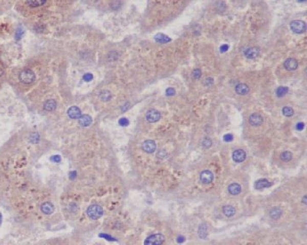
IHC (Immunohistochemistry)
(Glutamine Synthetase on Liver Cancer)
IHC (Immunohistochemistry)
(Glutamine Synthetase on Liver Cancer)
Related Product Information for anti-Glutamine Synthetase antibody
Glutamine Synthetase (GS) catalyzes the conversion of glutamate and ammonia to glutamine in the liver, and is expressed in pericentral hepatocytes, but not in periportal hepatocytes or in the mid-zonal. Detection of Glutamine Synthetase by immunohistochemistry provides the information of Glutamine Synthetase expression level in testing tissue samples.
Customer Reviews
Loading reviews...
Share Your Experience
Similar Products


Product Notes
The Glutamine Synthetase (Catalog #AAA58781) is an Antibody produced from Mouse and is intended for research purposes only. The product is available for immediate purchase. The Glutamine Synthetase reacts with Human and may cross-react with other species as described in the data sheet. AAA Biotech's Glutamine Synthetase can be used in a range of immunoassay formats including, but not limited to, IHC (Immunohistochemistry). Researchers should empirically determine the suitability of the Glutamine Synthetase for an application not listed in the data sheet. Researchers commonly develop new applications and it is an integral, important part of the investigative research process. It is sometimes possible for the material contained within the vial of "Glutamine Synthetase, Monoclonal Antibody" to become dispersed throughout the inside of the vial, particularly around the seal of said vial, during shipment and storage. We always suggest centrifuging these vials to consolidate all of the liquid away from the lid and to the bottom of the vial prior to opening. Please be advised that certain products may require dry ice for shipping and that, if this is the case, an additional dry ice fee may also be required.Precautions
All products in the AAA Biotech catalog are strictly for research-use only, and are absolutely not suitable for use in any sort of medical, therapeutic, prophylactic, in-vivo, or diagnostic capacity. By purchasing a product from AAA Biotech, you are explicitly certifying that said products will be properly tested and used in line with industry standard. AAA Biotech and its authorized distribution partners reserve the right to refuse to fulfill any order if we have any indication that a purchaser may be intending to use a product outside of our accepted criteria.Disclaimer
Though we do strive to guarantee the information represented in this datasheet, AAA Biotech cannot be held responsible for any oversights or imprecisions. AAA Biotech reserves the right to adjust any aspect of this datasheet at any time and without notice. It is the responsibility of the customer to inform AAA Biotech of any product performance issues observed or experienced within 30 days of receipt of said product. To see additional details on this or any of our other policies, please see our Terms & Conditions page.Item has been added to Shopping Cart
If you are ready to order, navigate to Shopping Cart and get ready to checkout.