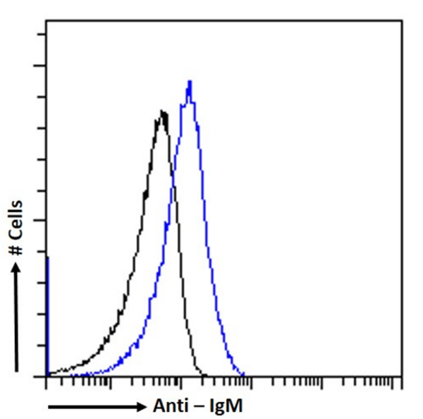
 FCM/FACS (Flow Cytometry)
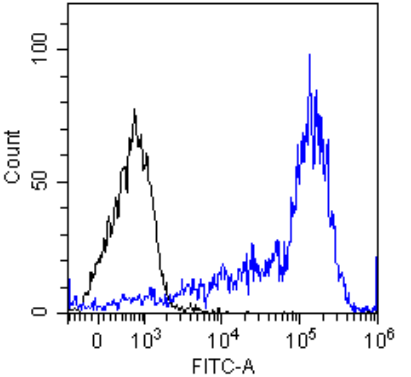
(Flow-cytometry using the anti-IgM M15/8 (AAA72065) Daudi cells were stained with unimmunized rabbit IgG antibody (black line) or the rabbit-chimeric version of M15/8 (, blue line) at a concentration of 10ug/ml for 30 mins at RT. After washing, bound antibody was detected using anti-rabbit IgG JK (FITC-conjugate) antibody (129936) at 2ug/ml and cells analyzed on a FACSCanto flow-cytometer.)
FCM/FACS (Flow Cytometry)
(Flow-cytometry using the anti-IgM M15/8 (AAA72065) Daudi cells were stained with unimmunized rabbit IgG antibody (black line) or the rabbit-chimeric version of M15/8 (, blue line) at a concentration of 10ug/ml for 30 mins at RT. After washing, bound antibody was detected using anti-rabbit IgG JK (FITC-conjugate) antibody (129936) at 2ug/ml and cells analyzed on a FACSCanto flow-cytometer.)
Rabbit anti-Human IgM Monoclonal Antibody | anti-IgM antibody
Anti-IgM [M15/8]
FCM/FACS (Flow Cytometry)
(Flow-cytometry using the anti-IgM M15/8 (AAA72065) Daudi cells were stained with unimmunized rabbit IgG antibody (black line) or the rabbit-chimeric version of M15/8 (, blue line) at a concentration of 10ug/ml for 30 mins at RT. After washing, bound antibody was detected using anti-rabbit IgG JK (FITC-conjugate) antibody (129936) at 2ug/ml and cells analyzed on a FACSCanto flow-cytometer.)
WB (Western Blot)
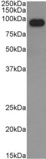
(Western Blot using anti-IgM antibody M15/8 (AAA72065) Human spleen lysate (35ug protein in RIPA buffer) was resolved on a 10% SDS PAGE gel and blots probed with the chimeric rabbit version of M15/8 () at 0.3ug/ml before detection using an anti-rabbit secondary antibody. A primary incubation of 1h was used and protein was detected by chemiluminescence. The expected band size for IgM is 180/900kDa for monomeric/pentameric IgM respectively. Here it is likely that the observed fragment is single glycosylated IgM heavy chain as AAA72065 is targetted to the Fc region of IgM (Cragg et al., PMID: 10070880). successfully detected IgM in human spleen lysate.)
WB (Western Blot)
(Western Blot using anti-IgM antibody M15/8 (AAA72065) Human spleen lysate (35ug protein in RIPA buffer) was resolved on a 10% SDS PAGE gel and blots probed with the chimeric rabbit version of M15/8 () at 0.3ug/ml before detection using an anti-rabbit secondary antibody. A primary incubation of 1h was used and protein was detected by chemiluminescence. The expected band size for IgM is 180/900kDa for monomeric/pentameric IgM respectively. Here it is likely that the observed fragment is single glycosylated IgM heavy chain as AAA72065 is targetted to the Fc region of IgM (Cragg et al., PMID: 10070880). successfully detected IgM in human spleen lysate.)
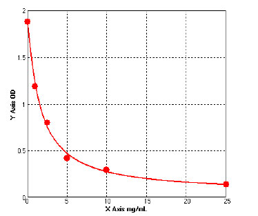
ELISA
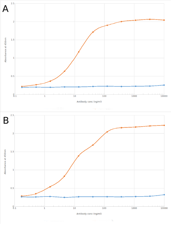
(ELISA of anti-IgM antibody on IgM Fc region peptide and a murine IgM antibody. Binding curves of the rabbit chimeric version of the anti-IgM antibody M15/8 (; red line) and isotype control (anti-Fluorescein atnibody; blue line) to an ELISA plate coated with either IgM Fc region peptide (Pr00108-15.5) at a concentration of 5ug/ml (A) or murine IgM antibody (B; Anti-CD41 antibody) again at 5ug/ml. A 3-fold serial dilution from 10,000 to 0.17 ng/ml was performed using . For signal detection, a 1:4000 dilution of HRP-labelled anti-rabbit IgG1 antibody was used. Both IgM Fc region peptide (monomeric ligand) and murine IgM antibody (multimeric ligand) were recognised by .)
ELISA
(ELISA of anti-IgM antibody on IgM Fc region peptide and a murine IgM antibody. Binding curves of the rabbit chimeric version of the anti-IgM antibody M15/8 (; red line) and isotype control (anti-Fluorescein atnibody; blue line) to an ELISA plate coated with either IgM Fc region peptide (Pr00108-15.5) at a concentration of 5ug/ml (A) or murine IgM antibody (B; Anti-CD41 antibody) again at 5ug/ml. A 3-fold serial dilution from 10,000 to 0.17 ng/ml was performed using . For signal detection, a 1:4000 dilution of HRP-labelled anti-rabbit IgG1 antibody was used. Both IgM Fc region peptide (monomeric ligand) and murine IgM antibody (multimeric ligand) were recognised by .)
FCM/FACS (Flow Cytometry)
(Flow-cytometry using the anti-IgM M15/8 (AAA72065) Daudi cells were stained with unimmunized rabbit IgG antibody (black line) or the rabbit-chimeric version of M15/8 (, blue line) at a concentration of 10ug/ml for 30 mins at RT. After washing, bound antibody was detected using anti-rabbit IgG JK (FITC-conjugate) antibody (129936) at 2ug/ml and cells analyzed on a FACSCanto flow-cytometer.)
BALB/c mice immunized with human IgM.
Note on publication: Describes the characterization of the anti-proliferative effect of this antibody on B-cell lymphoma cell lines when applied in a cross-linking F(ab')3 format.
NCBI and Uniprot Product Information
Customer Reviews
Loading reviews...
Share Your Experience
Similar Products


Product Notes
The IgM ighm (Catalog #AAA72065) is an Antibody produced from Rabbit and is intended for research purposes only. The product is available for immediate purchase. The Anti-IgM [M15/8] reacts with Human and may cross-react with other species as described in the data sheet. AAA Biotech's IgM can be used in a range of immunoassay formats including, but not limited to, Blocking, IP (Immunoprecipitation), IHC (Immunohistochemistry), FCM/FACS (Flow Cytometry), ELISA. Researchers should empirically determine the suitability of the IgM ighm for an application not listed in the data sheet. Researchers commonly develop new applications and it is an integral, important part of the investigative research process. It is sometimes possible for the material contained within the vial of "IgM, Monoclonal Antibody" to become dispersed throughout the inside of the vial, particularly around the seal of said vial, during shipment and storage. We always suggest centrifuging these vials to consolidate all of the liquid away from the lid and to the bottom of the vial prior to opening. Please be advised that certain products may require dry ice for shipping and that, if this is the case, an additional dry ice fee may also be required.Precautions
All products in the AAA Biotech catalog are strictly for research-use only, and are absolutely not suitable for use in any sort of medical, therapeutic, prophylactic, in-vivo, or diagnostic capacity. By purchasing a product from AAA Biotech, you are explicitly certifying that said products will be properly tested and used in line with industry standard. AAA Biotech and its authorized distribution partners reserve the right to refuse to fulfill any order if we have any indication that a purchaser may be intending to use a product outside of our accepted criteria.Disclaimer
Though we do strive to guarantee the information represented in this datasheet, AAA Biotech cannot be held responsible for any oversights or imprecisions. AAA Biotech reserves the right to adjust any aspect of this datasheet at any time and without notice. It is the responsibility of the customer to inform AAA Biotech of any product performance issues observed or experienced within 30 days of receipt of said product. To see additional details on this or any of our other policies, please see our Terms & Conditions page.Item has been added to Shopping Cart
If you are ready to order, navigate to Shopping Cart and get ready to checkout.