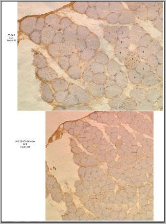
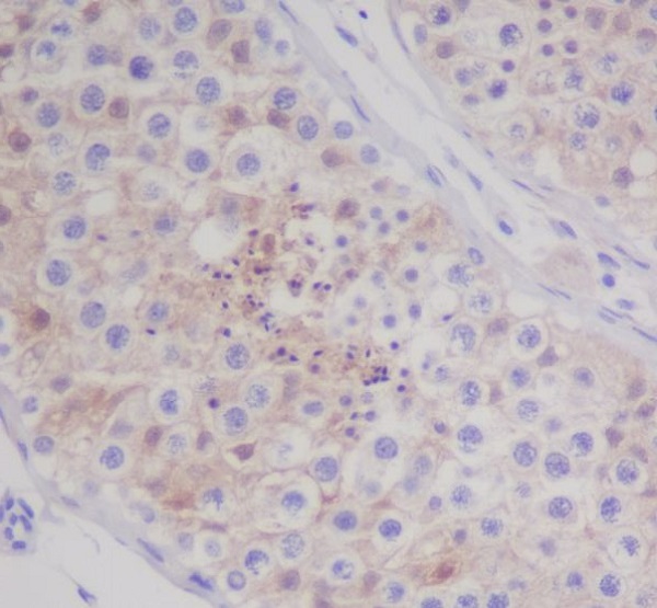
 IHC (Immunohistochemistry)
(10X(lower panel) and 20X (upper panel) immunohistochemistry images from muscle tissue of a diseased mouse off Dox after 5 weeks on regular food. Several fibers that have autophagic vesicles throughout are visible. Primary antibody used is. Data courtesy of Dr. Christy Caudill, Cincinnati Children's Hospital Medical Center.)
IHC (Immunohistochemistry)
(10X(lower panel) and 20X (upper panel) immunohistochemistry images from muscle tissue of a diseased mouse off Dox after 5 weeks on regular food. Several fibers that have autophagic vesicles throughout are visible. Primary antibody used is. Data courtesy of Dr. Christy Caudill, Cincinnati Children's Hospital Medical Center.)
Mouse LC3 Monoclonal Antibody | anti-MAP1LC3A antibody
LC3 Antibody (APG8)
IHC (Immunohistochemistry)
(10X(lower panel) and 20X (upper panel) immunohistochemistry images from muscle tissue of a diseased mouse off Dox after 5 weeks on regular food. Several fibers that have autophagic vesicles throughout are visible. Primary antibody used is. Data courtesy of Dr. Christy Caudill, Cincinnati Children's Hospital Medical Center.)
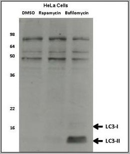
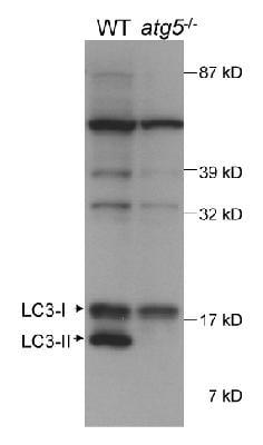
WB (Western Blot)
(Western blot analysis of anti-LC3 Mab Hela cell lysates, which were treated with rapamycin or bafilomycin overnight. Data courtesy of Dr. David Rubinsztein, Cambridge Institute for Medical Research.)
WB (Western Blot)
(Western blot analysis of anti-LC3 Mab Hela cell lysates, which were treated with rapamycin or bafilomycin overnight. Data courtesy of Dr. David Rubinsztein, Cambridge Institute for Medical Research.)
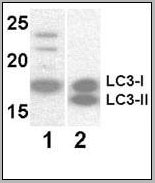
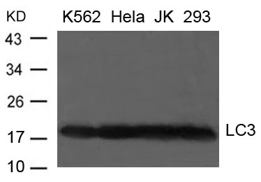
WB (Western Blot)
(Western blot analysis of anti-LC3 Mab at 8 ug/ml. Lane 1: Y79 (soluble fraction of cell extract); Lane 2: 293 transfected with human LC3 (whole cell extract).)
WB (Western Blot)
(Western blot analysis of anti-LC3 Mab at 8 ug/ml. Lane 1: Y79 (soluble fraction of cell extract); Lane 2: 293 transfected with human LC3 (whole cell extract).)
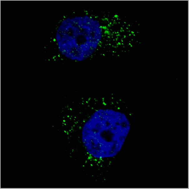
IF (Immunofluorescence)
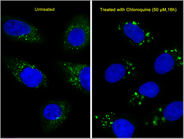
(Fluorescent image of U251 cells stained with AAA28636 LC3 (APG8) antibody.U251 cells were treated with Chloroquine (50 uM,16h), then fixed with 4% PFA (20 min), permeabilized with Triton X-100 (0.2%, 30 min). Cells were then incubated with AAA28636 LC3 (APG8) primary antibody (1:200, 2 h at room temperature). For secondary antibody, Alexa Fluor 488 conjugated donkey anti-mouse antibody (green) was used (1:1000, 1h). Nuclei were counterstained with Hoechst 33342 (blue) (10 ug/ml, 5 min). LC3 immunoreactivity is localized to autophagic vacuoles in the cytoplasm of U251 cells.)
IF (Immunofluorescence)
(Fluorescent image of U251 cells stained with AAA28636 LC3 (APG8) antibody.U251 cells were treated with Chloroquine (50 uM,16h), then fixed with 4% PFA (20 min), permeabilized with Triton X-100 (0.2%, 30 min). Cells were then incubated with AAA28636 LC3 (APG8) primary antibody (1:200, 2 h at room temperature). For secondary antibody, Alexa Fluor 488 conjugated donkey anti-mouse antibody (green) was used (1:1000, 1h). Nuclei were counterstained with Hoechst 33342 (blue) (10 ug/ml, 5 min). LC3 immunoreactivity is localized to autophagic vacuoles in the cytoplasm of U251 cells.)
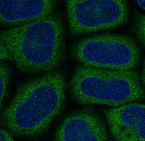
IF (Immunofluorescence)
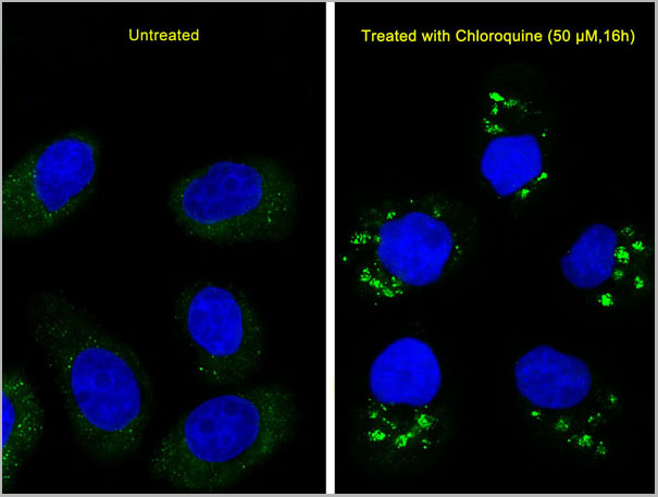
(Immunofluorescent analysis of U251 cells, using LC3 Antibody (APG8). U251 cells(right) were treated with Chloroquine (50 uM,16h). AAA28636 was diluted at 1:25 dilution. Dylight Fluor 488-conjugated goat anti-mouse lgG at 1:400 dilution was used as the secondary antibody (green). DAPI was used to stain the cell nuclear (blue).)
IF (Immunofluorescence)
(Immunofluorescent analysis of U251 cells, using LC3 Antibody (APG8). U251 cells(right) were treated with Chloroquine (50 uM,16h). AAA28636 was diluted at 1:25 dilution. Dylight Fluor 488-conjugated goat anti-mouse lgG at 1:400 dilution was used as the secondary antibody (green). DAPI was used to stain the cell nuclear (blue).)
WB (Western Blot)
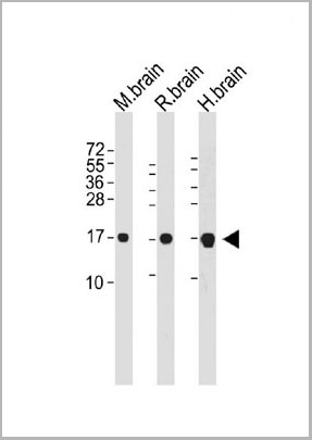
(All lanes : Anti-MAP1LC3A Antibody at dilutionLane 1: Mouse brain whole cell lysateLane 2:Rat brain whole cell lysateLane 3: Human brain whole cell lysateLysates/proteins at 20 ug per lane.Secondary Goat Anti-mouse IgG, (H+L), Peroxidase conjugated at 1/10000 dilution. Predicted band size : 14 kDaBlocking/Dilution buffer: 5% NFDM/TBST.)
WB (Western Blot)
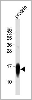
(Anti-MAP1LC3A Antibody at 1:5000 dilution + recombinant protein Lysates/proteins at 20 ug per lane. Secondary Goat Anti-mouse IgG, (H+L), Peroxidase conjugated at 1/10000 dilution. Predicted band size : 14 kDa. Blocking/Dilution buffer: 5% NFDM/TBST.)
WB (Western Blot)
(Anti-MAP1LC3A Antibody at 1:5000 dilution + recombinant protein Lysates/proteins at 20 ug per lane. Secondary Goat Anti-mouse IgG, (H+L), Peroxidase conjugated at 1/10000 dilution. Predicted band size : 14 kDa. Blocking/Dilution buffer: 5% NFDM/TBST.)
1.Autophagy negatively regulates Wnt signalling by promoting Dishevelled degradation. Gao C, et al. Nat Cell Biol, 2010 Aug. PMID 20639871.
2.The prolyl isomerase Pin1 induces LC-3 expression and mediates tamoxifen resistance in breast cancer. Namgoong GM, et al. J Biol Chem, 2010 Jul 30. PMID 20479004.
3.Protein kinase C inhibits autophagy and phosphorylates LC3. Jiang H, et al. Biochem Biophys Res Commun, 2010 May 14. PMID 20398630.
4. Processing of autophagic protein LC3 by the 20S proteasome. Gao Z, et al. Autophagy, 2010 Jan. PMID 20061800.
5.Defective autophagy associated with LC3 puncta in epothilone-resistant cancer cells. Shen S, et al. Cell Cycle, 2010 Jan 15. PMID 20023420.
References for U251 cell line:
1. Westermark B.; Pontén J.; Hugosson R. (1973)." Determinants for the establishment of permanent tissue culture lines from human gliomas". Acta Pathol Microbiol Scand A. 81:791-805. [PMID: 4359449].
2. Pontén, J.,Westermark B. (1978)." Properties of Human Malignant Glioma Cells in Vitro". Medical Biology 56: 184-193.[PMID: 359950].
3. Geng Y.;Kohli L.; Klocke B.J.; Roth K.A.(2010). "Chloroquine-induced autophagic vacuole accumulation and cell death in glioma cells is p53 independent". Neuro Oncol. 12(5): 473-481.[ PMID: 20406898].
NCBI and Uniprot Product Information
Customer Reviews
Loading reviews...
Share Your Experience
Similar Products

Product Notes
The MAP1LC3A map1lc3a (Catalog #AAA28636) is an Antibody produced from Mouse and is intended for research purposes only. The product is available for immediate purchase. The LC3 Antibody (APG8) reacts with Human, mouse, rat and may cross-react with other species as described in the data sheet. AAA Biotech's LC3 can be used in a range of immunoassay formats including, but not limited to, WB (Western Blot), ELISA. WB~~1:8000. Researchers should empirically determine the suitability of the MAP1LC3A map1lc3a for an application not listed in the data sheet. Researchers commonly develop new applications and it is an integral, important part of the investigative research process. It is sometimes possible for the material contained within the vial of "LC3, Monoclonal Antibody" to become dispersed throughout the inside of the vial, particularly around the seal of said vial, during shipment and storage. We always suggest centrifuging these vials to consolidate all of the liquid away from the lid and to the bottom of the vial prior to opening. Please be advised that certain products may require dry ice for shipping and that, if this is the case, an additional dry ice fee may also be required.Precautions
All products in the AAA Biotech catalog are strictly for research-use only, and are absolutely not suitable for use in any sort of medical, therapeutic, prophylactic, in-vivo, or diagnostic capacity. By purchasing a product from AAA Biotech, you are explicitly certifying that said products will be properly tested and used in line with industry standard. AAA Biotech and its authorized distribution partners reserve the right to refuse to fulfill any order if we have any indication that a purchaser may be intending to use a product outside of our accepted criteria.Disclaimer
Though we do strive to guarantee the information represented in this datasheet, AAA Biotech cannot be held responsible for any oversights or imprecisions. AAA Biotech reserves the right to adjust any aspect of this datasheet at any time and without notice. It is the responsibility of the customer to inform AAA Biotech of any product performance issues observed or experienced within 30 days of receipt of said product. To see additional details on this or any of our other policies, please see our Terms & Conditions page.Item has been added to Shopping Cart
If you are ready to order, navigate to Shopping Cart and get ready to checkout.