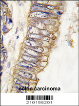
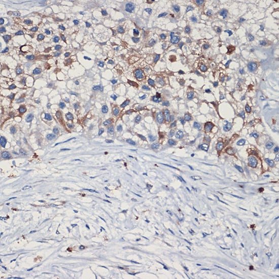
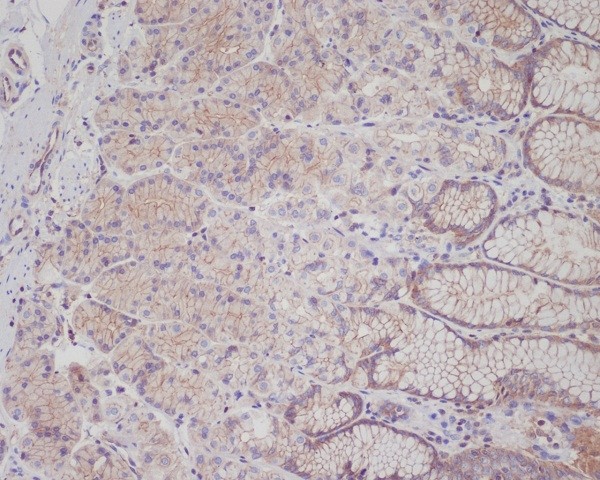
 IHC (Immunohistochemistry)
(Formalin-fixed and paraffin-embedded human colon carcinoma tissue reacted with MET/HGFR Antibody , which was peroxidase-conjugated to the secondary antibody, followed by DAB staining. This data demonstrates the use of this antibody for immunohistochemistry; clinical relevance has not been evaluated.)
IHC (Immunohistochemistry)
(Formalin-fixed and paraffin-embedded human colon carcinoma tissue reacted with MET/HGFR Antibody , which was peroxidase-conjugated to the secondary antibody, followed by DAB staining. This data demonstrates the use of this antibody for immunohistochemistry; clinical relevance has not been evaluated.)
Mouse anti-Human, mouse MET/HGFR Monoclonal Antibody | anti-MET antibody
MET/HGFR Antibody
IHC (Immunohistochemistry)
(Formalin-fixed and paraffin-embedded human colon carcinoma tissue reacted with MET/HGFR Antibody , which was peroxidase-conjugated to the secondary antibody, followed by DAB staining. This data demonstrates the use of this antibody for immunohistochemistry; clinical relevance has not been evaluated.)
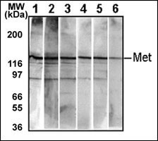
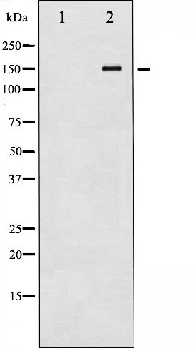
WB (Western Blot)
(Detection of endogenous Met in HepG2 cell line. 10 ug/lane of HepG2 cell lysate was used to examine the expression of human Met. Lanes 1-5 represent AAA Biotech ’s different anti-Met monoclonal antibodies that are. Lane 6 represents auto-phosohorylated-Met in HepG2 cell line detected by anti-phospho-Met Mab.)
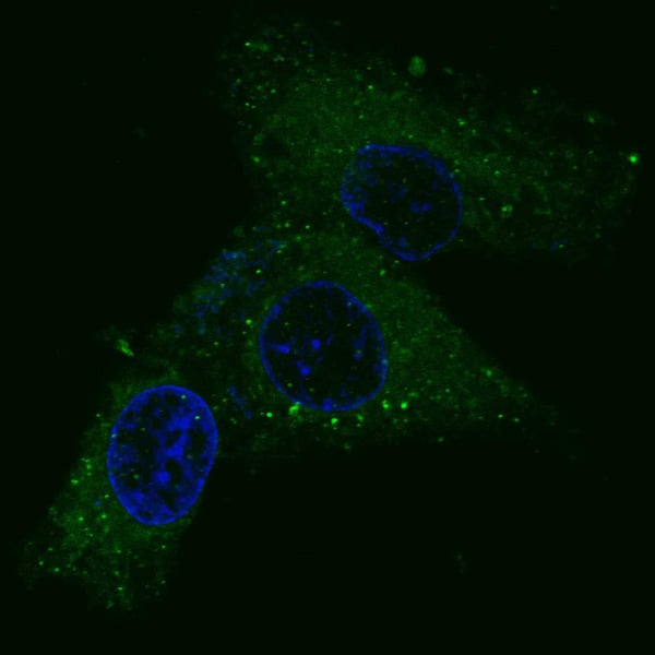
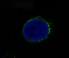
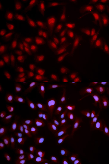
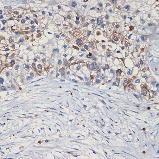
IF (Immunofluorescence)
(Fluorescent confocal image of HepG2 cells stained with MET/HGFR antibody. HepG2 cells were fixed with 4% PFA (20 min), permeabilized with Triton X-100 (0.2%, 30 min). Cells were then incubated with AAA283479 MET/HGFR primary antibody (1:100, 2 h at room temperature). For secondary antibody, Alexa Fluor 488 conjugated donkey anti-mouse antibody (green) was used (1:1000, 1h). Nuclei were counterstained with Hoechst 33342 (blue) (10 ug/ml, 5 min). Note the highly specific localization of the MET immunosignal to the cytoplasm, supported by Human Protein Atlas Data (http://www.proteinatlas.org/ENSG00000105976).)
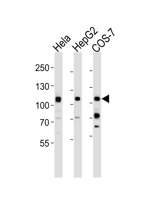
WB (Western Blot)
(Western blot analysis of lysates from Hela, HepG2, COS-7 cell line (from left to right), using MET/HGFR Antibody(4AT44). 4AT44 was diluted at 1:1000 at each lane. A goat anti-mouse IgG H&L(HRP) at 1:3000 dilution was used as the secondary antibody. Lysates at 35ug per lane.)
WB (Western Blot)
(Western blot analysis of lysates from Hela, HepG2, COS-7 cell line (from left to right), using MET/HGFR Antibody(4AT44). 4AT44 was diluted at 1:1000 at each lane. A goat anti-mouse IgG H&L(HRP) at 1:3000 dilution was used as the secondary antibody. Lysates at 35ug per lane.)
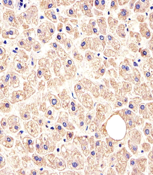
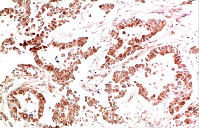
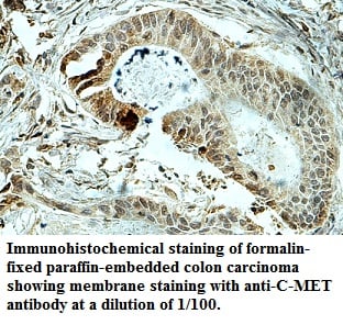
IHC (Immunohistochemistry)
(Immunohistochemical analysis of paraffin-embedded H.liver section using MET/HGFR Antibody. AAA283479 was diluted at 1:25 dilution. A peroxidase-conjugated goat anti-rabbit IgG at 1:400 dilution was used as the secondary antibody, followed by DAB staining.)
IHC (Immunohistochemistry)
(Immunohistochemical analysis of paraffin-embedded H.liver section using MET/HGFR Antibody. AAA283479 was diluted at 1:25 dilution. A peroxidase-conjugated goat anti-rabbit IgG at 1:400 dilution was used as the secondary antibody, followed by DAB staining.)
1.MET receptor sequence variants R970C and T992I lack transforming capacity. Tyner JW, et al. Cancer Res, 2010 Aug 1. PMID 20670955.
2.Further evidence for the role of MET in autism susceptibility. Thanseem I, et al. Neurosci Res, 2010 Oct. PMID 20615438.
3.Increased HGF and c-Met in muscle tissues of polymyositis and dermatomyositis patients: beneficial roles of HGF in muscle regeneration. Sugiura T, et al. Clin Immunol, 2010 Sep. PMID 20580899.
4.Correlation between hepatocyte growth factor receptor and vascular endothelial growth factor-A in breast carcinoma. Gisterek I, et al. Folia Histochem Cytobiol, 2010 Jan 1. PMID 20529820.
5.MET overexpressing chordomas frequently exhibit polysomy of chromosome 7 but no MET activation through sarcoma-specific gene fusions. Grabellus F, et al. Tumour Biol, 2010 Jun. PMID 20512480.
References for HepG2 cell line:
1. Knowles BB, et al. (1980). Human hepatocellular carcinoma cell lines secrete the major plasma proteins and hepatitis B surface antigen. Science 209: 497-499.[ PubMed: 6248960].
2. Darlington GJ, et al. (1987). Growth and hepatospecific gene expression of human hepatoma cells in a defined medium. In Vitro Cell. Dev. Biol. 23: 349-354.[PubMed: 3034851].
3. Ihrke, G; Neufeld, EB; Meads, T; Shanks, MR; Cassio, D; Laurent, M; Schroer, TA; Pagano, RE et al. (1993). "WIF-B cells: an in vitro model for studies of hepatocyte polarity". Journal of Cell Biology 123 (6): 1761-1775. [PubMed:7506266].
4. Mersch-Sundermann, V.; Knasmüller, S.; Wu, X. J.; Darroudi, F.; Kassie, F. (2004). "Use of a human-derived liver cell line for the detection of cytoprotective, antigenotoxic and cogenotoxic agents". Toxicology 198 (1-3): 329-340. [PubMed:15138059].
NCBI and Uniprot Product Information
Customer Reviews
Loading reviews...
Share Your Experience
Similar Products


Product Notes
The MET met (Catalog #AAA283479) is an Antibody produced from Mouse and is intended for research purposes only. The product is available for immediate purchase. The MET/HGFR Antibody reacts with Human, mouse and may cross-react with other species as described in the data sheet. AAA Biotech's MET/HGFR can be used in a range of immunoassay formats including, but not limited to, IHC (Immunohistochemistry), WB (Western Blot), ELISA, IF (Immunofluorescence). Researchers should empirically determine the suitability of the MET met for an application not listed in the data sheet. Researchers commonly develop new applications and it is an integral, important part of the investigative research process. It is sometimes possible for the material contained within the vial of "MET/HGFR, Monoclonal Antibody" to become dispersed throughout the inside of the vial, particularly around the seal of said vial, during shipment and storage. We always suggest centrifuging these vials to consolidate all of the liquid away from the lid and to the bottom of the vial prior to opening. Please be advised that certain products may require dry ice for shipping and that, if this is the case, an additional dry ice fee may also be required.Precautions
All products in the AAA Biotech catalog are strictly for research-use only, and are absolutely not suitable for use in any sort of medical, therapeutic, prophylactic, in-vivo, or diagnostic capacity. By purchasing a product from AAA Biotech, you are explicitly certifying that said products will be properly tested and used in line with industry standard. AAA Biotech and its authorized distribution partners reserve the right to refuse to fulfill any order if we have any indication that a purchaser may be intending to use a product outside of our accepted criteria.Disclaimer
Though we do strive to guarantee the information represented in this datasheet, AAA Biotech cannot be held responsible for any oversights or imprecisions. AAA Biotech reserves the right to adjust any aspect of this datasheet at any time and without notice. It is the responsibility of the customer to inform AAA Biotech of any product performance issues observed or experienced within 30 days of receipt of said product. To see additional details on this or any of our other policies, please see our Terms & Conditions page.Item has been added to Shopping Cart
If you are ready to order, navigate to Shopping Cart and get ready to checkout.