
 Application Data
(Published customer image: NL scheme and PLP staining. (A) Type-I NL with demyelination of the whole width of the cortex (#) and adjacent WM (*). (B) Type-II intracortical lesion evolving around a vessel. (C) Type-III subpial NL. Demyelination spreads from the pial surface until cortical layer 3. Scale bars represent 500 um.From:Yao B, Hametner S, van Gelderen P, Merkle H, Chen C, et al. (2014) 7 Tesla Magnetic Resonance Imaging to Detect Cortical Pathology in Multiple Sclerosis. PLoS ONE 9(10): e108863. doi:10.1371/journal.pone.0108863)
Application Data
(Published customer image: NL scheme and PLP staining. (A) Type-I NL with demyelination of the whole width of the cortex (#) and adjacent WM (*). (B) Type-II intracortical lesion evolving around a vessel. (C) Type-III subpial NL. Demyelination spreads from the pial surface until cortical layer 3. Scale bars represent 500 um.From:Yao B, Hametner S, van Gelderen P, Merkle H, Chen C, et al. (2014) 7 Tesla Magnetic Resonance Imaging to Detect Cortical Pathology in Multiple Sclerosis. PLoS ONE 9(10): e108863. doi:10.1371/journal.pone.0108863)
Mouse MYELIN PROTEOLIPID PROTEIN Monoclonal Antibody | anti-PLP1 antibody
MOUSE ANTI MYELIN PROTEOLIPID PROTEIN
Purified IgG - liquid
Preparation
Shelf Life: 18 months from date of despatch.
Application Data
(Published customer image: NL scheme and PLP staining. (A) Type-I NL with demyelination of the whole width of the cortex (#) and adjacent WM (*). (B) Type-II intracortical lesion evolving around a vessel. (C) Type-III subpial NL. Demyelination spreads from the pial surface until cortical layer 3. Scale bars represent 500 um.From:Yao B, Hametner S, van Gelderen P, Merkle H, Chen C, et al. (2014) 7 Tesla Magnetic Resonance Imaging to Detect Cortical Pathology in Multiple Sclerosis. PLoS ONE 9(10): e108863. doi:10.1371/journal.pone.0108863)
Application Data
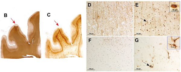
(Published customer image: Iron loss in NLs. PLP myelin staining (B) and iron staining (C) on two consecutive slides of tissue MS2 disclose iron loss together with myelin loss in a NL (red arrows). Higher magnification (D, E, F, G) of PLP (D, F) and iron stainings (E, G). In NAGM (D, E), iron is present in oligodendrocytes (black arrow and inset in E) and myelin sheaths. In a NL (F, G), iron is removed from the demyelinated cortical parenchyma and present in perivascular macrophages and microglia (black arrow and inset in G).From:Yao B, Hametner S, van Gelderen P, Merkle H, Chen C, et al. (2014) 7 Tesla Magnetic Resonance Imaging to Detect Cortical Pathology in Multiple Sclerosis. PLoS ONE 9(10): e108863. doi:10.1371/journal.pone.0108863)
Application Data
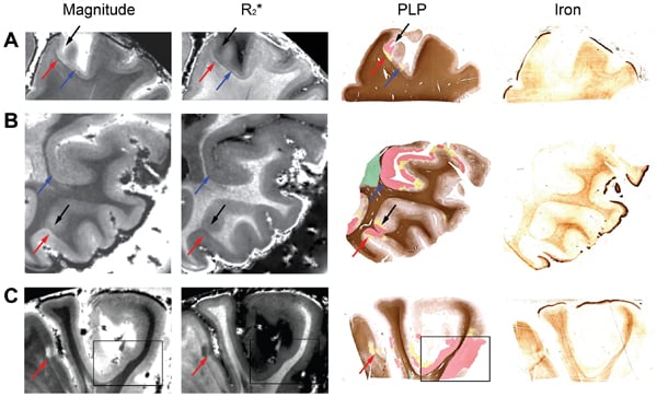
(Published customer image:NLs on MRI and tissue sections stained for PLP and iron. Examples Examples of NLs from tissue MS1 (A, B and C) identified on MRI magnitude (TE = 25.2 ms) and R2* images as well as color-coded PLP-staining and iron staining. Red arrows point towards NLs identified by MRI and confirmed to correspond to area of demyelination by the color-coded PLP staining. Blue arrows point towards NLs identified by the color-coded PLP staining and only retrospectively identified by MRI. Black arrows and black box point towards NLs identified by the color-coded PLP staining and not by MRI even upon a second retrospective image inspection. In the black box we include a large area of demyelination which goes entirely undetected by MRI. In the color-coded PLP staining of figure: green = complete WM demyelination, red = complete GM demyelination, yellow = areas of variably reduced myelin density.From:Yao B, Hametner S, van Gelderen P, Merkle H, Chen C, et al. (2014) 7 Tesla Magnetic Resonance Imaging to Detect Cortical Pathology in Multiple Sclerosis. PLoS ONE 9(10): e108863. doi:10.1371/journal.pone.0108863)
Application Data
(Published customer image:NLs on MRI and tissue sections stained for PLP and iron. Examples Examples of NLs from tissue MS1 (A, B and C) identified on MRI magnitude (TE = 25.2 ms) and R2* images as well as color-coded PLP-staining and iron staining. Red arrows point towards NLs identified by MRI and confirmed to correspond to area of demyelination by the color-coded PLP staining. Blue arrows point towards NLs identified by the color-coded PLP staining and only retrospectively identified by MRI. Black arrows and black box point towards NLs identified by the color-coded PLP staining and not by MRI even upon a second retrospective image inspection. In the black box we include a large area of demyelination which goes entirely undetected by MRI. In the color-coded PLP staining of figure: green = complete WM demyelination, red = complete GM demyelination, yellow = areas of variably reduced myelin density.From:Yao B, Hametner S, van Gelderen P, Merkle H, Chen C, et al. (2014) 7 Tesla Magnetic Resonance Imaging to Detect Cortical Pathology in Multiple Sclerosis. PLoS ONE 9(10): e108863. doi:10.1371/journal.pone.0108863)
NCBI and Uniprot Product Information
Customer Reviews
Loading reviews...
Share Your Experience
Similar Products


Product Notes
The PLP1 plp1 (Catalog #AAA49446) is an Antibody produced from Mouse and is intended for research purposes only. The product is available for immediate purchase. AAA Biotech's MYELIN PROTEOLIPID PROTEIN can be used in a range of immunoassay formats including, but not limited to, WB (Western Blot), IHC (Immunohistochemistry), IF (Immunofluorescence), FCM/FACS (Flow Cytometry), IHC (Immunohistochemistry). Researchers should empirically determine the suitability of the PLP1 plp1 for an application not listed in the data sheet. Researchers commonly develop new applications and it is an integral, important part of the investigative research process. It is sometimes possible for the material contained within the vial of "MYELIN PROTEOLIPID PROTEIN, Monoclonal Antibody" to become dispersed throughout the inside of the vial, particularly around the seal of said vial, during shipment and storage. We always suggest centrifuging these vials to consolidate all of the liquid away from the lid and to the bottom of the vial prior to opening. Please be advised that certain products may require dry ice for shipping and that, if this is the case, an additional dry ice fee may also be required.Precautions
All products in the AAA Biotech catalog are strictly for research-use only, and are absolutely not suitable for use in any sort of medical, therapeutic, prophylactic, in-vivo, or diagnostic capacity. By purchasing a product from AAA Biotech, you are explicitly certifying that said products will be properly tested and used in line with industry standard. AAA Biotech and its authorized distribution partners reserve the right to refuse to fulfill any order if we have any indication that a purchaser may be intending to use a product outside of our accepted criteria.Disclaimer
Though we do strive to guarantee the information represented in this datasheet, AAA Biotech cannot be held responsible for any oversights or imprecisions. AAA Biotech reserves the right to adjust any aspect of this datasheet at any time and without notice. It is the responsibility of the customer to inform AAA Biotech of any product performance issues observed or experienced within 30 days of receipt of said product. To see additional details on this or any of our other policies, please see our Terms & Conditions page.Item has been added to Shopping Cart
If you are ready to order, navigate to Shopping Cart and get ready to checkout.