


 IP (Immunoprecipitation)
(Immunoprecipitation analysis of 300ug extracts of HeLa cells using 3ug SQSTM1/p62 antibody . Western blot was performed from the immunoprecipitate using SQSTM1/p62 antibody at a dilition of 1:1000.)
IP (Immunoprecipitation)
(Immunoprecipitation analysis of 300ug extracts of HeLa cells using 3ug SQSTM1/p62 antibody . Western blot was performed from the immunoprecipitate using SQSTM1/p62 antibody at a dilition of 1:1000.)
Rabbit SQSTM1/p62 Monoclonal Antibody | anti-SQSTM1/p62 antibody
[KO Validated] SQSTM1/p62 Rabbit mAb
IHC: 1:50-1:200
IF: 1:50-1:200
IP: 1:50-1:200
Customer Validation:
WB: Mouse, Human
IHC: Mouse
IP (Immunoprecipitation)
(Immunoprecipitation analysis of 300ug extracts of HeLa cells using 3ug SQSTM1/p62 antibody . Western blot was performed from the immunoprecipitate using SQSTM1/p62 antibody at a dilition of 1:1000.)
IP (Immunoprecipitation)
(Immunoprecipitation analysis of 300ug extracts of HeLa cells using 3ug SQSTM1/p62 antibody . Western blot was performed from the immunoprecipitate using SQSTM1/p62 antibody at a dilition of 1:1000.)
IF (Immunofluorescence)
(Immunofluorescence analysis of L929 cells using SQSTM1/p62 antibody at dilution of 1:100. Blue: DAPI for nuclear staining.)
IF (Immunofluorescence)
(Immunofluorescence analysis of L929 cells using SQSTM1/p62 antibody at dilution of 1:100. Blue: DAPI for nuclear staining.)
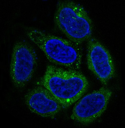
IF (Immunofluorescence)
(Immunofluorescence analysis of HeLa cells using SQSTM1/p62 antibody at dilution of 1:100. Blue: DAPI for nuclear staining.)
IF (Immunofluorescence)
(Immunofluorescence analysis of HeLa cells using SQSTM1/p62 antibody at dilution of 1:100. Blue: DAPI for nuclear staining.)
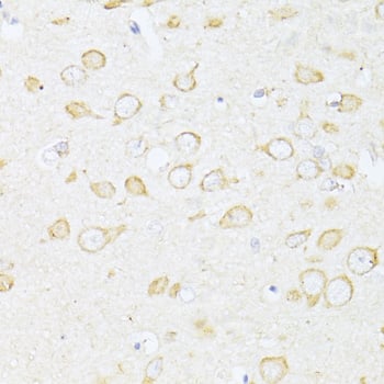
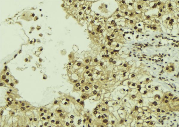
IHC (Immunohistochemistry)
(Immunohistochemistry of paraffin-embedded human lung cancer using SQSTM1/p62 antibody at dilution of 1:100 (40x lens).)
IHC (Immunohistochemistry)
(Immunohistochemistry of paraffin-embedded human lung cancer using SQSTM1/p62 antibody at dilution of 1:100 (40x lens).)
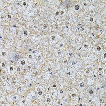
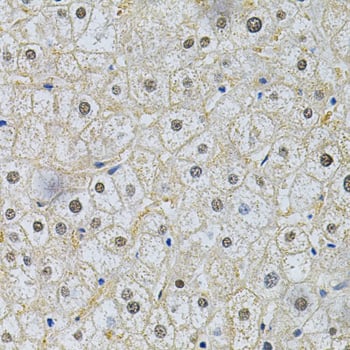
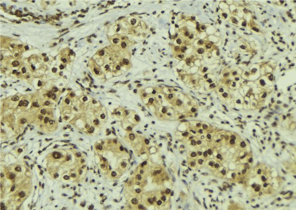
IHC (Immunohistochemistry)
(Immunohistochemistry of paraffin-embedded Rat spleen using SQSTM1/p62 antibody at dilution of 1:100 (40x lens).)
IHC (Immunohistochemistry)
(Immunohistochemistry of paraffin-embedded Rat spleen using SQSTM1/p62 antibody at dilution of 1:100 (40x lens).)
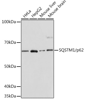
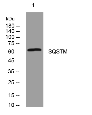
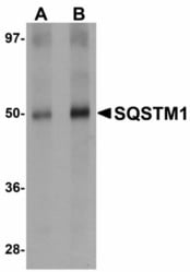
WB (Western Blot)
(Western blot analysis of extracts of various cell lines, using p62/SQSTM1 antibody at 1:1000 dilution.Secondary antibody: HRP Goat Anti-Rabbit IgG (H+L) (AS014) at 1:10000 dilution.Lysates/proteins: 25ug per lane.Blocking buffer: 3% nonfat dry milk in TBST.Detection: ECL Basic Kit (RM00020).Exposure time: 10s.)
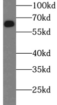
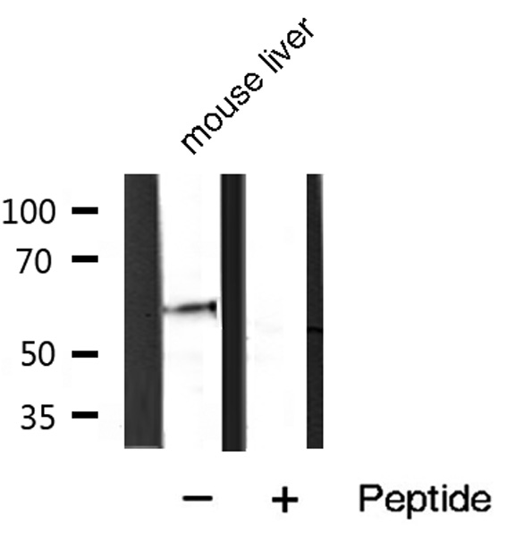
WB (Western Blot)
(Western blot analysis of extracts from normal (control) and SQSTM1/p62 knockout (KO) 293T cells, using SQSTM1/p62 antibody at 1:1000 dilution.Secondary antibody: HRP Goat Anti-Rabbit IgG (H+L) (AS014) at 1:10000 dilution.Lysates/proteins: 25ug per lane.Blocking buffer: 3% nonfat dry milk in TBST.Detection: ECL Basic Kit (RM00020).Exposure time: 3min.)
NCBI and Uniprot Product Information
Customer Reviews
Loading reviews...
Share Your Experience
Similar Products


Product Notes
The SQSTM1/p62 sqstm1 (Catalog #AAA28383) is an Antibody produced from Rabbit and is intended for research purposes only. The product is available for immediate purchase. The [KO Validated] SQSTM1/p62 Rabbit mAb reacts with Human, Mouse, Rat and may cross-react with other species as described in the data sheet. AAA Biotech's SQSTM1/p62 can be used in a range of immunoassay formats including, but not limited to, WB (Western Blot), IHC (Immunohistochemistry), IF (Immunofluorescence), IP (Immunoprecipitation). WB: 1:500-1:2000 IHC: 1:50-1:200 IF: 1:50-1:200 IP: 1:50-1:200 Customer Validation: WB: Mouse, Human IHC: Mouse. Researchers should empirically determine the suitability of the SQSTM1/p62 sqstm1 for an application not listed in the data sheet. Researchers commonly develop new applications and it is an integral, important part of the investigative research process. It is sometimes possible for the material contained within the vial of "SQSTM1/p62, Monoclonal Antibody" to become dispersed throughout the inside of the vial, particularly around the seal of said vial, during shipment and storage. We always suggest centrifuging these vials to consolidate all of the liquid away from the lid and to the bottom of the vial prior to opening. Please be advised that certain products may require dry ice for shipping and that, if this is the case, an additional dry ice fee may also be required.Precautions
All products in the AAA Biotech catalog are strictly for research-use only, and are absolutely not suitable for use in any sort of medical, therapeutic, prophylactic, in-vivo, or diagnostic capacity. By purchasing a product from AAA Biotech, you are explicitly certifying that said products will be properly tested and used in line with industry standard. AAA Biotech and its authorized distribution partners reserve the right to refuse to fulfill any order if we have any indication that a purchaser may be intending to use a product outside of our accepted criteria.Disclaimer
Though we do strive to guarantee the information represented in this datasheet, AAA Biotech cannot be held responsible for any oversights or imprecisions. AAA Biotech reserves the right to adjust any aspect of this datasheet at any time and without notice. It is the responsibility of the customer to inform AAA Biotech of any product performance issues observed or experienced within 30 days of receipt of said product. To see additional details on this or any of our other policies, please see our Terms & Conditions page.Frequently Asked Questions
What is p62/SQSTM1 used as a marker for in autophagy studies?
p62/SQSTM1 (sequestosome 1) is a multifunctional adaptor protein serving as a direct marker of autophagic flux. p62 associates with ubiquitinated protein aggregates and delivers them to autophagosomes via LC3 interaction, then undergoes degradation within autolysosomes. Therefore, p62 accumulation indicates impaired autophagy, while depletion indicates active autophagic progression.
Can a p62 antibody be used to detect protein aggregates in cells?
Yes, p62 specifically recognizes and binds ubiquitinated protein aggregates through its UBA (ubiquitin-binding associated) domain, making this antibody excellent for detecting aggregate pathology. In immunofluorescence, p62-positive puncta visualize intracellular aggregates. In Western blot, p62 levels quantify aggregate burden, making the antibody invaluable for studying neurodegenerative diseases.
Is the p62 antibody suitable for Western blot or immunofluorescence applications?
Yes, this polyclonal is validated for both applications. In Western blot, it detects p62 at its expected molecular weight (~62 kDa, though calculated MW is 48 kDa due to post-translational modifications). In immunofluorescence, it produces bright punctate staining reflecting p62-positive aggregates, allowing quantitative assessment of autophagy flux changes.
How does p62 expression change during autophagy or stress conditions?
Under normal conditions, p62 maintains relatively stable baseline levels. During acute autophagy induction (nutrient starvation), p62 accumulates transiently as autophagosomes form, then rapidly decreases as autophagic flux progresses. Under chronic stress or autophagy inhibition (Bafilomycin A1, ATG gene knockouts), p62 accumulates to high levels, marking blocked autophagic degradation.
Can p62 antibody be used to study neurodegenerative or cancer research?
Yes, p62 is extensively used in both fields. In neurodegeneration (Alzheimer's, Huntington's, ALS), p62-positive aggregates accumulate, marking pathological protein inclusions correlating with disease severity. In cancer, altered p62 levels influence cell survival, metastatic potential, and chemotherapy resistance through autophagy-dependent and independent mechanisms.
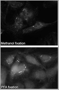
Does fixation method affect p62 staining quality?
Yes, the fixation method significantly impacts detection. Formalin or PFA fixation (4%, 15-30 minutes) provides optimal preservation of p62 protein structure and epitope accessibility. Prolonged fixation (>30 minutes) or cross-linking can reduce antibody penetration and signal intensity. Methanol or acetone may disrupt p62's membrane-associated organization; brief PFA fixation maintains p62 aggregate morphology.
Item has been added to Shopping Cart
If you are ready to order, navigate to Shopping Cart and get ready to checkout.