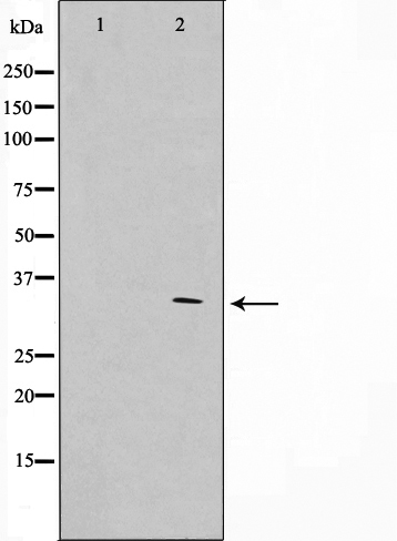

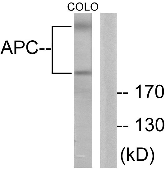
 WB (Western Blot)
(Western blot analysis on HuvEc cell lysate using Phospho-APC(Ser2054) Antibody. The lane on the left is treated with the antigen-specific peptide.)
WB (Western Blot)
(Western blot analysis on HuvEc cell lysate using Phospho-APC(Ser2054) Antibody. The lane on the left is treated with the antigen-specific peptide.)
Rabbit anti-Human APC Polyclonal Antibody | anti-APC antibody
Phospho-APC(Ser2054) Antibody
Phosphate buffered saline, pH 7.4, 150mM NaCl, 0.02% sodium azide and 50% glycerol.
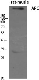
WB (Western Blot)
(Western blot analysis on HuvEc cell lysate using Phospho-APC(Ser2054) Antibody. The lane on the left is treated with the antigen-specific peptide.)
WB (Western Blot)
(Western blot analysis on HuvEc cell lysate using Phospho-APC(Ser2054) Antibody. The lane on the left is treated with the antigen-specific peptide.)
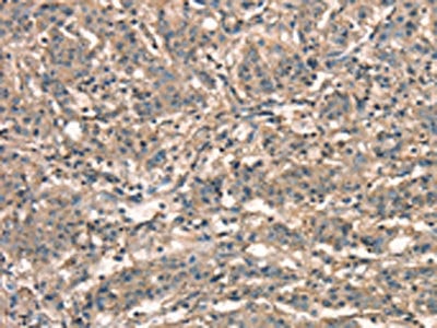
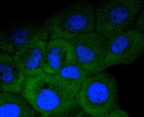
IHC (Immunohiostchemistry)
(AAA321246 at 1/100 staining human Rectum tissue sections by IHC-P. The tissue was formaldehyde fixed and a heat mediated antigen retrieval step in citrate buffer was performed. The tissue was then blocked and incubated with the antibody for 1.5 hours at 22 degree C. An HRP conjugated goat anti-rabbit antibody was used as the secondary.)
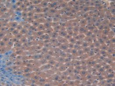
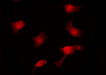
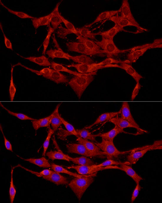
IF (Immunofluorescence)
(AAA321246 staining HuvEc by IF/ICC. The sample were fixed with PFA and permeabilized in 0.1% Triton X-100, then blocked in 10% serum for 45 minutes at 25 degree C. The primary antibody was diluted at 1/200 and incubated with the sample for 1 hour at 37 degree C. An Alexa Fluor 594 conjugated goat anti-rabbit IgG (H+L) Ab, diluted at 1/600, was used as the secondary antibody.)
Function: Tumor suppressor. Promotes rapid degradation of CTNNB1 and participates in Wnt signaling as a negative regulator. APC activity is correlated with its phosphorylation state. Activates the GEF activity of SPATA13 and ARHGEF4. Plays a role in hepatocyte growth factor (HGF)-induced cell migration. Required for MMP9 up-regulation via the JNK signaling pathway in colorectal tumor cells. Acts as a mediator of ERBB2-dependent stabilization of microtubules at the cell cortex. It is required for the localization of MACF1 to the cell membrane and this localization of MACF1 is critical for its function in microtubule stabilization.
Subunit Structure: Forms homooligomers and heterooligomers with APC2. Interacts with DIAPH1 and DIAPH2 (By similarity). Interacts with PDZ domains of DLG1 and DLG3. Associates with catenins. Binds axin. Interacts with ARHGEF4 (via N-terminus). Interacts with MAPRE1 (via C-terminus); probably required for APC targeting to the growing microtubule plus ends. Interacts with MAPRE2 and MAPRE3 (via C-terminus). Found in a complex consisting of ARHGEF4, APC and CTNNB1. Interacts with SCRIB; may mediate APC targeting to adherens junctions of epithelial cells. Interacts with SPATA13 (via N-terminus and SH3 domain). Interacts with ASAP1 (via SH3 domain). Found in a complex composed of MACF1, APC, AXIN1, CTNNB1 and GSK3B (By similarity). Interacts at the cell membrane with AMER1 and AMER2 (via ARM repeats). Interacts with KHDRBS1. The complex composed, at least, of APC, CTNNB1 and GSK3B interacts with JPT1; the interaction requires the inactive form of GSK3B (phosphorylated at 'Ser-9') (PubMed:25169422).
Post-translational Modifications: Phosphorylated by GSK3B. Ubiquitinated, leading to its degradation by the proteasome. Ubiquitination is facilitated by Axin. Deubiquitinated by ZRANB1/TRABID.
Similarity: The microtubule tip localization signal (MtLS) motif; mediates interaction with MAPRE1 and targeting to the growing microtubule plus ends. Belongs to the adenomatous polyposis coli (APC) family.
NCBI and Uniprot Product Information
Predicted: 312 kDa
Customer Reviews
Loading reviews...
Share Your Experience
Similar Products


Product Notes
The APC apc (Catalog #AAA321246) is an Antibody produced from Rabbit and is intended for research purposes only. The product is available for immediate purchase. The Phospho-APC(Ser2054) Antibody reacts with Human and may cross-react with other species as described in the data sheet. AAA Biotech's APC can be used in a range of immunoassay formats including, but not limited to, ELISA, ICC (Immunocytochemistry), IF (Immunofluorescence), IHC (Immunohistochemistry), WB (Western Blot). Researchers should empirically determine the suitability of the APC apc for an application not listed in the data sheet. Researchers commonly develop new applications and it is an integral, important part of the investigative research process. It is sometimes possible for the material contained within the vial of "APC, Polyclonal Antibody" to become dispersed throughout the inside of the vial, particularly around the seal of said vial, during shipment and storage. We always suggest centrifuging these vials to consolidate all of the liquid away from the lid and to the bottom of the vial prior to opening. Please be advised that certain products may require dry ice for shipping and that, if this is the case, an additional dry ice fee may also be required.Precautions
All products in the AAA Biotech catalog are strictly for research-use only, and are absolutely not suitable for use in any sort of medical, therapeutic, prophylactic, in-vivo, or diagnostic capacity. By purchasing a product from AAA Biotech, you are explicitly certifying that said products will be properly tested and used in line with industry standard. AAA Biotech and its authorized distribution partners reserve the right to refuse to fulfill any order if we have any indication that a purchaser may be intending to use a product outside of our accepted criteria.Disclaimer
Though we do strive to guarantee the information represented in this datasheet, AAA Biotech cannot be held responsible for any oversights or imprecisions. AAA Biotech reserves the right to adjust any aspect of this datasheet at any time and without notice. It is the responsibility of the customer to inform AAA Biotech of any product performance issues observed or experienced within 30 days of receipt of said product. To see additional details on this or any of our other policies, please see our Terms & Conditions page.Item has been added to Shopping Cart
If you are ready to order, navigate to Shopping Cart and get ready to checkout.