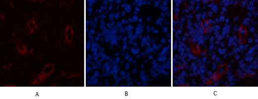
 IF (Immunofluorescence)
(Immunofluorescence analysis of human-stomach tissue. 1,AR Polyclonal Antibody(red) was diluted at 1:200(4 degree C,overnight). 2, Cy3 labled Secondary antibody was diluted at 1:300(room temperature, 50min).3, Picture B: DAPI(blue) 10min. Picture A:Target. Picture B: DAPI. Picture C: merge of A+B)
IF (Immunofluorescence)
(Immunofluorescence analysis of human-stomach tissue. 1,AR Polyclonal Antibody(red) was diluted at 1:200(4 degree C,overnight). 2, Cy3 labled Secondary antibody was diluted at 1:300(room temperature, 50min).3, Picture B: DAPI(blue) 10min. Picture A:Target. Picture B: DAPI. Picture C: merge of A+B)
Rabbit AR Polyclonal Antibody | anti-AR antibody
AR Rabbit Polyclonal Antibody
WB: 1:500-2000
ELISA: 1:10000-20000
IHC: 1:50-300
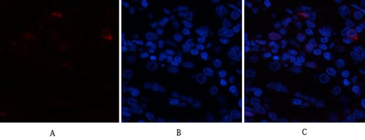
IF (Immunofluorescence)
(Immunofluorescence analysis of human-stomach tissue. 1,AR Polyclonal Antibody(red) was diluted at 1:200(4 degree C,overnight). 2, Cy3 labled Secondary antibody was diluted at 1:300(room temperature, 50min).3, Picture B: DAPI(blue) 10min. Picture A:Target. Picture B: DAPI. Picture C: merge of A+B)
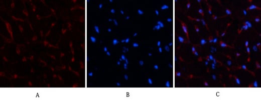
IF (Immunofluorescence)
(Immunofluorescence analysis of rat-heart tissue. 1,AR Polyclonal Antibody(red) was diluted at 1:200(4 degree C,overnight). 2, Cy3 labled Secondary antibody was diluted at 1:300(room temperature, 50min).3, Picture B: DAPI(blue) 10min. Picture A:Target. Picture B: DAPI. Picture C: merge of A+B)
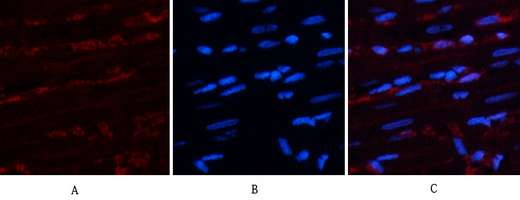
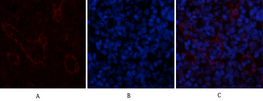
IF (Immunofluorescence)
(Immunofluorescence analysis of rat-spleen tissue. 1,AR Polyclonal Antibody(red) was diluted at 1:200(4 degree C,overnight). 2, Cy3 labled Secondary antibody was diluted at 1:300(room temperature, 50min).3, Picture B: DAPI(blue) 10min. Picture A:Target. Picture B: DAPI. Picture C: merge of A+B)
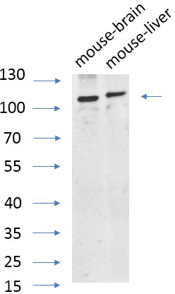
WB (Western Blot)
(Western Blot analysis of various cells using primary antibody diluted at 1:1000(4 degree C overnight). Secondary antibody:Goat Anti-rabbit IgG IRDye 800( diluted at 1:5000, 25 degree C, 1 hour). Cell lysate was extracted by Minute™ Plasma Membrane Protein Isolation and Cell Fractionation Kit(SM-005, Inventbiotech,MN,USA).)
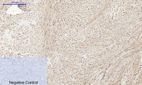
IHC (Immunohistochemistry)
(Immunohistochemical analysis of paraffin-embedded Human-uterus tissue. 1,AR Polyclonal Antibody was diluted at 1:200(4 degree C,overnight). 2, Sodium citrate pH 6.0 was used for antibody retrieval(>98 degree C,20min). 3,Secondary antibody was diluted at 1:200(room temperature, 30min). Negative control was used by secondary antibody only.)
IHC (Immunohistochemistry)
(Immunohistochemical analysis of paraffin-embedded Rat-heart tissue. 1,AR Polyclonal Antibody was diluted at 1:200(4 degree C,overnight). 2, Sodium citrate pH 6.0 was used for antibody retrieval(>98 degree C,20min). 3,Secondary antibody was diluted at 1:200(room temperature, 30min). Negative control was used by secondary antibody only.)
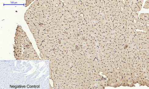
IHC (Immunohistochemistry)
(Immunohistochemical analysis of paraffin-embedded Mouse-liver tissue. 1,AR Polyclonal Antibody was diluted at 1:200(4 degree C,overnight). 2, Sodium citrate pH 6.0 was used for antibody retrieval(>98 degree C,20min). 3,Secondary antibody was diluted at 1:200(room temperature, 30min). Negative control was used by secondary antibody only.)
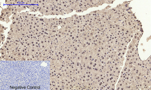
IHC (Immunohistochemistry)
(Immunohistochemical analysis of paraffin-embedded Mouse-kidney tissue. 1,AR Polyclonal Antibody was diluted at 1:200(4 degree C,overnight). 2, Sodium citrate pH 6.0 was used for antibody retrieval(>98 degree C,20min). 3,Secondary antibody was diluted at 1:200(room temperature, 30min). Negative control was used by secondary antibody only.)
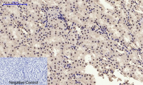
WB (Western Blot)
(Western Blot analysis of Hela cells using AR Polyclonal Antibody. Secondary antibody was diluted at 1:20000)
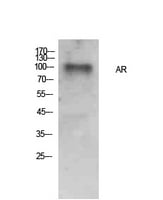
WB (Western Blot)
(Western Blot analysis of Hela cells using AR Polyclonal Antibody. Secondary antibody was diluted at 1:20000)
NCBI and Uniprot Product Information
Similar Products


Product Notes
The AR ar (Catalog #AAA30774) is an Antibody produced from Rabbit and is intended for research purposes only. The product is available for immediate purchase. The AR Rabbit Polyclonal Antibody reacts with Human, Mouse, Rat and may cross-react with other species as described in the data sheet. AAA Biotech's AR can be used in a range of immunoassay formats including, but not limited to, Immunofluorescence (IF), Immunocytochemistry (ICC), Western Blot (WB), Immunohistochemistry-Paraffin (IHC-P), ELISA (EIA). IF: 1:50-200 WB: 1:500-2000 ELISA: 1:10000-20000 IHC: 1:50-300. Researchers should empirically determine the suitability of the AR ar for an application not listed in the data sheet. Researchers commonly develop new applications and it is an integral, important part of the investigative research process. It is sometimes possible for the material contained within the vial of "AR, Polyclonal Antibody" to become dispersed throughout the inside of the vial, particularly around the seal of said vial, during shipment and storage. We always suggest centrifuging these vials to consolidate all of the liquid away from the lid and to the bottom of the vial prior to opening. Please be advised that certain products may require dry ice for shipping and that, if this is the case, an additional dry ice fee may also be required.Precautions
All products in the AAA Biotech catalog are strictly for research-use only, and are absolutely not suitable for use in any sort of medical, therapeutic, prophylactic, in-vivo, or diagnostic capacity. By purchasing a product from AAA Biotech, you are explicitly certifying that said products will be properly tested and used in line with industry standard. AAA Biotech and its authorized distribution partners reserve the right to refuse to fulfill any order if we have any indication that a purchaser may be intending to use a product outside of our accepted criteria.Disclaimer
Though we do strive to guarantee the information represented in this datasheet, AAA Biotech cannot be held responsible for any oversights or imprecisions. AAA Biotech reserves the right to adjust any aspect of this datasheet at any time and without notice. It is the responsibility of the customer to inform AAA Biotech of any product performance issues observed or experienced within 30 days of receipt of said product. To see additional details on this or any of our other policies, please see our Terms & Conditions page.Item has been added to Shopping Cart
If you are ready to order, navigate to Shopping Cart and get ready to checkout.