
 FCM (Flow Cytometry)
(Figure 7. Flow Cytometry analysis of K562 cells using anti-CASP3 antibody (AAA11633).Overlay histogram showing K562 cells stained with AAA11633 (Blue line).The cells were blocked with 10% normal goat serum. And then incubated with rabbit anti-CASP3 Antibody (AAA11633,1ug/1x10^6 cells) for 30 min at 20 degree C. DyLight ®488 conjugated goat anti-rabbit IgG (5-10ug/1x10^6 cells) was used as secondary antibody for 30 minutes at 20 degree C. Isotype control antibody (Green line) was rabbit IgG (1ug/1x106) used under the same conditions. Unlabelled sample (Red line) was also used as a control.)
FCM (Flow Cytometry)
(Figure 7. Flow Cytometry analysis of K562 cells using anti-CASP3 antibody (AAA11633).Overlay histogram showing K562 cells stained with AAA11633 (Blue line).The cells were blocked with 10% normal goat serum. And then incubated with rabbit anti-CASP3 Antibody (AAA11633,1ug/1x10^6 cells) for 30 min at 20 degree C. DyLight ®488 conjugated goat anti-rabbit IgG (5-10ug/1x10^6 cells) was used as secondary antibody for 30 minutes at 20 degree C. Isotype control antibody (Green line) was rabbit IgG (1ug/1x106) used under the same conditions. Unlabelled sample (Red line) was also used as a control.)
Rabbit Caspase-3 Polyclonal Antibody | anti-CASP3 antibody
Anti-Caspase-3 Antibody
Each vial contains 5mg BSA, 0.9mg NaCl, 0.2mg Na2HPO4, 0.05mg NaN3.
Concentration: 0.1-0.5mug/ml
Tested Species: Human, Rat
Immunohistochemistry (Paraffin-embedded Section):
Concentration: 0.5-1mug/ml
Tested Species: Human, Mouse, Rat
Antigen Retrieval: By Heat
WB: The detection limit for Caspase-3 is approximately 0.25ng/lane under reducing conditions.
Tested Species: In-house tested species with positive results.
By Heat: Boiling the paraffin sections in 10mM citrate buffer, pH6.0, for 20 mins is required for the staining of formalin/paraffin sections.
Other applications have not been tested.
Optimal dilutions should be determined by end users.
FCM (Flow Cytometry)
(Figure 7. Flow Cytometry analysis of K562 cells using anti-CASP3 antibody (AAA11633).Overlay histogram showing K562 cells stained with AAA11633 (Blue line).The cells were blocked with 10% normal goat serum. And then incubated with rabbit anti-CASP3 Antibody (AAA11633,1ug/1x10^6 cells) for 30 min at 20 degree C. DyLight ®488 conjugated goat anti-rabbit IgG (5-10ug/1x10^6 cells) was used as secondary antibody for 30 minutes at 20 degree C. Isotype control antibody (Green line) was rabbit IgG (1ug/1x106) used under the same conditions. Unlabelled sample (Red line) was also used as a control.)
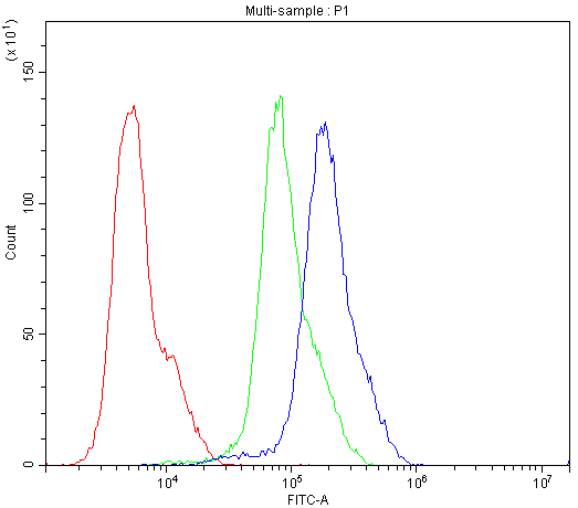
FCM (Flow Cytometry)
(Figure 6. Flow Cytometry analysis of K562 cells using anti-CASP3 antibody (AAA11633).Overlay histogram showing K562 cells stained with AAA11633 (Blue line).The cells were blocked with 10% normal goat serum. And then incubated with rabbit anti-CASP3 Antibody (AAA11633,1ug/1x10^6 cells) for 30 min at 20 degree C. DyLight ®488 conjugated goat anti-rabbit IgG (5-10ug/1x10^6 cells) was used as secondary antibody for 30 minutes at 20 degree C. Isotype control antibody (Green line) was rabbit IgG (1ug/1x106) used under the same conditions. Unlabelled sample (Red line) was also used as a control.)
WB (Western Blot)
(Figure 5. Western blot analysis of Caspase3 using anti-Caspase3 antibody (AAA11633).Electrophoresis was performed on a 5-20% SDS-PAGE gel at 70V (Stacking gel) / 90V (Resolving gel) for 2-3 hours. The sample well of each lane was loaded with 50ug of sample under reducing conditions.lane 1: rat liver tissue lysate,lane 2: rat thymus tissue lysate,lane 3: SMMC whole cell lysate.After Electrophoresis, proteins were transferred to a Nitrocellulose membrane at 150mA for 50-90 minutes. Blocked the membrane with 5% Non-fat Milk/ TBS for 1.5 hour at RT. The membrane was incubated with rabbit anti-Caspase3 antigen affinity purified polyclonal antibody at 0.5ug/mL overnight at 4 degree C, then washed with TBS-0.1%Tween 3 times with 5 minutes each and probed with a goat anti-rabbit IgG-HRP secondary antibody at a dilution of 1:10000 for 1.5 hour at RT. The signal is developed using an Enhanced Chemiluminescent detection (ECL) kit with Tanon 5200 system. A specific band was detected for Caspase3 at approximately 31KD. The expected band size for Caspase3 is at 31KD.)
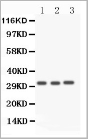
IHC (Immunohistochemistry)
(Figure 4. IHC analysis of Caspase3 using anti-Caspase3 antibody (AAA11633).Caspase3 was detected in paraffin-embedded section of human lung cancer tissue. Heat mediated antigen retrieval was performed in citrate buffer (pH6, epitope retrieval solution) for 20 mins. The tissue section was blocked with 10% goat serum. The tissue section was then incubated with 1ug/ml rabbit anti-Caspase3 Antibody (AAA11633) overnight at 4 degree C. Biotinylated goat anti-rabbit IgG was used as secondary antibody and incubated for 30 minutes at 37 degree C. The tissue section was developed using Strepavidin-Biotin-Complex (SABC) with DAB as the chromogen.)
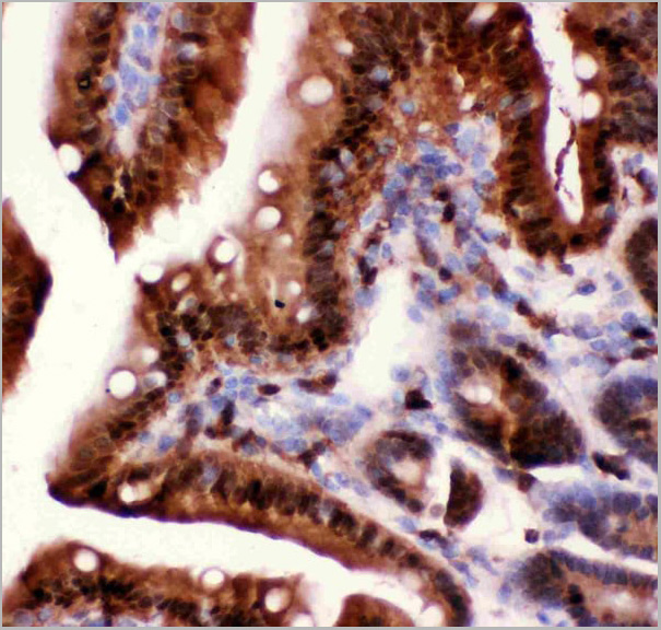
IHC (Immunohistochemistry)
(Figure 3. IHC analysis of Caspase3 using anti-Caspase3 antibody (AAA11633).Caspase3 was detected in paraffin-embedded section of rat intestine tissue. Heat mediated antigen retrieval was performed in citrate buffer (pH6, epitope retrieval solution) for 20 mins. The tissue section was blocked with 10% goat serum. The tissue section was then incubated with 1ug/ml rabbit anti-Caspase3 Antibody (AAA11633) overnight at 4 degree C. Biotinylated goat anti-rabbit IgG was used as secondary antibody and incubated for 30 minutes at 37 degree C. The tissue section was developed using Strepavidin-Biotin-Complex (SABC) with DAB as the chromogen.)
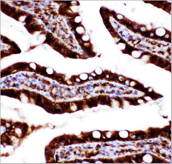
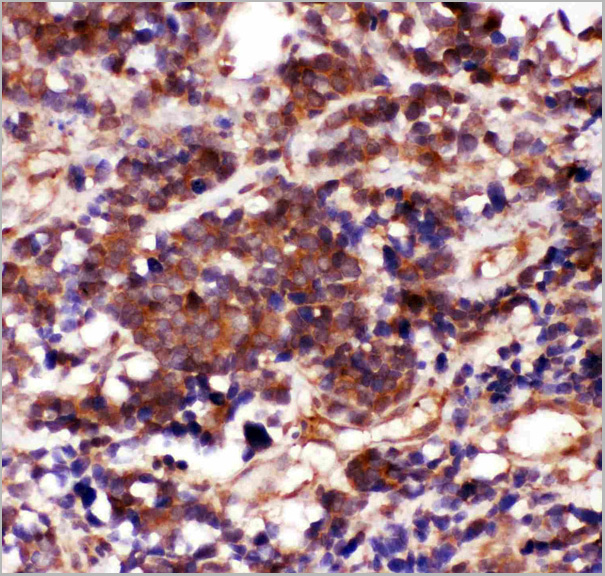
IHC (Immunohistochemistry)
(Figure 2. IHC analysis of Caspase3 using anti-Caspase3 antibody (AAA11633).Caspase3 was detected in paraffin-embedded section of mouse intestine tissue. Heat mediated antigen retrieval was performed in citrate buffer (pH6, epitope retrieval solution) for 20 mins. The tissue section was blocked with 10% goat serum. The tissue section was then incubated with 1ug/ml rabbit anti-Caspase3 Antibody (AAA11633) overnight at 4 degree C. Biotinylated goat anti-rabbit IgG was used as secondary antibody and incubated for 30 minutes at 37 degree C. The tissue section was developed using Strepavidin-Biotin-Complex (SABC) with DAB as the chromogen.)
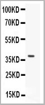
WB (Western Blot)
(Figure 1. Western blot analysis of Caspase3 using anti-Caspase3 antibody (AAA11633).Electrophoresis was performed on a 5-20% SDS-PAGE gel at 70V (Stacking gel) / 90V (Resolving gel) for 2-3 hours.lane 1: recombinant human Caspase3 protein 0.5ng.After Electrophoresis, proteins were transferred to a Nitrocellulose membrane at 150mA for 50-90 minutes. Blocked the membrane with 5% Non-fat Milk/ TBS for 1.5 hour at RT. The membrane was incubated with rabbit anti-Caspase3 antigen affinity purified polyclonal antibody at 0.5ug/mL overnight at 4 degree C, then washed with TBS-0.1%Tween 3 times with 5 minutes each and probed with a goat anti-rabbit IgG-HRP secondary antibody at a dilution of 1:10000 for 1.5 hour at RT. The signal is developed using an Enhanced Chemiluminescent detection (ECL) kit with Tanon 5200 system. A specific band was detected for Caspase3 at approximately 39KD. The expected band size for Caspase3 is at 39KD.)
Background: Caspase 3 is a caspase protein which interacts with Survivin, XIAP, CFLAR, Caspase 8, HCLS1, Deleted in Colorectal Cancer, TRAF3 and GroEL. This gene which is located on 4q35 encodes a protein that is a member of the cysteine-aspartic acid protease (caspase) family. Sequential activation of caspases plays a central role in the execution-phase of cell apoptosis. Caspases exist as inactive proenzymes that undergo proteolytic processing at conserved aspartic residues to produce two subunits, large and small, that dimerize to form the active enzyme. It is the predominant caspase involved in the cleavage of amyloid-beta 4A precursor protein, which is associated with neuronal death in Alzheimer's disease. And the caspase-3 activation in heart failure sequentially cleaves SRF and generates a truncated SRF that appears to function as a dominant-negative transcription factor. Additionally, the caspase-3 influence on bone mineral density should be considered in any in vivo application of caspase-3 inhibitors to the treatment of human disease. In erythroid precursors undergoing terminal differentiation, Hsp70 prevents active CASP3 from cleaving GATA1 and inducing apoptosis.
NCBI and Uniprot Product Information
Similar Products


Product Notes
The CASP3 casp3 (Catalog #AAA11633) is an Antibody produced from Rabbit and is intended for research purposes only. The product is available for immediate purchase. The Anti-Caspase-3 Antibody reacts with Human, Mouse, Rat. No cross reactivity with other proteins. and may cross-react with other species as described in the data sheet. AAA Biotech's Caspase-3 can be used in a range of immunoassay formats including, but not limited to, Western Blot (WB), Immunohistochemistry (IHC-P). Western Blot: Concentration: 0.1-0.5mug/ml Tested Species: Human, Rat Immunohistochemistry (Paraffin-embedded Section): Concentration: 0.5-1mug/ml Tested Species: Human, Mouse, Rat Antigen Retrieval: By Heat WB: The detection limit for Caspase-3 is approximately 0.25ng/lane under reducing conditions. Tested Species: In-house tested species with positive results. By Heat: Boiling the paraffin sections in 10mM citrate buffer, pH6.0, for 20 mins is required for the staining of formalin/paraffin sections. Other applications have not been tested. Optimal dilutions should be determined by end users. Researchers should empirically determine the suitability of the CASP3 casp3 for an application not listed in the data sheet. Researchers commonly develop new applications and it is an integral, important part of the investigative research process. It is sometimes possible for the material contained within the vial of "Caspase-3, Polyclonal Antibody" to become dispersed throughout the inside of the vial, particularly around the seal of said vial, during shipment and storage. We always suggest centrifuging these vials to consolidate all of the liquid away from the lid and to the bottom of the vial prior to opening. Please be advised that certain products may require dry ice for shipping and that, if this is the case, an additional dry ice fee may also be required.Precautions
All products in the AAA Biotech catalog are strictly for research-use only, and are absolutely not suitable for use in any sort of medical, therapeutic, prophylactic, in-vivo, or diagnostic capacity. By purchasing a product from AAA Biotech, you are explicitly certifying that said products will be properly tested and used in line with industry standard. AAA Biotech and its authorized distribution partners reserve the right to refuse to fulfill any order if we have any indication that a purchaser may be intending to use a product outside of our accepted criteria.Disclaimer
Though we do strive to guarantee the information represented in this datasheet, AAA Biotech cannot be held responsible for any oversights or imprecisions. AAA Biotech reserves the right to adjust any aspect of this datasheet at any time and without notice. It is the responsibility of the customer to inform AAA Biotech of any product performance issues observed or experienced within 30 days of receipt of said product. To see additional details on this or any of our other policies, please see our Terms & Conditions page.Item has been added to Shopping Cart
If you are ready to order, navigate to Shopping Cart and get ready to checkout.