

Rabbit anti-Human Caspase-9/CASP9 Polyclonal Antibody | anti-CASP9 antibody
Anti-Caspase-9/CASP9 Antibody
IHC-P: 2-5ug/ml|Human|
FC/FACS/FCM: 1-3ug/1x106 cells|Human|
Direct ELISA: 0.1-0.5ug/ml|Human|
FCM (Flow Cytometry)
(Figure 6. Flow Cytometry analysis of Hela cells using anti-Caspase-9/CASP9 antibody (AAA19215).Overlay histogram showing Hela cells stained with AAA19215 (Blue line). The cells were blocked with 10% normal goat serum. And then incubated with rabbit anti-Caspase-9/CASP9 Antibody (AAA19215, 1 μg/1x106 cells) for 30 min at 20 degree C. DyLight®488 conjugated goat anti-rabbit IgG (5-10 μg/1x106 cells) was used as secondary antibody for 30 minutes at 20 degree C. Isotype control antibody (Green line) was rabbit IgG (1 μg/1x106) used under the same conditions. Unlabelled sample (Red line) was also used as a control.)
IHC (Immunohistochemistry)
(Figure 5. IHC analysis of Caspase-9/CASP9 using anti-Caspase-9/CASP9 antibody (AAA19215).Caspase-9/CASP9 was detected in a paraffin-embedded section of human lung cancer tissue. Heat mediated antigen retrieval was performed in EDTA buffer (pH 8. 0, epitope retrieval solution). The tissue section was blocked with 10% goat serum. The tissue section was then incubated with 2 μg/ml rabbit anti-Caspase-9/CASP9 Antibody (AAA19215) overnight at 4 degree C. Biotinylated goat anti-rabbit IgG was used as secondary antibody and incubated for 30 minutes at 37 degree C. The tissue section was developed using Strepavidin-Biotin-Complex (SABC) (Catalog # with DAB as the chromogen.)
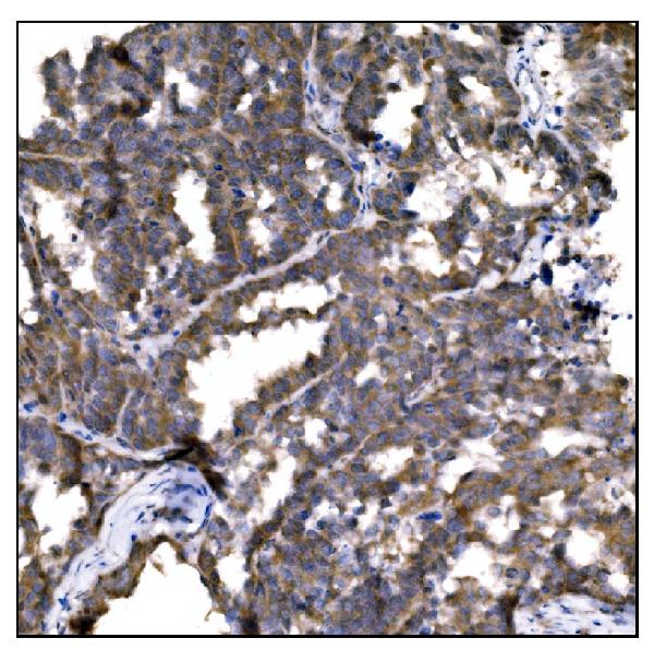
IHC (Immunohistochemistry)
(Figure 4. IHC analysis of Caspase-9/CASP9 using anti-Caspase-9/CASP9 antibody (AAA19215).Caspase-9/CASP9 was detected in a paraffin-embedded section of human gallbladder adenocarcinoma tissue. Heat mediated antigen retrieval was performed in EDTA buffer (pH 8. 0, epitope retrieval solution). The tissue section was blocked with 10% goat serum. The tissue section was then incubated with 2 μg/ml rabbit anti-Caspase-9/CASP9 Antibody (AAA19215) overnight at 4 degree C. Biotinylated goat anti-rabbit IgG was used as secondary antibody and incubated for 30 minutes at 37 degree C. The tissue section was developed using Strepavidin-Biotin-Complex (SABC) (Catalog # with DAB as the chromogen.)
IHC (Immunohistochemistry)
(Figure 3. IHC analysis of Caspase-9/CASP9 using anti-Caspase-9/CASP9 antibody (AAA19215).Caspase-9/CASP9 was detected in a paraffin-embedded section of human ovarian cancer tissue. Heat mediated antigen retrieval was performed in EDTA buffer (pH 8. 0, epitope retrieval solution). The tissue section was blocked with 10% goat serum. The tissue section was then incubated with 2 μg/ml rabbit anti-Caspase-9/CASP9 Antibody (AAA19215) overnight at 4 degree C. Biotinylated goat anti-rabbit IgG was used as secondary antibody and incubated for 30 minutes at 37 degree C. The tissue section was developed using Strepavidin-Biotin-Complex (SABC) (Catalog # with DAB as the chromogen.)
IHC (Immunohistochemistry)
(Figure 2. IHC analysis of Caspase-9/CASP9 using anti-Caspase-9/CASP9 antibody (AAA19215).Caspase-9/CASP9 was detected in a paraffin-embedded section of human breast cancer tissue. Heat mediated antigen retrieval was performed in EDTA buffer (pH 8. 0, epitope retrieval solution). The tissue section was blocked with 10% goat serum. The tissue section was then incubated with 2 μg/ml rabbit anti-Caspase-9/CASP9 Antibody (AAA19215) overnight at 4 degree C. Biotinylated goat anti-rabbit IgG was used as secondary antibody and incubated for 30 minutes at 37 degree C. The tissue section was developed using Strepavidin-Biotin-Complex (SABC) (Catalog # with DAB as the chromogen.)
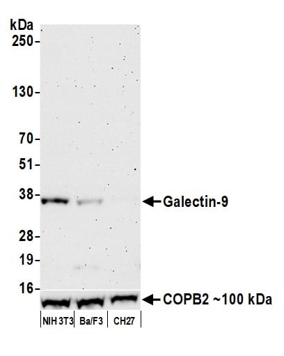
WB (Western Blot)
(Figure 1. Western blot analysis of Caspase-9/CASP9 using anti-Caspase-9/CASP9 antibody (AAA19215).Electrophoresis was performed on a 5-20% SDS-PAGE gel at 70V (Stacking gel) / 90V (Resolving gel) for 2-3 hours. The sample well of each lane was loaded with 30 ug of sample under reducing conditions.Lane 1: human Jurkat whole cell lysates.After electrophoresis, proteins were transferred to a nitrocellulose membrane at 150 mA for 50-90 minutes. Blocked the membrane with 5% non-fat milk/TBS for 1. 5 hour at RT. The membrane was incubated with rabbit anti-Caspase-9/CASP9 antigen affinity purified polyclonal antibody (Catalog # AAA19215) at 0. 5 μg/mL overnight at 4 degree C, then washed with TBS-0. 1%Tween 3 times with 5 minutes each and probed with a goat anti-rabbit IgG-HRP secondary antibody at a dilution of 1:5000 for 1. 5 hour at RT. The signal is developed using an Enhanced Chemiluminescent detection (ECL) kit (Catalog # with Tanon 5200 system. A specific band was detected for Caspase-9/CASP9 at approximately 46 kDa. The expected band size for Caspase-9/CASP9 is at 46 kDa.)
2. Xu D, et al. Apoptotic block in colon cancer cells may be rectified by lentivirus mediated overexpression of caspase-9. Acta Gastroenterol Belg, 2013 Dec.